. 2017 Nov 1;56(11):1970-1981.
doi: 10.1093/rheumatology/kex280.

Scleroderma keratinocytes promote fibroblast activation independent of transforming growth factor beta

Affiliations

Scleroderma keratinocytes promote fibroblast activation independent of transforming growth factor beta

Sara S McCoy et al. Rheumatology (Oxford). .

Abstract

Objectives: SSc is a devastating disease that results in fibrosis of the skin and other organs. Fibroblasts are a key driver of the fibrotic process through deposition of extracellular matrix. The mechanisms by which fibroblasts are induced to become pro-fibrotic remain unclear. Thus, we examined the ability of SSc keratinocytes to promote fibroblast activation and the source of this effect.

Methods: Keratinocytes were isolated from skin biopsies of 9 lcSSc, 10 dcSSc and 13 control patients. Conditioned media was saved from the cultures. Normal fresh primary fibroblasts were exposed to healthy control and SSc keratinocyte conditioned media in the presence or absence of neutralizing antibodies for TGF-β. Gene expression was assessed by microarrays and real-time PCR. Immunocytochemistry was performed for α-smooth muscle actin (α-SMA), collagen type 1 (COL1A1) and CCL5 expression.

Results: SSc keratinocyte conditioned media promoted fibroblast activation, characterized by increased α-SMA and COL1A1 mRNA and protein expression. This effect was independent of TGF-β. Microarray analysis identified upregulation of nuclear factor κB (NF-κB) and downregulation of peroxisome proliferator-activated receptor γ (PPAR-γ) pathways in both SSc subtypes. Scleroderma keratinocytes exhibited increased expression of NF-κB-regulated cytokines and chemokines and lesional skin staining confirmed upregulation of CCL5 in basal keratinocytes.

Conclusion: Scleroderma keratinocytes promote the activation of fibroblasts in a TGF-β-independent manner and demonstrate an imbalance in NF-κB1 and PPAR-γ expression leading to increased cytokine and CCL5 production. Further study of keratinocyte mediators of fibrosis, including CCL5, may provide novel targets for skin fibrosis therapy.

Keywords: CCL5; NF-κB; PPAR-γ; TGF-β; fibroblast; keratinocyte; scleroderma.


Figures

F<sc>ig</sc>. 1
Fig. 1
Fibroblast to myofibroblast conversion is increased in normal fibroblasts stimulated with SSc keratinocyte conditioned media (A) Fresh fibroblasts were cultured with 50% keratinocyte conditioned media and 50% RPMI with 1% FBS for 24 h or 72 h and the cells were either harvested for RNA or fixed for immunocytochemistry. (B) Real-time PCR of fibroblasts stimulated with SSc or normal keratinocyte conditioned media for 24 h. Five lcSSc, five dcSSc and five normal samples were studied in triplicate on two separate fresh fibroblast cultures. Each dot represents the average of the triplicate. Error bars represent s.e.m.. α-SMA expression reported as a fold change compared with normal with significance (P < 0.05) calculated by comparison of ΔCTs via unpaired Student’s t-test. (C) Immunocytochemistry of fibroblasts stimulated with SSc or normal keratinocyte conditioned media for 72 h. Three lcSSc, three dcSSc and five normal samples were studied in triplicate. Fibroblasts were fixed and stained with anti-α-SMA antibody and a secondary FITC-labelled antibody. Microscopy was performed and at least 20 images were taken per slide. α-SMA fluorescence was quantified with MetaMorph software as described in the Methods section. (D) Representative microscopy demonstrating α-SMA staining after exposure to SSc keratinocyte conditioned media (left) or normal keratinocyte conditioned media (right). Size bar = 50 mm. *P < 0.05, **P < 0.01, ***P < 0.001, ****P < 0.0001.
F<sc>ig</sc>. 2
Fig. 2
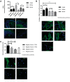
Keratinocyte-induced myofibroblast transformation is independent of TGF-β (A) Immunocytochemistry of TGF-β expression of three lcSSc, four dcSSc and four normal skin samples in triplicate demonstrate that there was no difference between dermis and epidermis in limited and normal samples. Greater TGF-β expression was seen in dcSSc dermis compared with epidermis. Representative biopsies of limited (upper left), diffuse (upper right), normal slides (lower left) and negative isotype control (lower right) stained with TGF-β. (B) Fibroblasts were incubated with normal or SSc conditioned media for 24 h with and without a neutralizing TGF-β antibody (two lcSSc, two dcSSc and three normal, in triplicate). At least 20 images were taken per slide using fluorescent microscopy. α-SMA expression was quantified with MetaMorph software as described in the Methods section. Representative images of fibroblasts stained for α-SMA are shown: lcSSc fibroblast α-SMA expression (upper left), lcSSc fibroblast + TGF-β neutralizing antibody (upper right), dcSSc fibroblast α-SMA expression (middle left), dcSSc fibroblast + TGF-β neutralizing antibody (middle right) and healthy control (bottom). (C) To validate the efficacy of our neutralizing antibody, fibroblasts were incubated with 10 ng/ml rhTGF-β in the presence or absence of TGF-β neutralizing antibody and α-SMA expression was evaluated as in (B). Representative images of normal fibroblasts treated with rhTGF-β and TGF-β neutralizing antibody (Ab) stained for α-SMA incubated in normal culture media are shown: stimulated with rhTGF-β (upper left), treated with rhTGF-β and TGF-β neutralizing Ab (upper right), negative control (lower left) and negative control treated with TGF-β neutralizing Ab (lower right). Size bar = 50 mm. *P < 0.05, **P < 0.01, ***P < 0.001, ****P < 0.0001.
F<sc>ig</sc>. 3
Fig. 3
Transcriptional networks from dcSSc and lcSSc identify overlapping and unique pathways There were 123 genes regulated in the same direction in the 135 genes commonly regulated in both diseases. The pictures display the 100 best-connected genes (most relevant genes/interactions) co-cited in PubMed abstracts in the same sentence linked to a function word (e.g. gene A activates gene B). Orange represents the genes that are upregulated and green represents the genes that are downregulated in disease compared with normal.
F<sc>ig</sc>. 4
Fig. 4
NF-κB-regulated cytokines are increased in SSc keratinocytes (A) Real-time PCR was completed on RNA from nine SSc and five healthy control keratinocyte cultures for the indicated genes. Data are expressed as fold change (FC) in the SSc sample vs the average of healthy controls. Graphs represent the mean (s.e.m.). Significance was analysed via Student’s unpaired t-test. *P < 0.05, **P < 0.01. (B) FCs of indicated genes were plotted vs the FC of NF-κB and linear regression was completed (indicated lines). (D) FCs of indicated genes were plotted vs MRSS for each patient. Linear regression was performed. *P < 0.05, **P < 0.01. NS, not significant. (C) Representative CCL5 immunohistochemistry from three healthy controls and seven SSc patients. Scale bar = 100 μm.

References

    1. Lawrence RC, Helmick CG, Arnett FC. et al. Estimates of the prevalence of arthritis and selected musculoskeletal disorders in the United States. Arthritis Rheum 1998;41:778–99. - PubMed
    1. Chifflot H, Fautrel B, Sordet C, Chatelus E, Sibilia J.. Incidence and prevalence of systemic sclerosis: a systematic literature review. Semin Arthritis Rheum 2008;37:223–35. - PubMed
    1. Al-Dhaher FF, Pope JE, Ouimet JM.. Determinants of morbidity and mortality of systemic sclerosis in Canada. Semin Arthritis Rheum 2010;39:269–77. - PubMed
    1. Shand L, Lunt M, Nihtyanova S. et al. Relationship between change in skin score and disease outcome in diffuse cutaneous systemic sclerosis: application of a latent linear trajectory model. Arthritis Rheum 2007;56:2422–31. - PubMed
    1. Wynn TA. Common and unique mechanisms regulate fibrosis in various fibroproliferative diseases. J Clin Invest 2007;117:524–9. - PMC - PubMed

MeSH terms