. 2017 Dec 5;216(10):1308-1317.
doi: 10.1093/infdis/jix474.

A Link Between a Common Mutation in CFTR and Impaired Innate and Adaptive Viral Defense

Affiliations

A Link Between a Common Mutation in CFTR and Impaired Innate and Adaptive Viral Defense

Emma Svedin et al. J Infect Dis. .

Abstract

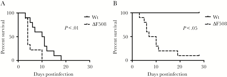
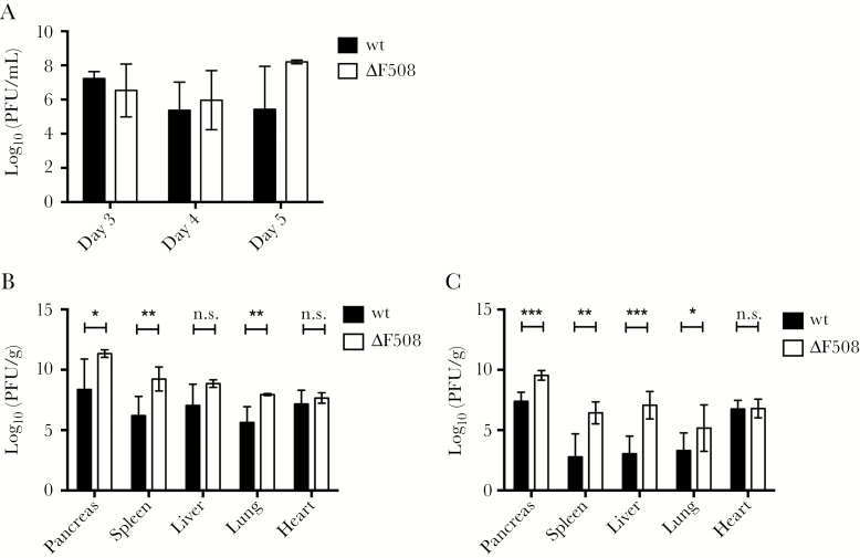
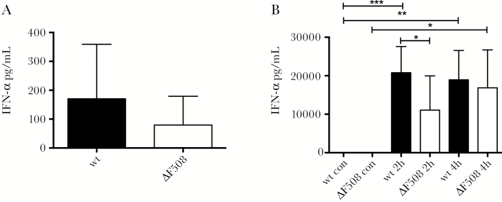
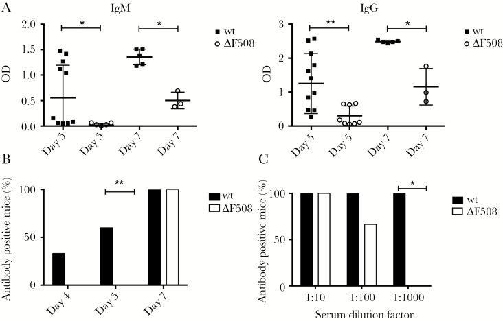
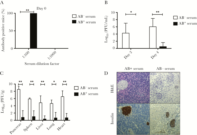
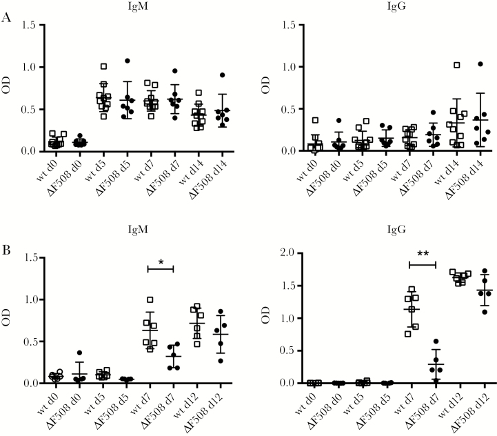
Acute respiratory virus infections predispose the cystic fibrosis (CF) lung to chronic bacterial colonization, which contributes to high mortality. For reasons unknown, respiratory virus infections have a prolonged duration in CF. Here, we demonstrate that mice carrying the most frequent cystic fibrosis transmembrane conductance regulator (CFTR) mutation in humans, ΔF508, show increased morbidity and mortality following infection with a common human enterovirus. ΔF508 mice demonstrated impaired viral clearance, a slower type I interferon response and delayed production of virus-neutralizing antibodies. While the ΔF508 mice had a normal immune cell repertoire, unchanged serum immunoglobulin concentrations and an intact immune response to a T-cell-independent antigen, their response to a T-cell-dependent antigen was significantly delayed. Our studies reveal a novel function for CFTR in antiviral immunity and demonstrate that the ΔF508 mutation in cftr is coupled to an impaired adaptive immune response. This important insight could open up new approaches for patient care and treatment.

Keywords: Coxsackievirus; antibody response; antiviral defense; cystic fibrosis; immunity.


Figures

Figure 1.
Figure 1.
ΔF508 mice demonstrate an increased mortality rate after infection with Coxsackievirus. ΔF508 mice (dotted lines) and wild-type (wt) littermate controls (solid lines) were infected with Coxsackievirus B3 (CVB3) and monitored for 28 days. A, Wt (n = 10) and ΔF508 (n = 9) mice infected with 104 plaque-forming units (PFU) CVB3/mouse. B, wt (n = 3) and ΔF508 (n = 10) infected with 102 PFU CVB3/mouse. Log-rank (Mantel–Cox) test.
Figure 2.
Figure 2.
Sustained high level viral replication in ΔF508 mice after infection with Coxsackievirus. Wild-type (wt; black bars) and littermate ΔF508 mice (white bars) were infected with Coxsackievirus B3 (102 plaque-forming units [PFU]/mouse). A, Blood was harvested on days 3 (n = 3–5 animals per genotype), 4 (n = 9–12 animals per genotype), and 5 (n = 3–4 animals per genotype) postinfection for viremia measurements using plaque assay. B, Titers of replicating virus particles in organs harvested on day 5 postinfection were measured by plaque assay (n = 4–7 animals per genotype). C, Titers of replicating virus particles in organs harvested on day 7 postinfection were measured by plaque assay (n = 6–8 animals per genotype). A–C, Plaque assay results are presented as log10(PFU/g wet tissue or mL blood) and represent means ± standard deviation *P < .05, **P < .01, ***P < .001 and n.s, nonsignificant, Mann–Whitney test.
Figure 3.
Figure 3.
ΔF508 mice show a delayed interferon-alpha (IFN-α) response after stimulation with poly(I:C). A, Wild-type (wt; n = 6) and ΔF508 (n = 8) mice were infected with Coxsackievirus B3 (104 plaque-forming units [PFU]/mouse) and serum was drawn on day 2 postinfection. B, Wt (n = 8–14 per time point) and ΔF508 (n = 6–8 per time point) mice were stimulated with poly(I:C) (100 μg/mouse, intraperitoneal) and serum was drawn at 2 hours and 4 hours after stimulation. IFN-α levels in serum from wt mice (black bars) and ΔF508 mice (white bars) from infected mice (A) or mice stimulated with Poly(I:C) (B) were measured using enzyme-linked immunosorbent assay. Data is presented as means ± SD *P < .05, **P < .01, and ***P < .001, two-way analysis of variance with Holm–Sidak’s correction.
Figure 4.
Figure 4.
Mice carrying the ΔF508 mutation have a delayed antibody response to Coxsackievirus B3 (CVB3). A, Wt and ΔF508 mice were infected with 102 PFU CVB3/mouse. Virus-specific IgM and IgG antibodies were measured in serum samples harvested from infected animals on day 5 (n = 8–12 per genotype) and 7 (n = 3–5 per genotype) postinfection using a CVB3 virus like particles-specific enzyme-linked immunosorbent assay. B, C, Serum drawn on days 4 (n = 4–6 animals per genotype), 5 (n = 7–15 animals per genotype), and 7 (n = 3–8 animals per genotype) postinfection were evaluated for the presence of neutralizing antibodies, as described in Supplementary Material Methods. B, The percentage of antibody-positive wt (black bars) and ΔF508 (white bars) mice at the indicated days postinfection is shown. C, The percentage of antibody-positive wt mice (black bars, n = 8) and ΔF508 mice (white bars, n = 3) at indicated serum dilutions on day 7 postinfection. Data in A is presented as mean ± SD *P < .05, **P < .01. Mann–Whitney test (A) and Fisher exact test (B, C). Abbreviation: OD, optical density.
Figure 5.
Figure 5.
Passive immunization protects ΔF508 mice from Coxsackievirus B3 (CVB3) infection and tissue damage. Antibody-positive (immune) and negative (nonimmune) sera was prepared from previously infected wt mice, as described in the Supplementary Material Methods section. Passive immunizations were performed 24 hours prior to CVB3 challenge by the administration of 400 µl (intraperitoneal) immune or nonimmune sera to the ΔF508 mice. A, The neutralizing capacity of sera drawn at 24 hours after passive immunization was measured at 2 separate dilutions, 1:100 and 1:1000, as described in the Supplementary Material Methods. The percentage of animals that had sera with virus-neutralizing capacity is shown as the percentage of all animals in each respective group. Black and white bars indicate animals receiving immune (AB+; n = 5) and nonimmune (AB; n = 5) sera, respectively. B, C, Passively immunized ΔF508 mice were infected with CVB3 (102 plaque-forming units [PFU]/mouse). Blood was drawn on days 3 and 4 postinfection for viremia measurements (B). C, On day 7 postinfection, the mice were sacrificed and the indicated organs were retrieved for histological analysis and measurements of replicating virus particles. Note that in the group receiving nonimmune serum, one mouse had to be sacrificed on day 6 postinfection due to CVB3-induced illness. The results are presented as log10(PFU/g wet tissue or mL blood) and represents means ± SD. D, Representative images of pancreas sections from CVB3-infected ΔF508 mice receiving antibody-negative (AB; n = 5) or antibody-positive (AB+; n = 5) sera, respectively. Upper panels, H&E staining. Lower panels, immunohistochemistry using primary antibody detecting insulin positive cells within the pancreas parenchyma. Original magnification × 10. *P < .05, **P < .01. Fisher exact test (A) and Mann–Whitney test (B, C).
Figure 6.
Figure 6.
ΔF508 mice have an intact T-cell–independent antibody response but show a delayed response to a T-cell–dependent antigen. A, Wild-type (wt; n = 10; black circles) and ΔF508 (n = 7; open squares) mice were injected with the T-cell–independent antigen 2,4,6-trinitrophenyl (TNP)-Ficoll (10 µg/mouse, intraperitoneal [IP]). B, Wt (n = 6; black circles) and ΔF508 (n = 5; open squares) mice were challenged with the T-cell–dependent antigen recombinant Semliki Forest virus expressing beta-galactosidase (rSFV-βGal; 1 × 10⁵ Infectious units [IU]/mouse, intravenous [IV]). Serum was drawn at the indicated time points (days 0, 5, 7, and 12 postinjection) and levels of nitrophenyl (NP)-specific antibodies in serum (A) and βGal (B) specific IgM and IgG were measured using enzyme-linked immunosorbent assay. Open squares and black circles represents wt and ΔF508 mice, respectively. Data are presented as mean ± standard deviation *P < .05, **P < .01. Mann−Whitney test. Abbreviation: OD, optical density.

References

    1. Elborn JS. Cystic fibrosis. Lancet 2016; 388:2519–31. - PubMed
    1. Billard L, Le Berre R, Pilorge L, Payan C, Hery-Arnaud G, Vallet S. Viruses in cystic fibrosis patients’ airways. Crit Rev Microbiol 2017; 43:1–19. - PubMed
    1. Flight W, Jones A. The diagnosis and management of respiratory viral infections in cystic fibrosis. Expert Rev Respir Med 2017; 11:221–7. - PubMed
    1. Dijkema JS, van Ewijk BE, Wilbrink B, Wolfs TF, Kimpen JL, van der Ent CK. Frequency and duration of rhinovirus infections in children with cystic fibrosis and healthy controls: a longitudinal cohort study. Pediatr Infect Dis J 2016; 35:379–83. - PubMed
    1. van Ewijk BE, van der Zalm MM, Wolfs TF et al. . Prevalence and impact of respiratory viral infections in young children with cystic fibrosis: prospective cohort study. Pediatrics 2008; 122:1171–6. - PubMed

Publication types

MeSH terms