. 2017 Jul 12;8(38):63949-63962.
doi: 10.18632/oncotarget.19213. eCollection 2017 Sep 8.

Isothiocyanates suppress the invasion and metastasis of tumors by targeting FAK/MMP-9 activity

Affiliations

Isothiocyanates suppress the invasion and metastasis of tumors by targeting FAK/MMP-9 activity

Yun-Jeong Jeong et al. Oncotarget. .

Abstract

Isothiocyanates, which are present as glucosinolate precursors in cruciferous vegetables, have strong activity against various cancers. Here, we compared the anti-metastatic effects of isothiocyanates (benzyl isothiocyanate (BITC), phenethyl isothiocyanate (PEITC), and sulforaphane (SFN)) by examining how they regulate MMP-9 expression. Isothiocyanates, particularly PEITC, suppressed 12-O-tetradecanoylphorbol-13-acetate (TPA)-induced MMP-9 activity and invasion in various cancer cell lines. By contrast, N-methyl phenethylamine, a PEITC analog without an isothiocyanate functional group, had no effect. A reporter gene assay demonstrated that BITC, PEITC, and SFN suppressed TAP-induced MMP-9 expression by inhibiting AP-1 and NF-κB in U20S osteosarcoma cells. All three compounds reduced phosphorylation of FAK, ERK1/2, and Akt. In addition, MMP-9 expression was downregulated by inhibiting FAK, ERK1/2, and Akt. Isothiocyanates-mediated inhibition of FAK phosphorylation suppressed phosphorylation of ERK1/2 and Akt in U2OS and A549 cells, along with the translocation of p65 and c-Fos, suggesting that isothiocyanates inhibit MMP-9 expression and cell invasion by blocking phosphorylation of FAK. Furthermore, isothiocyanates, abolished MMP-9 expression and tumor metastasis in vivo with the following efficacy: PEITC>BITC>SFN. Thus, isothiocyanates act as anti-metastatic compounds that suppress MMP-9 activity/expression by inhibiting NF-κB and AP-1 via suppression of the FAK/ERK and FAK/Akt signaling pathways.

Keywords: FAK; MMP-9; cancer invasion; isothiocyanates; metastasis.


Conflict of interest statement

CONFLICTS OF INTEREST The authors have declared that there are no conflicts of interest.

Figures

Figure 1
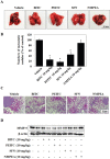
Figure 1. PEITC inhibits MMP-9 activity and tumor invasion of various cancer cells
(A) Cells in serum-free medium were treated with PEITC followed by TPA and incubated for 24 h. MMP-9 activity was analyzed by a zymography assay. (B and C) Cells were loaded into the upper chamber of Matrigel-coated Transwells and treated with PEITC and cultured for 24 h. (D and E) U2OS cells were treated with TPA plus isothiocyanates for 24 h. MMP-9 activity and expression were analyzed in a zymography assay and by western blotting, respectively. β-actin was used as a control. (F) U2OS cells were cultured on a dish, scratched, and then treated with isothiocyanates followed by TPA for 24 h. Migrating cells were photographed by phase contrast microscopy. (G) C57BL/6N mice (n = 4) were injected subcutaneously with a mixture of Matrigel (500 μL) and U2OS cells (3 × 10⁶ cells), along with the indicated drugs. Mice were euthanized 7 days after implantation, and the Matrigel plugs were removed and photographed. The amount of hemoglobin in the Matrigel plugs was quantified using Drabkin's reagent. Data represent the mean ± S.E. of three independent experiments. *p<0.05 vs. TPA.
Figure 2
Figure 2. Isothiocyanates inhibit the activity of AP-1 and NF-κB in the MMP-9 promoter in U2OS cells
(A) U2OS cells were treated with TPA and/or isothiocyanates for 24 h. Expression of mRNA encoding MMP-9 and TIMP-1 was then analyzed by RT-PCR. (B) U2OS cells were treated with TPA and isothiocyanates for 24 h, and the expression of p65, c-Fos, and c-Jun, and Lamin B (internal control) was analyzed by western blotting following nuclear extraction. (C–E) U2OS cells were transfected with the pGL2-MMP-9-WT, pGL2-MMP-9-mAP-1, and pGL2-MMP-9-mNF-κB reporter plasmids and then treated with TPA and isothiocyanates for 24 h. Relative luciferase activity in the cell extract was then measured.
Figure 3
Figure 3. Isothiocyanates inhibit phosphorylation of ERK, Akt, and FAK in U2OS cells
(A and C) U2OS cells were incubated with isothiocyanates and TPA for 24 h, and expression of the indicated proteins was determined by western blotting. (B) U2OS cells were treated with PD98059 or wortmannin for 24 h. Expression of MMP-9 and total and phosphorylated forms of ERK1/2 and Akt were measured by western blotting. β-actin is shown as a control. (D) Immunofluorescence microscopy analysis of changes in FAK phosphorylation after isothiocyanate treatment. U2OS cells were co-stained with an anti-p-FAK antibody and Hoechst 33342 and observed under a fluorescence microscope. Data are representative of three independent experiments.
Figure 4
Figure 4. Isothiocyanates inhibit MMP-9 activity/expression and tumor invasion by suppressing phosphorylation of FAK in U2OS cells
(A and C) U2OS cells were incubated with isothiocyanates and TPA for 24 h, and expression of the indicated proteins was determined by western blotting. (B) U2OS cells were plated onto a dish, scratched with a yellow pipette tip, and treated with PF573228 and TPA for 24 h. Migrating cells were photographed by phase contrast microscopy. (D–F) U2OS cells were transfected with the pGL2-MMP-9-WT, pGL2-MMP-9-mAP-1, and pGL2-MMP-9-mNF-κB reporter plasmids. The cells were then cultured with TPA and PF573228. Relative luciferase activity in the cell extract was then measured. (G) U2OS cells were transfected with negative control siRNA, siFAK1, or siFAK2, and then treated with TPA. Protein expression was determined by western blotting. (H) U2OS cells were transfected with the pGL2-MMP-9-WT reporter plasmid and siFAK2, and then treated with TPA for 24 h. Relative luciferase activity in the cell extract was then determined. (I) Cells were scratched, transfected with siFAK2, and then treated with TPA for 24 h. Migrating cells were photographed by phase contrast microscopy. Data represent the mean ± S.E. of three independent experiments. *p < 0.05 vs. control and #p < 0.05 vs. TPA.
Figure 5
Figure 5. Isothiocyanates inhibit MMP-9 activity and tumor invasion by suppressing phosphorylation of FAK in A549 cells
(A, B, and D) A549 cells were treated with TPA and isothiocyanates for 24 h. MMP-9 activity and expression were then analyzed in a zymography assay and by western blotting, respectively. β-actin is shown as a control. (C) A549 cells were plated onto a dish, scratched, and then treated with isothiocyanates followed by TPA for 24 h. Migrating cells were photographed by phase contrast microscopy. (E) A549 cells were treated with TPA, isothiocyanates, or PF573228 for 24 h, and expression of p65, c-Fos, c-Jun, and Lamin B (internal control) was analyzed by western blotting following nuclear extraction. Data represent the mean ± S.E. of three independent experiments. *p < 0.05 vs. TPA.
Figure 6
Figure 6. Isothiocyanates inhibit tumor growth in a phoenix A-transfected A549 cell xenograft mouse model
(A and B) Mice were inoculated subcutaneously into the right flank with 1 × 10⁷ phoenix A-transfected A549 cells. After the formation of palpable tumors (∼5 mm by Day 14), the mice were randomized into five groups (n = 4/group) and injected intraperitoneally (or not) with 5 mg/kg/100 μL BITC, PEITC, or SFN. Injections were administered daily. Mice were sacrificed on Day 35, and tumor size was measured. Bioluminescence imaging of tumor xenografts was performed following intraperitoneal injection of D-luciferin into mice previously injected with phoenix A-transfected A549 cells. Tumors were imaged using an in vivo imaging system. (C) Inset images show the sizes of representative tumors. (D) Average tumor weight in vehicle- and isothiocyanate-treated mice. Data represent the mean ± S.E. *p < 0.05 vs. vehicle. (E) Tumors were fixed, sectioned, and stained with hematoxylin and eosin (H&E) to examine tumor cell morphology. An anti-Ki67 antibody was used to assess tumor cell proliferation.
Figure 7
Figure 7. Isothiocyanates inhibit tumor metastasis in an A549 xenograft mouse model
(A) BALB/c nude mice (n = 5 per group) received a tail vein injection of A549 cells. The number of surface lung nodules was then quantified statistically. (B) The number of metastatic lung nodules in individual mice was counted under a dissection microscope. Data represent the mean ± S.E. *p < 0.05 vs. vehicle. (C) Representative images of surface lung nodules in sections stained with H&E. (D) Expression of MMP-9 in vehicle- and isothiocyanate-treated tumor tissue was determined by western blotting. β-actin is shown as a control. Data are representative of three independent experiments.
Figure 8
Figure 8. Schematic model for suppression of MMP-9 activity/expression by isothiocyanates
Isothiocyanates exert anti-metastatic activity by inhibiting MMP-9 with the following efficacy: PEITC>BITC>SFN. They suppress MMP-9 activity/expression by inhibiting NF-κB and AP-1 via suppression of the FAK/ERK and FAK/Akt signaling pathways.

Similar articles

Cited by

References

    1. Lu P, Takai K, Weaver VM, Werb Z. Extracellular matrix degradation and remodeling in development and disease. Cold Spring Harb Perspect Biol. 2011:3. - PMC - PubMed
    1. Gialeli C, Theocharis AD, Karamanos NK. Roles of matrix metalloproteinases in cancer progression and their pharmacological targeting. FEBS. 2011;278:16–27. - PubMed
    1. Chen YJ, Wei YY, Chen HT, Fong YC, Hsu CJ, Tsai CH, Hsu HC, Liu SH, Tang CH. Osteopontin increases migration and MMP-9 up-regulation via alphavbeta3 integrin, FAK, ERK, and NF-kappaB-dependent pathway in human chondrosarcoma cells. J Cel Physiol. 2009;221:98–108. - PubMed
    1. Jablonska-Trypuc A, Matejczyk M, Rosochacki S. Matrix metalloproteinases (MMPs), the main extracellular matrix (ECM) enzymes in collagen degradation, as a target for anticancer drugs. J Enzyme Inhib Med Chem. 2016;31:177–183. - PubMed
    1. Nyman JS, Lynch CC, Perrien DS, Thiolloy S, O'Quinn EC, Patil CA, Bi X, Pharr GM, Mahadevan-Jansen A, Mundy GR. Differential effects between the loss of MMP-2 and MMP-9 on structural and tissue-level properties of bone. J Bone Miner Res. 2011;26:1252–1260. - PMC - PubMed