Review
. 2017 Jun 28;8(38):64579-64590.
doi: 10.18632/oncotarget.18719. eCollection 2017 Sep 8.

Mechanisms of phosphenes in irradiated patients

Affiliations
Review

Mechanisms of phosphenes in irradiated patients

Thibaud Mathis et al. Oncotarget. .

Abstract

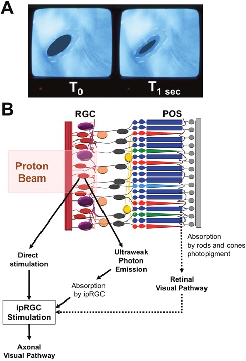
Anomalous visual perceptions have been reported in various diseases of the retina and visual pathways or can be experienced under specific conditions in healthy individuals. Phosphenes are perceptions of light in the absence of ambient light, occurring independently of the physiological and classical photonic stimulation of the retina. They are a frequent symptom in patients irradiated in the region of the central nervous system (CNS), head and neck and the eyes. Phosphenes have historically been attributed to complex physical phenomena such as Cherenkov radiation. While phosphenes are related to Cherenkov radiation under high energy photon/electron irradiation conditions, physical phenomena are unlikely to be responsible for light flashes at energies used for ocular proton therapy. Phosphenes may involve a direct role for ocular photoreceptors and possible interactions between cones and rods. Other mechanisms involving the retinal ganglion cells or ultraweak biophoton emission and rhodopsin bleaching after exposure to free radicals are also likely to be involved. Despite their frequency as shown in our preliminary observations, phosphenes have been underreported probably because their mechanism and impact are poorly understood. Recently, phosphenes have been used to restore the vision and whether they might predict vision loss after therapeutic irradiation is a current field of investigation. We have reviewed and also investigated here the mechanisms related to the occurrence of phosphenes in irradiated patients and especially in patients irradiated by proton therapy for ocular tumors.

Keywords: choroidal melanoma; eye tumors; phosphenes; proton beam therapy; radiation therapy.


Conflict of interest statement

CONFLICTS OF INTEREST No conflicting relationship exists for any author.

Figures

Figure 1
Figure 1. Characterization of phosphenes in patient irradiated by proton beam for eye tumor
(A) Color of phosphenes. (B) Occurrence of phosphenes depending on distance to optic nerve (left) and macula (right).
Figure 2
Figure 2. Role of the Cherenkov radiation in the occurrence of phosphenes in patients irradiated by proton therapy
(A) Schematic diagram showing the irradiation of a choroidal tumor (in red) by the proton beam. (B) Cherenkov effect. The charged particle is moving faster than the waves it emits. (C) Representation of a Compton scattering. Inelastic scattering of a photon by an electron results in decreasing energy of the photon; A part of the energy is transferred to the electron. (D) Representation of a Coulomb interaction of a proton with an atomic electron. The opposite charges of protons and electrons cause an attraction of the atomic electron (in red) by the proton (in blue).
Figure 3
Figure 3. Hypothesis of direct stimulation of the retina
(A) Wavelengths absorbed by rod and cone chromophores. Short, Medium and Long wavelengths are respectively absorbed by blue (S-cone), green (M-cone) and red (L-cone) cones. (B) Distribution of cones and rods in the retina. Cones are mainly located in the foveal area while rods are predominantly located in the optic nerve area and the retinal periphery. (C) Mechanisms of phosphenes according to the irradiated area. If cones are stimulated (macular area), colored phosphenes are predominant. If rods are stimulated (optic nerve and retinal periphery), white phosphenes are predominant.
Figure 4
Figure 4. Role of ultraweak bioluminescent photon emissions
(A) Direct irradiation of photoreceptors by the proton beam. Photoreceptor outer segments are directly irradiated by the proton beam. (B) Lipid peroxidation of the photoreceptor outer segments and production of free radicals. Free radicals (OH°) are produced secondary to ionizing irradiation near or within the retina. Free radicals react with lipids of the phospholipidic membrane and result in lipid peroxidation. Chemical interactions result in ultraweak bioluminescent photons by free radicals. (C) Absorption of ultraweak bioluminescent photons by retinal chromophores leading to the perception of phosphenes. The ultraweak bioluminescent photons bleach the retinal chromophores activating rods and cones. The subsequent phototransduction cascade results in the perception of phosphenes.
Figure 5
Figure 5. Role of intrinsically photosensitive retinal ganglion cells in radiotherapy-induced phosphenes
(A) In-room camera view shows pupillary constriction between left (T0) and right (T1 sec after initiation of the proton beam) inserts. (B) Schematic diagram showing the interactions between retinal ganglion cells, intrinsically photosensitive Retinal Ganglion Cell (ipRGC) and the proton beam. There are 3 hypotheses concerning the activation of ipRGC: (1) Direct stimulation by the proton beam. (2) Production of ultraweak bioluminescent photons which are then absorbed by the ipRGC. (3) Stimulation of rods and cones photopigments in the photoreceptor outer segments (POS) and activation of ipRGC by the normal retinal visual pathway. When excited, ipRGC discharges nerve impulses which travel through neuronal axons to specific brain targets such as the center for pupillary control.

Similar articles

Cited by

References

    1. Cervetto L, Demontis GC, Gargini C. Cellular mechanisms underlying the pharmacological induction of phosphenes. Br J Pharmacol. 2007;150:383–90. doi: 10.1038/sj.bjp.0706998. - DOI - PMC - PubMed
    1. Brown GC, Brown MM, Fischer DH. Photopsias: a key to diagnosis. Ophthalmology. 2015;122:2084–94. doi: 10.1016/j.ophtha.2015.06.025. - DOI - PubMed
    1. Merabet LB, Theoret H, Pascual-Leone A. Transcranial magnetic stimulation as an investigative tool in the study of visual function. Optom Vis Sci. 2003;80:356–68. - PubMed
    1. Lewis PM, Ackland HM, Lowery AJ, Rosenfeld JV. Restoration of vision in blind individuals using bionic devices: a review with a focus on cortical visual prostheses. Brain Res. 2015;1595:51–73. doi: 10.1016/j.brainres.2014.11.020. - DOI - PubMed
    1. Kar K, Krekelberg B. Transcranial electrical stimulation over visual cortex evokes phosphenes with a retinal origin. J Neurophysiol. 2012;108:2173–8. doi: 10.1152/jn.00505.2012. - DOI - PMC - PubMed

LinkOut - more resources