. 2017 Oct 1;140(10):2691-2705.
doi: 10.1093/brain/awx191.

Glymphatic MRI in idiopathic normal pressure hydrocephalus

Affiliations

Glymphatic MRI in idiopathic normal pressure hydrocephalus

Geir Ringstad et al. Brain. .

Abstract

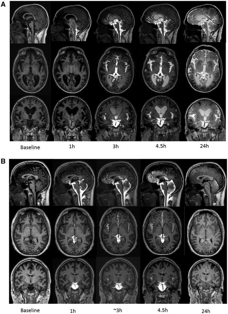
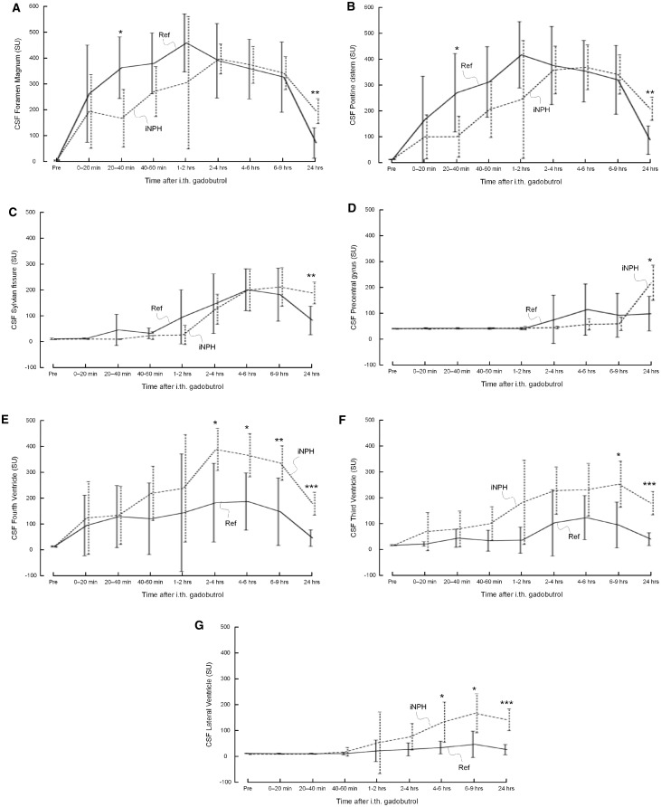
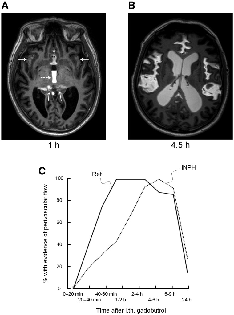
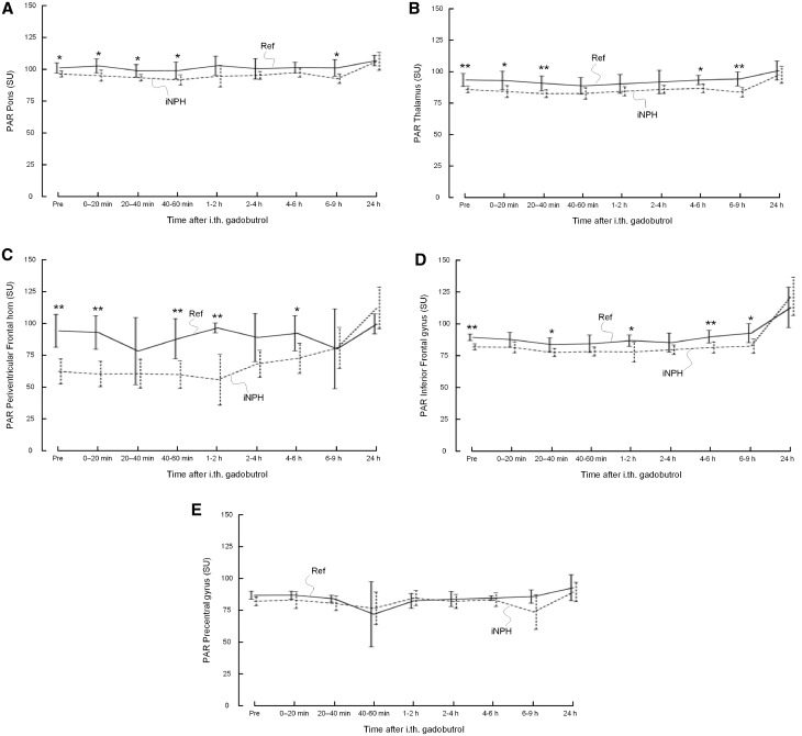
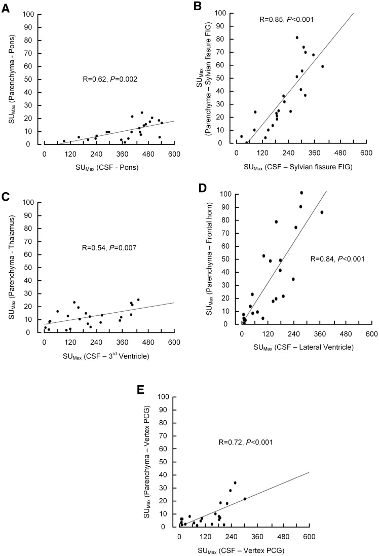
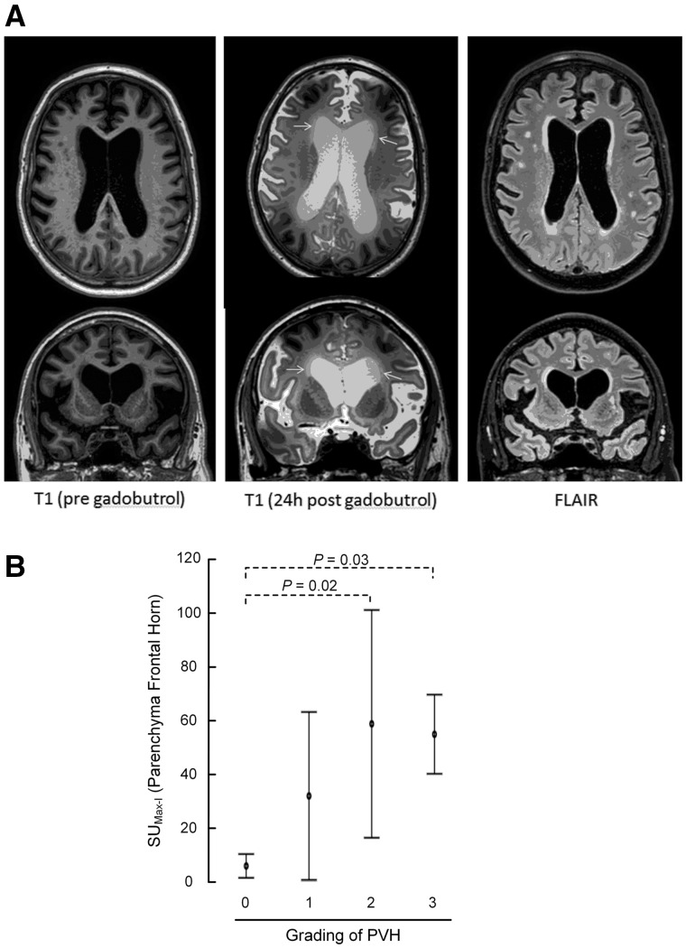
The glymphatic system has in previous studies been shown as fundamental to clearance of waste metabolites from the brain interstitial space, and is proposed to be instrumental in normal ageing and brain pathology such as Alzheimer's disease and brain trauma. Assessment of glymphatic function using magnetic resonance imaging with intrathecal contrast agent as a cerebrospinal fluid tracer has so far been limited to rodents. We aimed to image cerebrospinal fluid flow characteristics and glymphatic function in humans, and applied the methodology in a prospective study of 15 idiopathic normal pressure hydrocephalus patients (mean age 71.3 ± 8.1 years, three female and 12 male) and eight reference subjects (mean age 41.1 + 13.0 years, six female and two male) with suspected cerebrospinal fluid leakage (seven) and intracranial cyst (one). The imaging protocol included T1-weighted magnetic resonance imaging with equal sequence parameters before and at multiple time points through 24 h after intrathecal injection of the contrast agent gadobutrol at the lumbar level. All study subjects were kept in the supine position between examinations during the first day. Gadobutrol enhancement was measured at all imaging time points from regions of interest placed at predefined locations in brain parenchyma, the subarachnoid and intraventricular space, and inside the sagittal sinus. Parameters demonstrating gadobutrol enhancement and clearance in different locations were compared between idiopathic normal pressure hydrocephalus and reference subjects. A characteristic flow pattern in idiopathic normal hydrocephalus was ventricular reflux of gadobutrol from the subarachnoid space followed by transependymal gadobutrol migration. At the brain surfaces, gadobutrol propagated antegradely along large leptomeningeal arteries in all study subjects, and preceded glymphatic enhancement in adjacent brain tissue, indicating a pivotal role of intracranial pulsations for glymphatic function. In idiopathic normal pressure hydrocephalus, we found delayed enhancement (P < 0.05) and decreased clearance of gadobutrol (P < 0.05) at the Sylvian fissure. Parenchymal (glymphatic) enhancement peaked overnight in both study groups, possibly indicating a crucial role of sleep, and was larger in normal pressure hydrocephalus patients (P < 0.05 at inferior frontal gyrus). We interpret decreased gadobutrol clearance from the subarachnoid space, along with persisting enhancement in brain parenchyma, as signs of reduced glymphatic clearance in idiopathic normal hydrocephalus, and hypothesize that reduced glymphatic function is instrumental for dementia in this disease. The study shows promise for glymphatic magnetic resonance imaging as a method to assess human brain metabolic function and renders a potential for contrast enhanced brain extravascular space imaging.

Keywords: MRI; cerebrospinal fluid; gadobutrol; glymphatic function; idiopathic normal pressure hydrocephalus.


Figures

Figure 1
Figure 1
CSF contrast enhancement at multiple time points. Reconstructed T1-weighted images in sagittal (top row), axial (middle row) and coronal (bottom row) planes from MRI at baseline (before contrast agent administration) and at four of the subsequent imaging time points demonstrating time-dependent contrast enhancement of subarachnoid and intraventricular spaces in iNPH patient (A) and reference subject (B). Reflux of gadobutrol to the lateral ventricles was a typical feature of iNPH. In B, retrodural contrast enhancement can be seen on sagittal images (top row) at time points 1 h, 3 h and 4.5 h as sign of a CSF leakage (reference Subject 5).
Figure 2
Figure 2
Trend plots of CSF tracer (gadobutrol) enhancement depending on location. Gadobutrol enhancement was delayed in iNPH patients as compared to reference subjects in (A) foramen magnum, (B) nearby pons, (C) Sylvian fissure, and (D) precentral sulcus. On the other hand, enhancement was significantly stronger within CSF of (E) fourth ventricle, (F) third ventricle, and (G) lateral ventricles. Reference subjects: continuous lines; iNPH patients: dotted lines. Differences between groups at individual time points were determined by independent samples t-test (*P < 0.05, **P < 0.01, ***P < 0.001).
Figure 3
Figure 3
Perivascular enhancement. Gadobutrol enhancement in sulci traversed by the major cerebral arteries (anterior, middle and posterior) was identified and categorized as present/absent for each time point. (A) MRI in axial plane 1 h after contrast agent administration shows gadobutrol distributed to the interhemispheric fissure, Sylvian fissure and ambient cistern (arrows), and reflux to the third ventricle (dotted arrow). (B) At later time points, even though contrast subsequently distributed more freely in the subarachnoid spaces, there was a clear tendency for enhancement in cerebral fissures traversed by the anterior, middle and posterior arteries in both iNPH and reference subjects. (C) Evidence of perivascular gadolinium enhancement was categorized as present/absent for each time point. The percentage of individuals in whom a perivascular contrast enhancement was visualized, is plotted for each time point, showing significant differences between reference subjects and iNPH cohorts (20–40 min: P = 0.049; 40–60 min: P = 0.01; Pearson chi-square test). The plot in C suggests delayed perivascular flow at the brain surface in iNPH patients.
Figure 4
Figure 4
Glymphatic enhancement. Trend plots of gadobutrol enhancement in parenchyma depending on location, including (A) pons, (B) thalamus, (C) periventricular frontal horn, (D) inferior frontal gyrus (IFG), and (E) precentral gyrus. The T1 signal (signal unit) was significantly lower in iNPH cases at various locations in pons, thalamus, periventricular frontal horn, and IFG. In iNPH cases, the change was significantly higher in periventricular frontal horn and IFG between last daytime exam and at 24 h (Supplementary Table 3), and remained at a higher level after 24 h, indicative of delayed clearance. Reference subjects (Ref): continuous lines; iNPH patients: dotted lines. Differences between groups at individual time points were determined by independent samples t-test (*P < 0.05, **P < 0.01, ***P < 0.001).
Figure 5
Figure 5
Glymphatic enhancement as function of CSF enhancement. The association between maximum enhancement within the CSF spaces and nearby brain parenchyma was determined for different regions of interest. For all locations (A–E), there was a highly significant correlation between contrast agent availability within the CSF space and enhancement of gadobutrol within nearby parenchyma.
Figure 6
Figure 6
Transependymal flow of MRI contrast agent. (A) Periventricular hyperintensity (PVH) was identified and graded into four grades from FLAIR images (right column). The maximum increase in signal unit within the periventricular brain parenchyma between pre-contrast MRI (left column) and 24 h post-contrast MRI (middle column) was related to the periventricular hyperintensity categories. Top row: Axial sections; bottom row: coronal sections. (B) ANOVA with Bonferroni corrected post hoc tests revealed that periventricular enhancement was significantly larger in periventricular lucency grades 2 and 3 than grade 0.

Comment in

  • Gadobutrol and the End of the Tap Test.
    Welling LC, Welling MS, Figueiredo EG. Welling LC, et al. World Neurosurg. 2018 Feb;110:442. doi: 10.1016/j.wneu.2017.12.101. Epub 2017 Dec 21. World Neurosurg. 2018. PMID: 29275054 No abstract available.

References

    1. Adams RD, Fisher CM, Hakim S, Ojemann RG, Sweet WH. Symptomatic occult hydrocephalus with "Normal" cerebrospinal-fluid pressure. A treatable syndrome. N Eng J Med 1965; 273: 117–26. - PubMed
    1. Akbar JJ, Luetmer PH, Schwartz KM, Hunt CH, Diehn FE, Eckel LJ. The role of MR myelography with intrathecal gadolinium in localization of spinal CSF leaks in patients with spontaneous intracranial hypotension. AJNR Am J Neuroradiol 2012; 33: 535–40. - PMC - PubMed
    1. Asgari M, de Zelicourt D, Kurtcuoglu V. Glymphatic solute transport does not require bulk flow. Sci Rep 2016; 6: 38635. - PMC - PubMed
    1. Aydin K, Guven K, Sencer S, Jinkins JR, Minareci O. MRI cisternography with gadolinium-containing contrast medium: its role, advantages and limitations in the investigation of rhinorrhoea. Neuroradiology 2004; 46: 75–80. - PubMed
    1. Bakker EN, Bacskai BJ, Arbel-Ornath M, Aldea R, Bedussi B, Morris AW, et al.Lymphatic clearance of the brain: perivascular, paravascular and significance for neurodegenerative diseases. Cell Mol Neurobiol 2016; 36: 181–94. - PMC - PubMed