. 2017 Oct 1;140(10):2610-2622.
doi: 10.1093/brain/awx203.

Mutations of AKT3 are associated with a wide spectrum of developmental disorders including extreme megalencephaly

Affiliations

Mutations of AKT3 are associated with a wide spectrum of developmental disorders including extreme megalencephaly

Diana Alcantara et al. Brain. .

Abstract

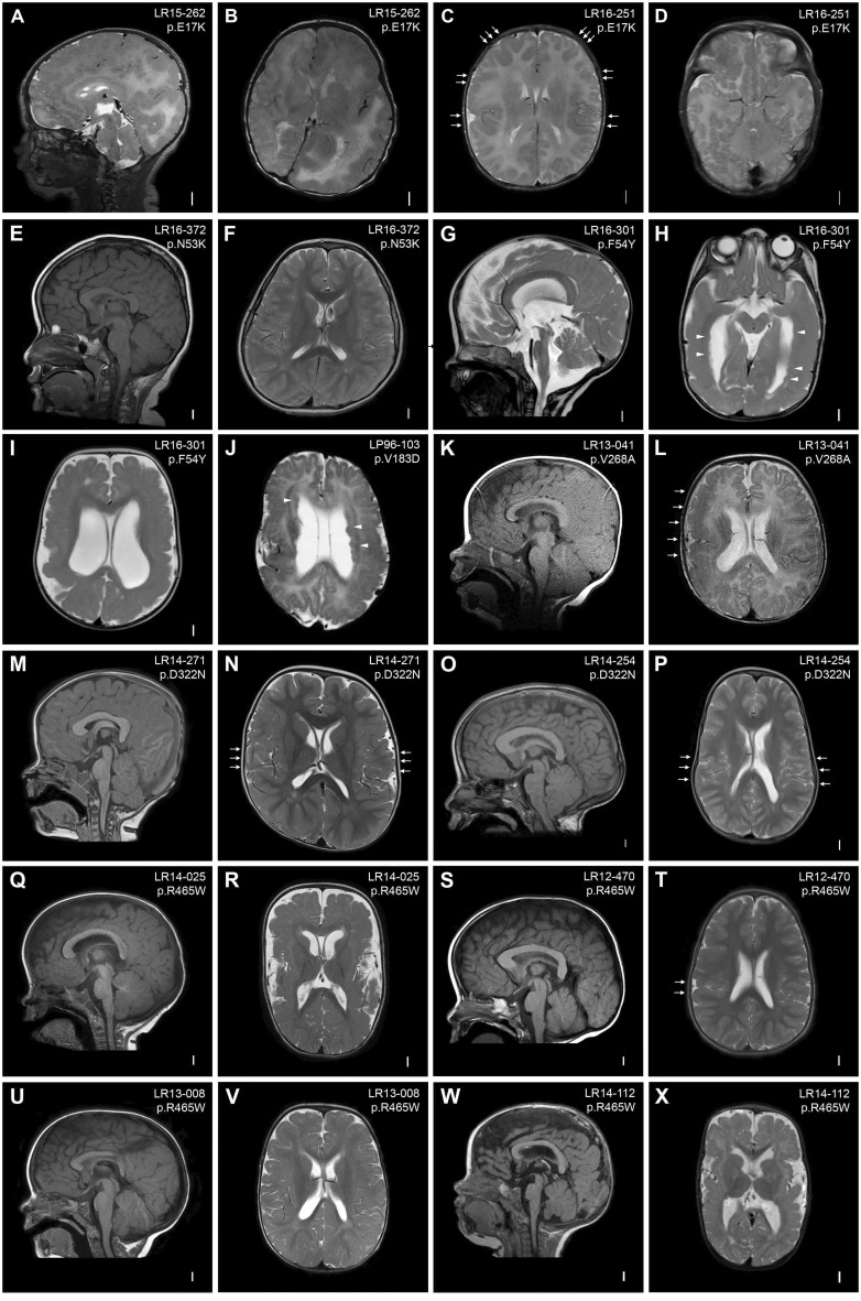
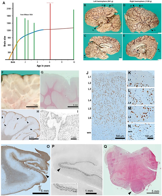
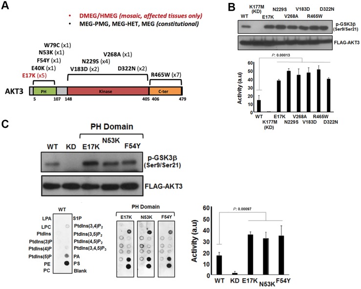
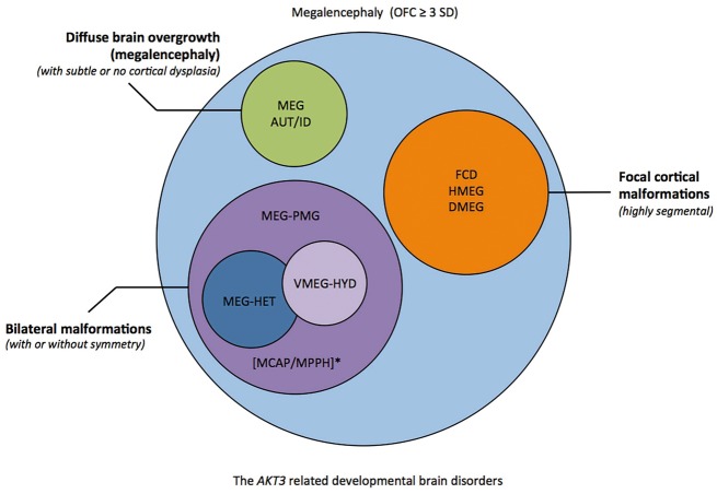
Mutations of genes within the phosphatidylinositol-3-kinase (PI3K)-AKT-MTOR pathway are well known causes of brain overgrowth (megalencephaly) as well as segmental cortical dysplasia (such as hemimegalencephaly, focal cortical dysplasia and polymicrogyria). Mutations of the AKT3 gene have been reported in a few individuals with brain malformations, to date. Therefore, our understanding regarding the clinical and molecular spectrum associated with mutations of this critical gene is limited, with no clear genotype-phenotype correlations. We sought to further delineate this spectrum, study levels of mosaicism and identify genotype-phenotype correlations of AKT3-related disorders. We performed targeted sequencing of AKT3 on individuals with these phenotypes by molecular inversion probes and/or Sanger sequencing to determine the type and level of mosaicism of mutations. We analysed all clinical and brain imaging data of mutation-positive individuals including neuropathological analysis in one instance. We performed ex vivo kinase assays on AKT3 engineered with the patient mutations and examined the phospholipid binding profile of pleckstrin homology domain localizing mutations. We identified 14 new individuals with AKT3 mutations with several phenotypes dependent on the type of mutation and level of mosaicism. Our comprehensive clinical characterization, and review of all previously published patients, broadly segregates individuals with AKT3 mutations into two groups: patients with highly asymmetric cortical dysplasia caused by the common p.E17K mutation, and patients with constitutional AKT3 mutations exhibiting more variable phenotypes including bilateral cortical malformations, polymicrogyria, periventricular nodular heterotopia and diffuse megalencephaly without cortical dysplasia. All mutations increased kinase activity, and pleckstrin homology domain mutants exhibited enhanced phospholipid binding. Overall, our study shows that activating mutations of the critical AKT3 gene are associated with a wide spectrum of brain involvement ranging from focal or segmental brain malformations (such as hemimegalencephaly and polymicrogyria) predominantly due to mosaic AKT3 mutations, to diffuse bilateral cortical malformations, megalencephaly and heterotopia due to constitutional AKT3 mutations. We also provide the first detailed neuropathological examination of a child with extreme megalencephaly due to a constitutional AKT3 mutation. This child has one of the largest documented paediatric brain sizes, to our knowledge. Finally, our data show that constitutional AKT3 mutations are associated with megalencephaly, with or without autism, similar to PTEN-related disorders. Recognition of this broad clinical and molecular spectrum of AKT3 mutations is important for providing early diagnosis and appropriate management of affected individuals, and will facilitate targeted design of future human clinical trials using PI3K-AKT pathway inhibitors.

Keywords: AKT3; epilepsy; hemimegalencephaly; megalencephaly; polymicrogyria.


Figures

Figure 1
Figure 1
Brain MRIs of AKT3 mutation-positive children. (A and B) Brain MRI of Patient LR15-262 showing markedly enlarged and dysplastic right cerebral hemisphere with diffuse cortical dysplasia and dysmyelination consistent with hemimegalencephaly. The contralateral hemisphere is markedly decreased in size with areas of cortical dysplasia (hemimicroencephaly). (C and D) Images of Patient LR16-251 showing multifocal areas of dysplastic cortex in the perisylvian, frontal, temporal and occipital regions (arrows). (E and F) Images of Patient LR16-372 showing a thick and dysplastic corpus callosum and deeply infolded perisylvian regions. (G–I) Images of Patient LR16-301 showing striking megalencephaly, ventriculomegaly, stretched but thick corpus callosum, diffuse polymicrogyria with deep infolding in the right occipital lobe, and bilateral periventricular nodular heterotopia (arrowheads). (J) Image of Patient LP96-103 showing diffuse bilateral perisylvian polymicrogyria, ventriculomegaly, cavum septum pellucidum et vergae and diffuse periventricular nodular heterotopia (arrowheads). (K and L) Images of Patient LR13-041 showing a large cerebellum with cerebellar tonsillar ectopia, bilateral polymicrogyria predominantly in the perisylvian region (more severe on the right, arrows) with dysmyelination. (M and N) Images of Patient LR14-271 showing diffuse megalencephaly with a thick corpus callosum and deep infolding in the perisylvian region suggestive of polymicrogyria (arrows). (O and P) Images of Patient LR14-254 showing diffuse megalencephaly, thick corpus callosum and bilaterally diffuse infolding of the perisylvian region suggestive of polymicrogyria (arrows). (Q and R) Images of Patient LR14-025 showing megalencephaly, thick corpus callosum and bilateral diffuse polymicrogyria with increased extra-axial space. (S and T) Images of Patient LR12-470 showing megalencephaly and thick corpus callosum. This patient also had deep infolding in the right perisylvian region suspicious for polymicrogyria, with very limited involvement (arrows). (U and V) Images of Patient LR13-008 showing diffuse megalencephaly and possible area of cortical dysplasia in the right perisylvian region. (W and X) Images of Patient LR14-112 showing diffuse megalencephaly, bilateral perisylvian polymicrogyria and bilateral ventriculomegaly.
Figure 2
Figure 2
Pathologic examination of the brain of Patient LR08-018 (p.R465W). (A) Brain size. Graph depicting the largest paediatric brain sizes (in grams) previously reported in the literature (green bars) relative to the brain size of Patient LR08-018 (with the p.R465W mutation, red bar) demonstrating that brain size for this patient is markedly larger than these patients (Wilson, 1934). The graph is an adaptation of the human brain growth diagram from the Smithsonian Institute (http://humanorigins.si.edu/human-characteristics/brains). The graph shows the periods of rapid brain growth (in orange) plus the period of decreased brain growth (in blue) followed by the plateau in brain growth. (B–E) Cerebral hemispheres. The massive brain (2313 g; approximately twice normal weight for age) was asymmetrically enlarged. The left hemisphere (B and C) weighed 941 g, and the right (D and E) weighed 1128 g. Primary fissures such as the Sylvian (sy), central (ce), postcentral (pc), and calcarine (ca) sulci were recognizable, but secondary and tertiary sulci were abnormal. Gyri appeared irregular and overall hyperconvoluted. The corpus callosum was present including genu (ge), body, and splenium (sp). The anterior commissure (ac) was present but small. White asterisks: artefactual disruption of hemispheres due to brain removal and transport. Black asterisks: torn junction of hemispheres and midbrain. All panels are at the same magnification. (F–I) Hyperconvolution and polymicrogyria in cerebral cortex. (F) Brain slice through parietal cortex showed redundant folds of cortex extending deep into white matter. (G) Histological section (haematoxylin and eosin) through the same region showed relatively sharp grey-white junctions. (H) Inferior temporal cortex exhibited features of polymicrogyria, including anomalous branching and fusion of gliotic layer 1 (GFAP immunohistochemistry). (I) Layer 1 fusion and branching were confirmed by NeuN immunohistochemistry. (J–N) Abnormal layering of cerebral cortex, and excessive white matter neurons. In foci not involved with polymicrogyria, such as right posterior perisylvian cortex, cortical layering was moderately disorganized. (J) Cortical layers were identified based on cell size and density. NeuN immunohistochemistry. (K) Layer 1 was cell-sparse and contained only small neurons. (L) Layer 4 contained typical small (granular) neurons. (M) Layer 6 neurons were particularly disorganized and maloriented. (N) Increased neurons in white matter (wm). Interestingly, neurons in this case were not strikingly enlarged or dysplastic, nor were any balloon cells present. (O–Q) Hippocampal and brainstem abnormalities of Patient LR08-018. (O) The left hippocampus was very small and gliotic, and the hippocampal sulcus (arrowhead) was open, suggesting a deficiency of perforant pathway fibres, which would normally cross the fused sulcus. GFAP immunohistochemistry. (P) Histologically, the dentate gyrus exhibited focal ‘tram-track’ splitting of the granule cell layer (arrowhead; enlarged 2× in inset), a finding usually associated with chronic epilepsy. (Q) The upper medulla showed marked asymmetry of the pyramidal tract, essentially limited to one side (arrowhead). The adjacent inferior olives were moderately hypoconvoluted (haematoxylin and eosin).
Figure 3
Figure 3
Analysis of AKT3 activity in vitro. (A) The primary structure of AKT3 showing the relative positions of the pleckstrin homology (PH) domain for lipid binding the catalytic kinase domain and C-terminal (C-ter) region. Mutations identified to date are shown along with the numbers of patients with these mutations in brackets. (B) Catalytic kinase domain and C-terminal localizing patient-derived AKT3 mutations are associated with elevated kinase activity. Ectopically expressed wild-type (WT) AKT, a kinase dead variant K177M, the E17K activating pleckstrin homology domain mutant and various patient mutants were assessed for kinase activity using a GSK3β peptide as a substrate in an ex vivo kinase assay. The upper panel shows immune detection of phosphorylated GSK3β peptide following western blotting with anti-phospho-GSK3β (Ser9/Ser21) antibody. The patient mutants all exhibit elevated phospho-activity compared to wild-type. The graph depicts quantitation of phospho-GSK3β (Ser9/Ser21) signal (a.u. = arbitrary units). Error bars represent mean ± SD (n = 4), P-values were determined using Student’s t-test. (C) Pleckstrin homology domain localizing patient mutations are associated with elevated kinase activity and altered phospholipid-binding profile. Left panels show western blot analysis of phospho-GSK3β (Ser9/Ser21) of ectopically expressed wild-type, K177M kinase dead and three pleckstrin homology domain patient mutants; E17K, N53K and F54Y. The graph depicts quantitation of phospho-GSK3β (Ser9/Ser21) signal. Error bars represent mean ± SD (n = 4), P-values were determined using Student’s t-test. The bottom panels depict PIP-membranes seeded with various lipids and phospholipids for dot blot binding analysis. Ectopically expressed FLAG-tagged wild-type and AKT3 pleckstrin homology domain mutants were incubated with the PIP Strips and bound protein detected by western blotting using anti-FLAG. All three pleckstrin homology domain mutants exhibit altered and elevated binding to specific phospholipids compared to wild-type. DMEG = dysplastic megalencephaly; HMEG = hemimegalencephaly; LPA = lysophophatidic acid; LPC = lysophosphocholine; MEG = megalencephaly; P = phosphate; PA = phosphatidic acid; PC = phosphatidylcholine; PE = phosphatidylethanolamine; PMG = polymicrogryria; PS = phosphatidylserine; PtdIns = phosphatidylinositol; S1P = sphingosine-1-phosphate.
Figure 4
Figure 4
Diagram showing the spectrum of AKT3-associated phenotypes. Several groups of partially overlapping developmental brain disorders are associated with AKT3 mutations that include the following phenotypes (i) focal malformations of cortical development that are highly segmental [e.g. focal cortical dysplasia (FCD), hemimegalencephaly (HMEG), dysplastic megalencephaly DMEG; orange]; (ii) bilateral polymicrogyria (PMG) (purple) with or without ventriculomegaly or hydrocephalus (VMEG-HYD; light purple), and heterotopia (HET; blue); (iii) diffuse megalencephaly with intellectual disability (ID) and/or autistic features (AUT) with subtle or no cortical dysplasia (green). *Of note, MCAP and MPPH fit within the second group of AKT3-related disorders, from the brain phenotype perspective. MCAP can be further clinically distinguished by somatic findings (somatic overgrowth, vascular or lymphatic abnormalities), and MPPH by the occurrence of polydactyly in a subset of affected individuals. A general molecular diagnostic workflow for megalencephaly has been proposed (Supplementary Fig. 1). OFC = orbitofrontal cortex.

References

    1. Arya VB, Flanagan SE, Schober E, Rami-Merhar B, Ellard S, Hussain K. Activating AKT2 mutation: hypoinsulinemic hypoketotic hypoglycemia. J Clin Endocrinol Metab 2014; 99: 391–4. - PubMed
    1. Baek ST, Copeland B, Yun EJ, Kwon SK, Guemez-Gamboa A, Schaffer AE, et al.An AKT3-FOXG1-reelin network underlies defective migration in human focal malformations of cortical development. Nat Med 2015; 21: 1445–54. - PMC - PubMed
    1. Barkovich AJ, Guerrini R, Kuzniecky RI, Jackson GD, Dobyns WB. A developmental and genetic classification for malformations of cortical development: update 2012. Brain 2012; 135: 1348–69. - PMC - PubMed
    1. Carpten JD, Faber AL, Horn C, Donoho GP, Briggs SL, Robbins CM, et al.A transforming mutation in the pleckstrin homology domain of AKT1 in cancer. Nature 2007; 448: 439–44. - PubMed
    1. Cho H, Mu J, Kim JK, Thorvaldsen JL, Chu Q, Crenshaw EB 3rd, et al.Insulin resistance and a diabetes mellitus-like syndrome in mice lacking the protein kinase Akt2 (PKB beta). Science 2001; 292: 1728–31. - PubMed

MeSH terms

Substances