. 2017 Oct 2;7(1):12492.
doi: 10.1038/s41598-017-12666-z.

Mast Cell-Intervertebral disc cell interactions regulate inflammation, catabolism and angiogenesis in Discogenic Back Pain

Affiliations

Mast Cell-Intervertebral disc cell interactions regulate inflammation, catabolism and angiogenesis in Discogenic Back Pain

Matthew G Wiet et al. Sci Rep. .

Abstract

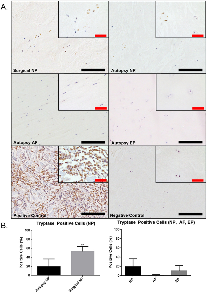
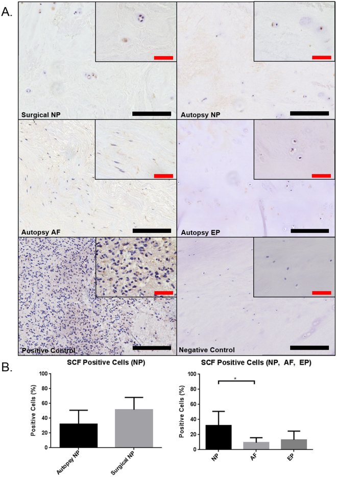
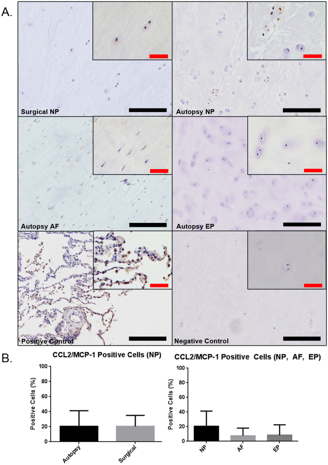
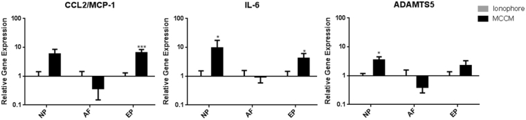
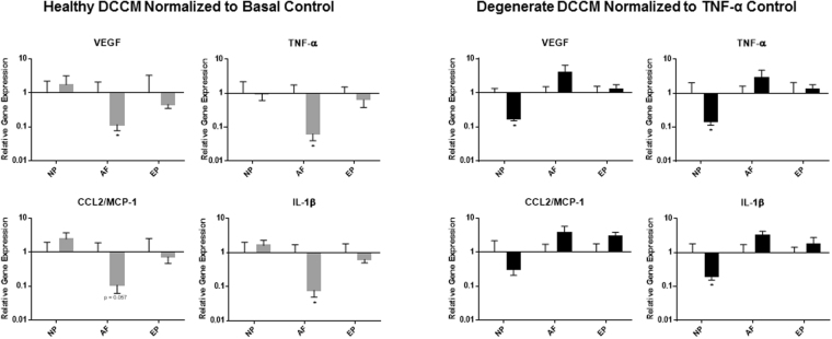
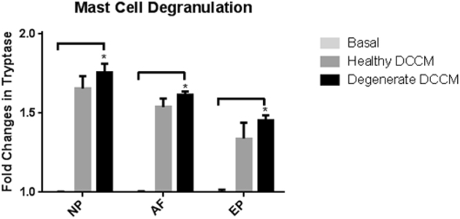
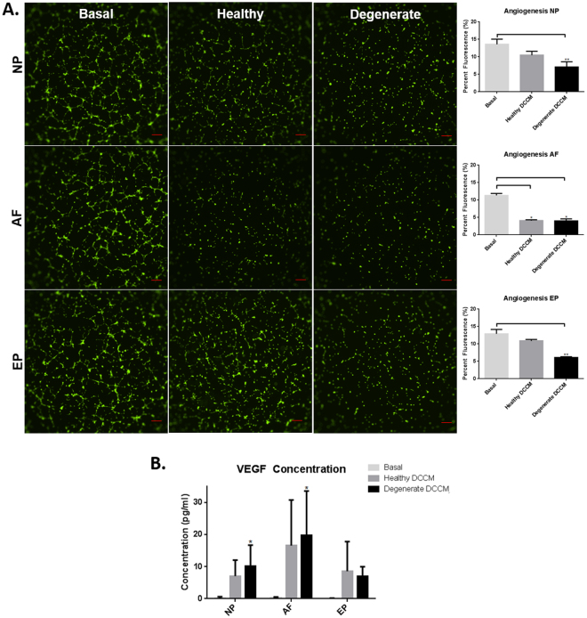
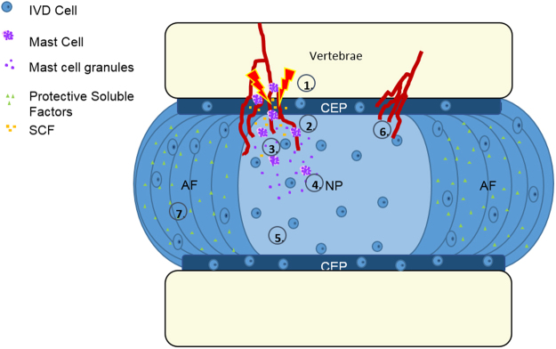
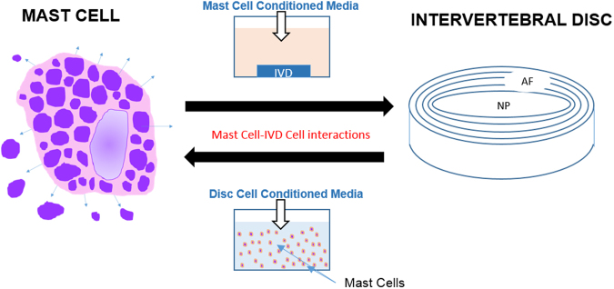
Low back pain (LBP) is a widespread debilitating disorder of significant socio-economic importance and intervertebral disc (IVD) degeneration has been implicated in its pathogenesis. Despite its high prevalence the underlying causes of LBP and IVD degeneration are not well understood. Recent work in musculoskeletal degenerative diseases such as osteoarthritis have revealed a critical role for immune cells, specifically mast cells in their pathophysiology, eluding to a potential role for these cells in the pathogenesis of IVD degeneration. This study sought to characterize the presence and role of mast cells within the IVD, specifically, mast cell-IVD cell interactions using immunohistochemistry and 3D in-vitro cell culture methods. Mast cells were upregulated in painful human IVD tissue and induced an inflammatory, catabolic and pro-angiogenic phenotype in bovine nucleus pulposus and cartilage endplate cells at the gene level. Healthy bovine annulus fibrosus cells, however, demonstrated a protective role against key inflammatory (IL-1β and TNFα) and pro-angiogenic (VEGFA) genes expressed by mast cells, and mitigated neo-angiogenesis formation in vitro. In conclusion, mast cells can infiltrate and elicit a degenerate phenotype in IVD cells, enhancing key disease processes that characterize the degenerate IVD, making them a potential therapeutic target for LBP.


Conflict of interest statement

The authors declare that they have no competing interests.

Figures

Figure 1
Figure 1
Immunohistochemical staining for mast cell specific tryptase in human IVD tissue (A). The positive control was tissue taken from a human mast cell tumor. Quantification of percent positive cells for tryptase demonstrated significant upregulation of tryptase in human surgical tissue samples compared to autopsy controls (B) (p = 0.0047) (Autopsy N = 7, Surgical N = 6). Black scale bar = 200 µm, Red scale bar = 50 µm.
Figure 2
Figure 2
Immunohistochemical staining for mast cell chemoattractant stem cell factor (SCF) in human IVD (SCF) (A). The positive control was tissue taken from human brain tissue. Quantification of percent positive cells for SCF demonstrated no significant differences between surgical and autopsy specimens (p = 0.081) however regional differences were observed between NP and AF (B) (p = 0.017) (Autopsy N = 7, Surgical N = 6). Black scale bar = 200 µm, Red scale bar = 50 µm.
Figure 3
Figure 3
Immunohistochemical staining for immune cell chemoattractant CCL2/MCP-1 (A). The positive control was taken from human lung tissue. Quantification of percent positive cells for CCL2/MCP-1 demonstrated no significant differences between surgical and autopsy (p = 0.17) or between regions (p = 0.87) (B) (Autopsy N = 7, Surgical N = 6). Black scale bar = 200 µm, Red scale bar = 50 µm.
Figure 4
Figure 4
Gene expression for CCL2/MCP-1, IL-6, ADAMTS5 in bovine disc cells in response to mast cell conditioned media (MCCM). Significant upregulation of ADAMTS5 (p = 0.023), and IL-6 (p = 0.023) in NP as well as upregulation of IL-6 (p = 0.0244) and MCP-1 (p = 0.0003) in CEP relative to the ionophore control (N = 11 for NP/AF and N = 10 for EP).
Figure 5
Figure 5
Gene expression of mast cells in response to disc cell conditioned media (DCCM). Significant decreases in mast cell expression of key inflammatory/angiogenic markers (VEGF, TNF-α, and IL-1β) (p = 0.015, p = 0.013, p = 0.013) when exposed to healthy AF DCCM relative to mast cells cultured in basal control conditions. Degenerate NP DCCM also decreased expression of TNF-α (p = 0.049) in mast cells relative to mast cells cultured with TNF-α (TNF-α control) (N = 4).
Figure 6
Figure 6
Mast cell activation as measured by the mast cell degranulation assay and release of tryptase into the media upon exposure to IVD DCCM. All three regions (NP, AF EP) of the degenerate IVD increased degranulation of mast cells relative to basal control conditions (p = 0.011, p = 0.0048, p = 0.0048 respectively). Healthy DCCM had a trend of increased degranulation (N = 4).
Figure 7
Figure 7
(A) Endothelial tubular formation in the presence of media from mast cells cultured in the IVD DCCM. Healthy and degenerate AF DCCM (p = 0.015), degenerate NP DCCM (p = 0.0002), and degenerate EP DCCM (p = 0.0002) all significantly down regulated endothelial tubular formation (N = 4). (B) Significant upregulation of VEGF secretion in mast cells exposed to AF DCCM and NP DCCM was observed compared to basal controls (p = 0.0066 and p = 0.046 respectively) (N = 3).
Figure 8
Figure 8
Hypothetical model wherein the disc undergoes injury/degeneration (1), followed by mast cell recruitment by SCF (2 & 3). Mast cells are then activated (4) in the degenerate environment and elicit catabolic changes in the IVD microenvironment (5) promoting neurovascular ingrowth and pain (6). In the healthy IVD, the AF produces protective soluble factors that help to maintain an immune-privileged microenvironment, inhibiting mast cell activation and angiogenesis (7).
Figure 9
Figure 9
Experimental model to characterize the effect of mast cells in the IVD microenvironment. Conditioned media from activated mast cells was applied to 3D in vitro constructs containing NP, AF, or EP cells. Healthy and degenerate IVD cell conditioned media (NP, AF, EP) was applied to mast cells in suspension.

References

    1. Katz JN. Lumbar disc disorders and low-back pain: socioeconomic factors and consequences. J. Bone Joint Surg. Am. 2006;88(Suppl 2):21–24. - PubMed
    1. Luoma K, et al. Low Back Pain in Relation to Lumbar Disc Degeneration. Spine (Phila Pa 1976) 2000;25:487–492. doi: 10.1097/00007632-200002150-00016. - DOI - PubMed
    1. Hagiyama M, et al. Enhanced Nerve-Mast Cell Interaction by a Neuronal Short Isoform of Cell Adhesion Molecule-1. J. Immunol. 2011;186:5983–5992. doi: 10.4049/jimmunol.1002244. - DOI - PubMed
    1. Adams M, Roughley P. What is intervertebral disc degeneration, and what causes it? Spine (Phila. Pa. 1976). 2006;31:2151–2161. doi: 10.1097/01.brs.0000231761.73859.2c. - DOI - PubMed
    1. Humzah MD, Soames RW. Human intervertebral disc: structure and function. Anat. Rec. 1988;220:337–356. doi: 10.1002/ar.1092200402. - DOI - PubMed

MeSH terms

LinkOut - more resources