

Salt Adaptation and Evolutionary Implication of a Nah-related PAHs Dioxygenase cloned from a Halophilic Phenanthrene Degrading Consortium
- PMID: 28970580
- PMCID: PMC5624874
- DOI: 10.1038/s41598-017-12979-z
Salt Adaptation and Evolutionary Implication of a Nah-related PAHs Dioxygenase cloned from a Halophilic Phenanthrene Degrading Consortium
Abstract
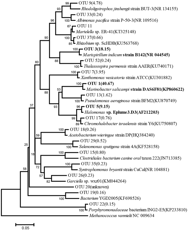
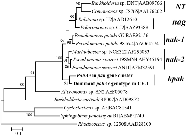
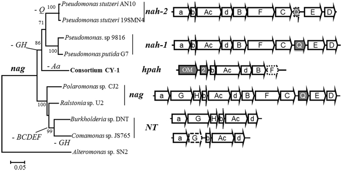
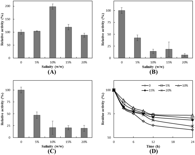
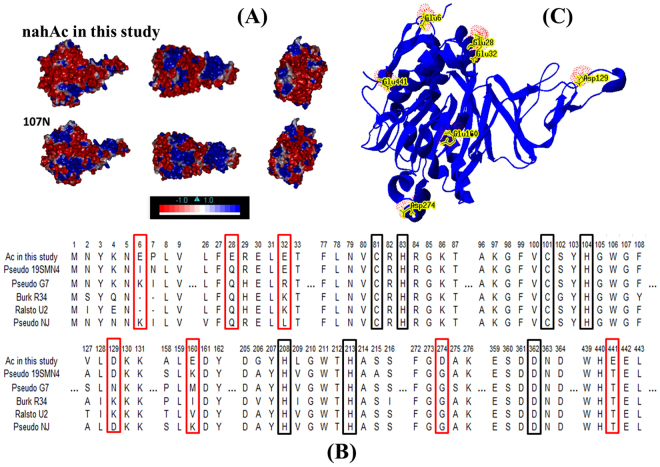
Polycyclic aromatic hydrocarbons (PAHs) pollutions often occur in marine and other saline environment, largely due to anthropogenic activities. However, study of the PAHs-degradation genotypes in halophiles is limited, compared with the mesophilic terrestrial PAHs degraders. In this study, a bacterial consortium (CY-1) was enriched from saline soil contaminated with crude oil using phenanthrene as the sole carbon source at 10% salinity. CY-1 was dominated by the moderate halophilic Marinobacter species, and its dominant PAHs ring-hydroxylating dioxygenase (RHD) genotypes shared high identity to the classic nah-related RHDs found in the mesophilic species. Further cloning of a 5.6-kb gene cluster from CY-1 unveiled the existence of a new type of PAHs degradation gene cluster (hpah), which most probably evolves from the nah-related gene clusters. Expression of the RHD in this gene cluster in E. coli lead to the discovery of its prominent salt-tolerant properties compared with two RHDs from mesophiles. As a common structural feature shared by all halophilic and halotolerant enzymes, higher abundance of acidic amino acids was also found on the surface of this RHD than its closest nah-related alleles. These results suggest evolution towards saline adaptation occurred after horizontal transfer of this hpah gene cluster into the halophiles.
Conflict of interest statement
The authors declare that they have no competing interests.
Figures
Similar articles
-
Degradation of n-alkanes and PAHs from the heavy crude oil using salt-tolerant bacterial consortia and analysis of their catabolic genes.Environ Sci Pollut Res Int. 2017 Apr;24(12):11392-11403. doi: 10.1007/s11356-017-8446-2. Epub 2017 Mar 17. Environ Sci Pollut Res Int. 2017. PMID: 28315056
-
Heterologous expression of polycyclic aromatic hydrocarbon ring-hydroxylating dioxygenase genes from a novel pyrene-degrading betaproteobacterium.Appl Environ Microbiol. 2012 May;78(10):3552-9. doi: 10.1128/AEM.00173-12. Epub 2012 Mar 16. Appl Environ Microbiol. 2012. PMID: 22427500 Free PMC article.
-
Co-metabolic degradation of benzo(e)pyrene by halophilic bacterial consortium at different saline conditions.J Environ Biol. 2014 May;35(3):445-52. J Environ Biol. 2014. PMID: 24812998
-
Polyaromatic Hydrocarbon Specific Ring Hydroxylating Dioxygenases: Diversity, Structure, Function, and Protein Engineering.Curr Protein Pept Sci. 2023;24(1):7-21. doi: 10.2174/1389203724666221108114537. Curr Protein Pept Sci. 2023. PMID: 36366847 Review.
-
Recent studies in microbial degradation of petroleum hydrocarbons in hypersaline environments.Front Microbiol. 2014 Apr 23;5:173. doi: 10.3389/fmicb.2014.00173. eCollection 2014. Front Microbiol. 2014. PMID: 24795705 Free PMC article. Review.
Cited by
-
Insights Into Mechanism of the Naphthalene-Enhanced Biodegradation of Phenanthrene by Pseudomonas sp. SL-6 Based on Omics Analysis.Front Microbiol. 2021 Nov 17;12:761216. doi: 10.3389/fmicb.2021.761216. eCollection 2021. Front Microbiol. 2021. PMID: 34867892 Free PMC article.
-
Salinity effect on the metabolic pathway and microbial function in phenanthrene degradation by a halophilic consortium.AMB Express. 2018 Apr 25;8(1):67. doi: 10.1186/s13568-018-0594-3. AMB Express. 2018. PMID: 29696463 Free PMC article.
References
-
- Tingting, et al. Effect of salinity on community structure and naphthalene dioxygenase gene diversity of a halophilic bacterial consortium. Frontiers of Environmental Science & Engineering. 2016;10:16.
Publication types
MeSH terms
Substances
LinkOut - more resources
Full Text Sources
Other Literature Sources
Research Materials
