. 2017 Oct 2;7(1):12537.
doi: 10.1038/s41598-017-12880-9.

Levels of S100B protein drive the reparative process in acute muscle injury and muscular dystrophy

Affiliations

Levels of S100B protein drive the reparative process in acute muscle injury and muscular dystrophy

Francesca Riuzzi et al. Sci Rep. .

Abstract

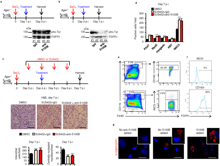
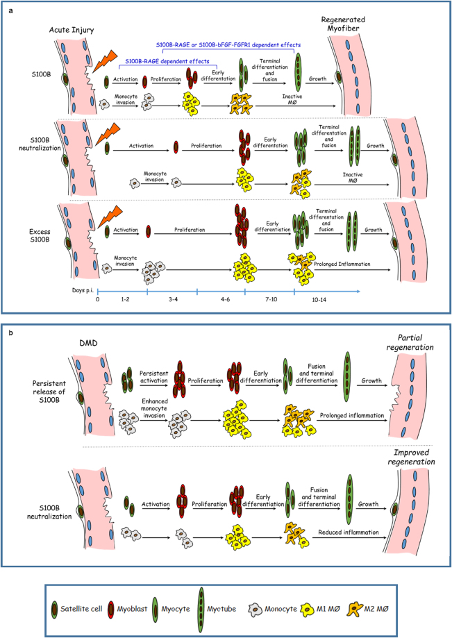
Regeneration of injured skeletal muscles relies on a tightly controlled chain of cellular and molecular events. We show that appropriate levels of S100B protein are required for timely muscle regeneration after acute injury. S100B released from damaged myofibers and infiltrating macrophages expands the myoblast population, attracts macrophages and promotes their polarization into M2 (pro-regenerative) phenotype, and modulates collagen deposition, by interacting with RAGE (receptor for advanced glycation end-products) or FGFR1 (fibroblast growth factor receptor 1) depending on the muscle repair phase and local conditions. However, persistence of high S100B levels compromises the regeneration process prolonging myoblast proliferation and macrophage infiltration, delaying M1/M2 macrophage transition, and promoting deposition of fibrotic tissue via RAGE engagement. Interestingly, S100B is released in high abundance from degenerating muscles of mdx mice, an animal model of Duchenne muscular dystrophy (DMD), and blocking S100B ameliorates histopathology. Thus, levels of S100B differentially affect skeletal muscle repair upon acute injury and in the context of muscular dystrophy, and S100B might be regarded as a potential molecular target in DMD.


Conflict of interest statement

The authors declare that they have no competing interests.

Figures

Figure 1
Figure 1
Blocking S100B early after acute muscle injury delays regeneration. (a) BaCl2-injured TA muscles were injected with IgG or a polyclonal anti-S100B antibody (Abcam No. ab41548) at d1 p.i. Treated muscles were excised at d3, d7 or d14 p.i. (b) Histology of muscles and counts of interstitial cells and centrally nucleated myofibers. (c) Counts of PAX7+, MyoD+, myogenin+, Ki67+, and RAGE+ cells (also see Fig. S1d,e). (d) Western blots of the indicated proteins in homogenates of IgG- and anti-S100B-treated muscles. Immunoblots of GAPDH and α-actinin are included as loading controls. (e) Myofiber size distribution at d14 p.i. in uninjured muscles and IgG- and anti-S100B-treated injured muscles. (f) Migration of primary mouse myoblasts in Boyden chambers in the absence or presence of S100B. Full-length blots are presented in Supplementary Fig. S7 “Fig. 1”. Results are means ± SEM (n = 6). **p < 0.01, ***p < 0.001 vs. control. The scale bar represents 50 µm in (b) and 200 µm in (f).
Figure 2
Figure 2
S100B affects macrophages early after acute muscle injury. (a) TA muscles were treated as described in the legend to Fig. 1a. Muscles were excised at d3 or d7 p.i. (b) Counts of MAC3+ cells. (cf,h) Macrophages were isolated from IgG- and anti-S100B-treated injured muscles and either counted (c), analyzed by real-time PCR (d,f,h), or subjected to western blotting (e) (also see Fig. S2c). (g) Peritoneal macrophages were subjected to a migration assay using Boyden chambers in the presence of increasing S100B doses. (i) IgG- and anti-S100B-treated injured muscles excised at d7 p.i. and subjected to collagen IV detection by immunohistochemistry and western blotting. (j) Macrophages were isolated from IgG- and anti-S100B-treated injured muscles at d3 p.i., cultured for 24 h in the absence or presence of 200 ng S100B/ml, and analyzed by real-time PCR. (k) Macrophages were treated with IFN-γ, IL-10 or IL-4 in the absence or presence of the NF-κB inhibitor, Bay11-7085, and analyzed by real-time PCR for S100b levels. (l) Western blot analysis of S100B in conditioned media of IFN-γ-, IL-10- or IL-4-stimulated peritoneal macrophages. (m) Proliferation assay of C2C12 myoblasts cultured in the presence of IgG- or anti-S100B-treated conditioned media from vehicle- or IFN-γ-stimulated peritoneal macrophages (left) and differentiation assay of C2C12 myoblasts cultured in the presence of IgG- or anti-S100B-treated conditioned media from vehicle- or IL-10-stimulated peritoneal macrophages (right). Immunoblots of α-tubulin are included as loading controls in western blots in e,m. Full-length blots are presented in Supplementary Fig. S7 “Fig. 2”. Results are means ± SEM (n = 6). * p < 0.05, **p < 0.01, ***p < 0.001 vs. control. # p < 0.01, ## p < 0.001 (d7 vs. d3 p.i., b and c; anti-S100B antibody- vs. IgG-treated, j; BAY11-7085-treated vs. control, k; IFN-γ-treated or IL-10-treated vs. control, m). The scale bar in i represents 50 µm.
Figure 3
Figure 3
S100B is required during the macrophage M2 phase for efficient regeneration. (a) Injured TA muscles were injected with IgG or anti-S100B antibody at d4 p.i. Treated muscle were excised at d7 or d14 p.i. (b) Histology of muscle tissue (upper panel) and counts of interstitial cells and centrally nucleated myofibers (lower panel). (c) PAX7+, MyoD+, myogenin+, MAC3+ and Ki67+ cell counts (also see Fig. S4b). (d) Western blots of the indicated proteins in homogenates of IgG- and anti-S100B-treated muscles. Immunoblots of GAPDH and α-actinin are included as loading controls. (e) Myofiber size distribution at d14 p.i. of uninjured muscles and IgG- and anti-S100B-treated injured muscles. (f,g) Macrophages isolated from IgG- and anti-S100B-treated injured muscles and analyzed by real-time PCR. Full-length blots are presented in Supplementary Fig. S7 “Fig. 3”. Results are means ± SEM (n = 6). **p < 0.01, ***p < 0.001 vs. control. The scale bar in (b,c and f) represents 50 μm.
Figure 4
Figure 4
S100B’s ability to promote regeneration of acutely injured skeletal muscles requires RAGE at early, but not mid-late regeneration phase. (a) Injured Ager −/− TA muscles were injected with IgG or anti-S100B antibody at d4 p.i. and excised at d7 p.i. (b) Counts of interstitial cells and centrally nucleated myofibers (also see Fig. S5h). (c) PAX7+, MyoD+, myogenin+, MAC3+ and Ki67+ cell counts (also see Fig. S5i). (d) Western blots of the indicated proteins in homogenates of IgG- and anti-S100B-treated Ager –/– muscles. Immunoblots of GAPDH and α-actinin are included as loading controls. Full-length blots are presented in Supplementary Fig. S8 “Fig. 4”. (e,f) Macrophages isolated at d7 p.i. from injured Ager −/− muscles and analyzed by real-time PCR to measure the indicated macrophage markers (e) and Tgfb (f). Results are means ± SEM (n = 6). *p < 0.05, **p < 0.01, ***p < 0.001 vs. control.
Figure 5
Figure 5
Late blockade of S100B results in altered bFGF/FGFR1 signaling. (a,b) Injured Ager −/− TA muscles were injected with IgG or anti-S100B antibody at d1 (a) or d4 (b) p.i. and excised at d3 or d7, respectively. Muscle homogenates were subjected to western blotting for detection of total and phosphorylated (pho-Tyr)-FGFR1. #Denotes experiment number. Full-length blots are presented in Supplementary Fig. S8 “Fig. 5”. (c) Conditions were as in (b) except that mice were treated with vehicle or SU5402 at d3, d4, d5 and d6 p.i. Muscles were excised at d7 p.i. and analyzed by histology for counts of interstitial cells and centrally nucleated myofibers. (d) PAX7+, MyoD+, myogenin+, MAC3+ and Ki67+ cell counts (also see Fig. S6b). (e) Flow cytometry analysis of CD163 and iNOS on CD11b+-F4/80+ cells isolated at d4 p.i. from Ager –/– muscles. Numbers above the gates indicate the frequency of positive cells. (f) Flow cytometry analysis of FGFR1 on CD11b+-F4/80+-CD163+ and CD11b+-F4/80+-iNOS+ cells isolated at d4 p.i. from Ager −/− muscles. Numbers above the gates indicate the frequency of positive cells. (g) Macrophages isolated at d4 p.i. from injured Ager −/− muscles and analyzed by proximity ligation assay for detection of S100B-bFGF-FGFR1 complexes. Results are means ± SEM (n = 6). **p < 0.01, ***p < 0.001 vs. control. The scale bar represents 50 µm in (c) and 100 µm in (g).
Figure 6
Figure 6
Persistence of S100B at damage sites following acute muscle injury prolongs the M1 macrophage (inflammatory) phase and dampens muscle regeneration. (a) Injured wild-type or Ager −/− TA muscles were injected with vehicle or S100B at d1, d3 and d5 p.i., excised at d7 p.i. and analyzed as detailed in b-f for wild-type TA and in g for Ager −/− TA. (b) Histology and counts of interstitial cell and centrally nucleated myofiber numbers. (c) Muscle homogenates were subjected to western blotting for detection of the indicated proteins. Immunoblots of GAPDH and α-actinin are included as loading controls. Full-length blots are presented in Supplementary Fig. S8 “Fig. 6”. (d) Counts of cell types based on immunohistochemistry for the indicated antigens. (e) Macrophages were isolated from muscles and subjected to real-time PCR for determination of levels of the indicated genes. (f) Muscles were analyzed for MAC3 or collagen IV expression by immunohistochemistry and western blotting. Immunoblots of α-actinin are included as loading controls. Full-length blots are presented in Supplementary Fig. S8 “Fig. 6”. (g) Injured Ager −/− TA muscles were injected with S100B as described in (a) and analyzed as described in (b). Results are means ± SEM (n = 6). **p < 0.01, ***p < 0.001 vs. control. The scale bar represents 50 µm in (b,f and g).
Figure 7
Figure 7
High levels of released S100B in mdx muscles prolong the M1 macrophage inflammatory phase and dampen muscle regeneration. (a,b) Uninjured, untreated TA muscles of 4-wk-old wild-type and mdx mice were excised and incubated in PBS for 2 h at 4 °C. Conditioned media were collected, TCA precipitated and subjected to western blotting for detection of released S100B. M denotes purified S100B (10 ng). #Denotes experiment number. Immunoblots of GAPDH and α-actinin are included as loading controls. Full-length blots are presented in Supplementary Fig. S8 “Fig. 7”. (c) Conditions were as in (a,b) excepting that muscles were fixed and paraffin-included. Cross-sections were subjected to immunohistochemistry (left panels) for detection of S100B. Mdx TA muscles processed as above were subjected to double immunofluorescence for detection of S100B (green) and either MAC3 (red) or myogenin (red) (right panels). (di) Four-wk-old mdx mice were i.p. injected with either IgG or anti-S100B antibody (1 µg/mouse) for 3 days every other day and sacrificed two days after the last injection (d). TA and QF muscles were subjected to histology for counts of normal, regenerating and regenerated myofibers (e), QF muscles were analyzed for myofiber size distribution (f), TA muscle homogenates were subjected to western blotting for detection of the indicated proteins (g), TA and QF muscles were subjected to either MAC3 immunohistochemistry for measurement of MAC3+ areas (h) or to IgG staining for measurement of necrotic areas (i). Immunoblots of GAPDH and α-actinin are included as loading controls. Full-length blots are presented in Supplementary Fig. S8 “Fig. 7”. Results are means ± SEM (n = 6) (e,h,i). #Denotes experiment number (g). **p < 0.01, ***p < 0.001 vs. control. The scale bar represents 50 µm in (c,e,h,i).
Figure 8
Figure 8
Schematic of the proposed role of S100B in muscle regeneration following acute injury and in mdx muscles. (a) Top panel. S100B, released from damaged myofibers and infiltrating macrophages, attracts macrophages to damage sites, promotes M1/M2 macrophage switch and stimulates myoblast proliferation RAGE-dependently during the first 3–4 days p.i. Depending on local conditions (e.g., myoblast density, S100B levels and bFGF availability) S100B can stimulate myoblast proliferation and M1/M2 macrophage switch either by engaging RAGE or by enhancing bFGF-FGFR1 signaling in this time interval and thereafter. Effects of S100B on attraction of macrophages to damage sites are strictly RAGE-dependent. Middle panel. Blocking S100B early after injury delays macrophage infiltration and M1/M2 macrophage switch with resultant delayed regeneration. Bottom panel. Defective clearance or excess release of S100B resulting in persistence of S100B at damage sites prolongs the M1 (proinflammatory) macrophage phase via RAGE engagement with resultant delayed regeneration. (b) In muscular dystrophy (DMD) levels of released S100B (from damaged myofibers and infiltrating macrophages) are high, which might contribute to a state of unrestricted inflammation and resultant defective regeneration (top panel). Indeed, blocking S100B in this condition reduces macrophage infiltration and inflammation and improves muscle regeneration (bottom panel).

Similar articles

Cited by

References

    1. Chargé SB, Rudnicki MA. Cellular and molecular regulation of muscle regeneration. Physiol Rev. 2004;84:209–238. doi: 10.1152/physrev.00019.2003. - DOI - PubMed
    1. Dellavalle A, et al. Pericytes of human skeletal muscle are myogenic precursors distinct from satellite cells. Nat Cell Biol. 2007;9:255–267. doi: 10.1038/ncb1542. - DOI - PubMed
    1. Pannérec A, Marazzi G, Sassoon D. Stem cells in the hood: the skeletal muscle niche. Trends Mol Med. 2012;18:599–606. doi: 10.1016/j.molmed.2012.07.004. - DOI - PubMed
    1. Joe AW, et al. Muscle injury activates resident fibro/adipogenic progenitors that facilitate myogenesis. Nat Cell Biol. 2010;12:153–163. doi: 10.1038/ncb2015. - DOI - PMC - PubMed
    1. Heredia JE, et al. Type 2 innate signals stimulate fibro/adipogenic progenitors to facilitate muscle regeneration. Cell. 2013;153:376–388. doi: 10.1016/j.cell.2013.02.053. - DOI - PMC - PubMed

Publication types

MeSH terms

Substances