

Thoracic splenosis: History is the key
- PMID: 28971001
- PMCID: PMC5612806
- DOI: 10.1016/j.rmcr.2017.09.006
Thoracic splenosis: History is the key
Abstract
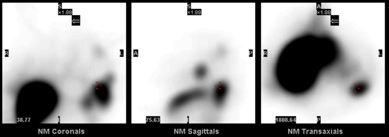
Splenosis is an acquired ectopic autotransplantation of splenic tissue; that occurs after traumatic splenic rupture and splenectomy [1]. Splenosis is a rare but benign disease, and the diagnosis can be challenging as the multiple incidentally found nodules could mimic malignancy [2]. Abdominopelvic Splenosis is thought to occur in as many as 65% of cases of splenic rupture [1]. However, Thoracic Splenosis is rare and usually involve the left parietal and visceral pleura [1,2]. Intraparenchymal lesions are less common but have been reported in cases of parenchymal and diaphragm laceration [1,2]. Taking a thorough history is of utmost importance, as these patients usually present more than two decades after the splenic traumatic rupture. The use of commonly available nuclear studies will further confirm the diagnosis [3]. This will help to avoid unnecessary procedures, like biopsies; and prevent the potential complications. We present a case of Thoracic Splenosis that highlights the importance of taking a detailed history; and the importance of using nuclear studies for the diagnosis. Further adding to its uniqueness, this case showed with multiple intraparenchymal nodules which is a less common presentation of Splenosis.
Figures


References
-
- O-Yurvati A.H., Thompson J.B., Woods T.N. Thoracic splenosis more than 40 years after thoracoabdominal trauma. J. Am. Osteopath Assoc. 2013 Nov;113(11):853–856. - PubMed
-
- Kwok C.M., Chen Y.T., Lin H.T. Portal vein entrance of splenic erythrocytic progenitor cells and local hypoxia of liver, two events cause intrahepatic splenosis. Med. Hypotheses. 2006;67:1330–1332. - PubMed
-
- Sánchez-Paniagua I., Baleato-González S., García-Figueiras R. Splenosis: non-invasive diagnosis of a great mimicker. RevEsp Enferm. Dig. 2016 Jan;108(1):40–41. - PubMed
Publication types
LinkOut - more resources
Full Text Sources
Other Literature Sources
