

Short-term mechanisms influencing volumetric brain dynamics
- PMID: 28971004
- PMCID: PMC5609861
- DOI: 10.1016/j.nicl.2017.09.002
Short-term mechanisms influencing volumetric brain dynamics
Abstract
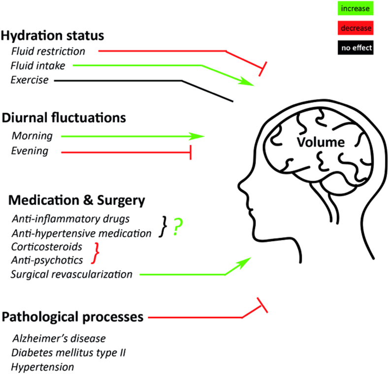
With the use of magnetic resonance imaging (MRI) and brain analysis tools, it has become possible to measure brain volume changes up to around 0.5%. Besides long-term brain changes caused by atrophy in aging or neurodegenerative disease, short-term mechanisms that influence brain volume may exist. When we focus on short-term changes of the brain, changes may be either physiological or pathological. As such determining the cause of volumetric dynamics of the brain is essential. Additionally for an accurate interpretation of longitudinal brain volume measures by means of neurodegeneration, knowledge about the short-term changes is needed. Therefore, in this review, we discuss the possible mechanisms influencing brain volumes on a short-term basis and set-out a framework of MRI techniques to be used for volumetric changes as well as the used analysis tools. 3D T1-weighted images are the images of choice when it comes to MRI of brain volume. These images are excellent to determine brain volume and can be used together with an analysis tool to determine the degree of volume change. Mechanisms that decrease global brain volume are: fluid restriction, evening MRI measurements, corticosteroids, antipsychotics and short-term effects of pathological processes like Alzheimer's disease, hypertension and Diabetes mellitus type II. Mechanisms increasing the brain volume include fluid intake, morning MRI measurements, surgical revascularization and probably medications like anti-inflammatory drugs and anti-hypertensive medication. Exercise was found to have no effect on brain volume on a short-term basis, which may imply that dehydration caused by exercise differs from dehydration by fluid restriction. In the upcoming years, attention should be directed towards studies investigating physiological short-term changes within the light of long-term pathological changes. Ultimately this may lead to a better understanding of the physiological short-term effects of pathological processes and may aid in early detection of these diseases.
Keywords: Brain volume; CVR, Cerebrovascular reactivity; Dehydration; FSL; Magnetic resonance imaging.
Figures
References
-
- Alsop D.C., Detre J.A., Golay X., Günther M., Hendrikse J., Hernandez-Garcia L., Lu H., Macintosh B.J., Parkes L.M., Smits M., Van Osch M.J.P., Wang D.J.J., Wong E.C., Zaharchuk G. Recommended implementation of arterial spin-labeled perfusion mri for clinical applications: a consensus of the ISMRM perfusion study group and the European consortium for ASL in dementia. Magn. Reson. Med. 2015;73:102–116. - PMC - PubMed
-
- Alzheimer A. Uber eine eigenartige Erkrankung der Hirnrinde. Allg Zeits Psychiatry Psych. Y Gerichtl. Med. 1907;64:146–148.
-
- Amato D., Beasley C.L., Hahn M.K., Vernon A.C. Neuroadaptations to antipsychotic drugs: insights from pre-clinical and human post-mortem studies. Neurosci. Biobehav. Rev. 2016 - PubMed
Publication types
MeSH terms
LinkOut - more resources
Full Text Sources
Other Literature Sources
Research Materials
