

Evaluation of striatonigral connectivity using probabilistic tractography in Parkinson's disease
- PMID: 28971007
- PMCID: PMC5608174
- DOI: 10.1016/j.nicl.2017.09.009
Evaluation of striatonigral connectivity using probabilistic tractography in Parkinson's disease
Abstract
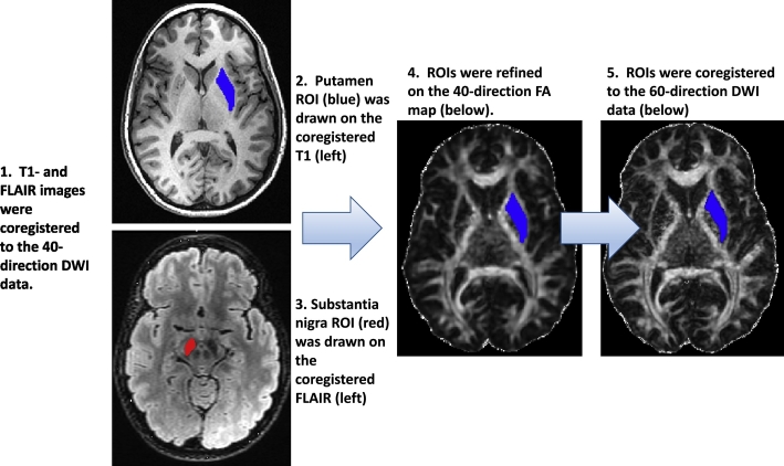
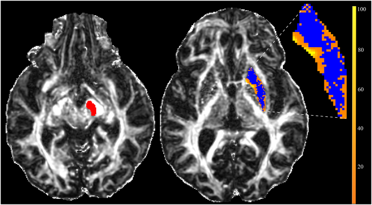
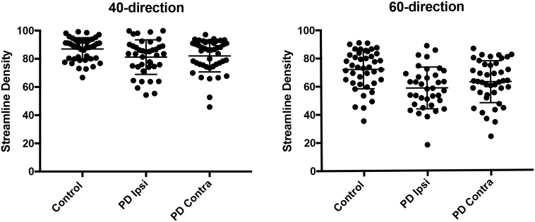
The cardinal movement abnormalities of Parkinson's disease (PD), including tremor, muscle rigidity, and reduced speed and frequency of movements, are caused by degeneration of dopaminergic neurons in the substantia nigra that project to the putamen, compromising information flow through frontal-subcortical circuits. Typically, the nigrostriatal pathway is more severely affected on the side of the brain opposite (contralateral) to the side of the body that manifests initial symptoms. Several studies have suggested that PD is also associated with changes in white matter microstructural integrity. The goal of the present study was to further develop methods for measuring striatonigral connectivity differences between PD patients and age-matched controls using diffusion weighted magnetic resonance imaging (MRI). In this cross-sectional study, 40 PD patients and 44 controls underwent diffusion weighted imaging (DWI) using a 40-direction MRI sequence as well as an optimized 60-direction sequence with overlapping slices. Regions of interest (ROIs) encompassing the putamen and substantia nigra were hand drawn in the space of the 40-direction data using high-contrast structural images and then coregistered to the 60-direction data. Probabilistic tractography was performed in the native space of each dataset by seeding the putamen ROI with an ipsilateral substantia nigra classification target. The effect of disease group (PD versus control) on mean putamen-SN connection probability and streamline density were then analyzed using generalized linear models controlling for age, gender, education, as well as seed and target region characteristics. Mean putamen-SN streamline density was lower in PD on both sides of the brain and in both 40- and 60-direction data. The optimized sequence provided a greater separation between PD and control means; however, individual values overlapped between groups. The 60-direction data also yielded mean connection probability values either trending (ipsilateral) or significantly (contralateral) lower in the PD group. There were minor between-group differences in average diffusion measures within the substantia nigra ROIs that did not affect the results of the GLM analyses when included as covariates. Based on these results, we conclude that mean striatonigral structural connectivity differs between PD and control groups and that use of an optimized 60-direction DWI sequence with overlapping slices increases the sensitivity of the technique to putative disease-related differences. However, overlap in individual values between disease groups limits its use as a classifier.
Keywords: ADRC, Alzheimer's Disease Research Center; AFNI, Analysis of Functional NeuroImages; Aged brain/metabolism/*pathology; BET, brain extraction tool; DWI, diffusion-weighted imaging; Diffusion tensor imaging/*methods; FA, fractional anisotropy; FLAIR, fluid attenuated inversion recovery; FOV, field of view; FSL, Oxford Centre for Functional MRI of the Brain Software Library; GE, general electric; HY, Hoehn and Yahr; Humans; ICC, interclass correlation coefficient; IRB, institutional review board; LMPD, longitudinal MRI biomarkers in Parkinson's disease study; MD, mean diffusivity; MRI, magnetic resonance imaging; PD, Parkinson's disease; PET, Positron Emission Tomography; Parkinson disease/classification/*pathology; RD, radial diffusivity; ROI, region of interest; SD, standard deviation; SN, substantia nigra; SNR, signal to noise ratio; SPECT, single photon emission tomography; SPM, Statistical Parametric Mapping software; Severity of illness index; TE, echo time; TFCE, threshold-free cluster enhancement; TI, inversion time; TR, repetition time; UPDRS, Unified Parkinson Disease Rating Scale; VA, Veterans Affairs.
Figures
Similar articles
-
Correlation of dopaminergic terminal dysfunction and microstructural abnormalities of the basal ganglia and the olfactory tract in Parkinson's disease.Brain. 2013 Oct;136(Pt 10):3028-37. doi: 10.1093/brain/awt234. Epub 2013 Sep 6. Brain. 2013. PMID: 24014521
-
Diffusion tensor imaging of the substantia nigra in Parkinson's disease revisited.Hum Brain Mapp. 2016 Jul;37(7):2547-56. doi: 10.1002/hbm.23192. Epub 2016 Mar 29. Hum Brain Mapp. 2016. PMID: 27029026 Free PMC article.
-
High-resolution diffusion tensor-imaging indicates asymmetric microstructural disorganization within substantia nigra in early Parkinson's disease.J Clin Neurosci. 2018 Apr;50:199-202. doi: 10.1016/j.jocn.2018.01.023. Epub 2018 Feb 1. J Clin Neurosci. 2018. PMID: 29366621
-
The role of diffusion tensor imaging and fractional anisotropy in the evaluation of patients with idiopathic normal pressure hydrocephalus: a literature review.Neurosurg Focus. 2016 Sep;41(3):E12. doi: 10.3171/2016.6.FOCUS16192. Neurosurg Focus. 2016. PMID: 27581308 Review.
-
Diffusion tensor imaging in Parkinson's disease: Review and meta-analysis.Neuroimage Clin. 2017 Jul 15;16:98-110. doi: 10.1016/j.nicl.2017.07.011. eCollection 2017. Neuroimage Clin. 2017. PMID: 28765809 Free PMC article. Review.
Cited by
-
Neuroimaging at 7 Tesla: a pictorial narrative review.Quant Imaging Med Surg. 2022 Jun;12(6):3406-3435. doi: 10.21037/qims-21-969. Quant Imaging Med Surg. 2022. PMID: 35655840 Free PMC article. Review.
-
Giant Tumefactive Perivascular Space: Advanced Fusion MR Imaging and Tractography Study-A Case Report and a Systematic Review.Diagnostics (Basel). 2023 Apr 30;13(9):1602. doi: 10.3390/diagnostics13091602. Diagnostics (Basel). 2023. PMID: 37174993 Free PMC article.
-
Feasibility of diffusion and probabilistic white matter analysis in patients implanted with a deep brain stimulator.Neuroimage Clin. 2020;25:102135. doi: 10.1016/j.nicl.2019.102135. Epub 2019 Dec 14. Neuroimage Clin. 2020. PMID: 31901789 Free PMC article.
-
Mesolimbic white matter connectivity mediates the preference for sweet food.Sci Rep. 2019 Mar 13;9(1):4349. doi: 10.1038/s41598-019-40935-6. Sci Rep. 2019. PMID: 30867529 Free PMC article.
-
The cortico-rubral and cerebello-rubral pathways are topographically organized within the human red nucleus.Sci Rep. 2019 Aug 20;9(1):12117. doi: 10.1038/s41598-019-48164-7. Sci Rep. 2019. PMID: 31431648 Free PMC article.
References
-
- Behrens T.E., Johansen-Berg H., Woolrich M.W., Smith S.M., Wheeler-Kingshott C.A., Boulby P.A., Barker G.J., Sillery E.L., Sheehan K., Ciccarelli O., Thompson A.J., Brady J.M., Matthews P.M. Non-invasive mapping of connections between human thalamus and cortex using diffusion imaging. Nat. Neurosci. 2003;6:750–757. - PubMed
-
- Behrens T.E., Woolrich M.W., Jenkinson M., Johansen-Berg H., Nunes R.G., Clare S., Matthews P.M., Brady J.M., Smith S.M. Characterization and propagation of uncertainty in diffusion-weighted MR imaging. Magn. Reson. Med. 2003;50:1077–1088. - PubMed
-
- Braak H., Del Tredici K., Rub U., de Vos R.A., Jansen Steur E.N., Braak E. Staging of brain pathology related to sporadic Parkinson's disease. Neurobiol. Aging. 2003;24:197–211. - PubMed
-
- Fahn S., Elton R.L. UPDRS Development Committee. The Unified Parkinson's Disease Rating Scale. In: Fahn S., Marsden C.D., Calne D.B., Goldstein M., editors. Recent Developments in Parkinson's Disease. 2nd edn. Macmillan Healthcare Information; Florham Park, NJ: 1987. pp. 153–163. (293–304)
MeSH terms
Grants and funding
LinkOut - more resources
Full Text Sources
Other Literature Sources
Medical
Miscellaneous
