. 2017 Nov 3;16(11):4177-4184.
doi: 10.1021/acs.jproteome.7b00527.

Hydroxylamine Chemical Digestion for Insoluble Extracellular Matrix Characterization

Affiliations

Hydroxylamine Chemical Digestion for Insoluble Extracellular Matrix Characterization

Alexander S Barrett et al. J Proteome Res. .

Abstract

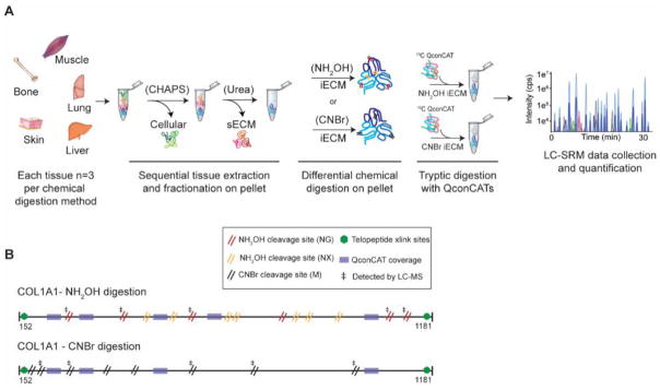
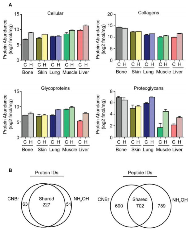
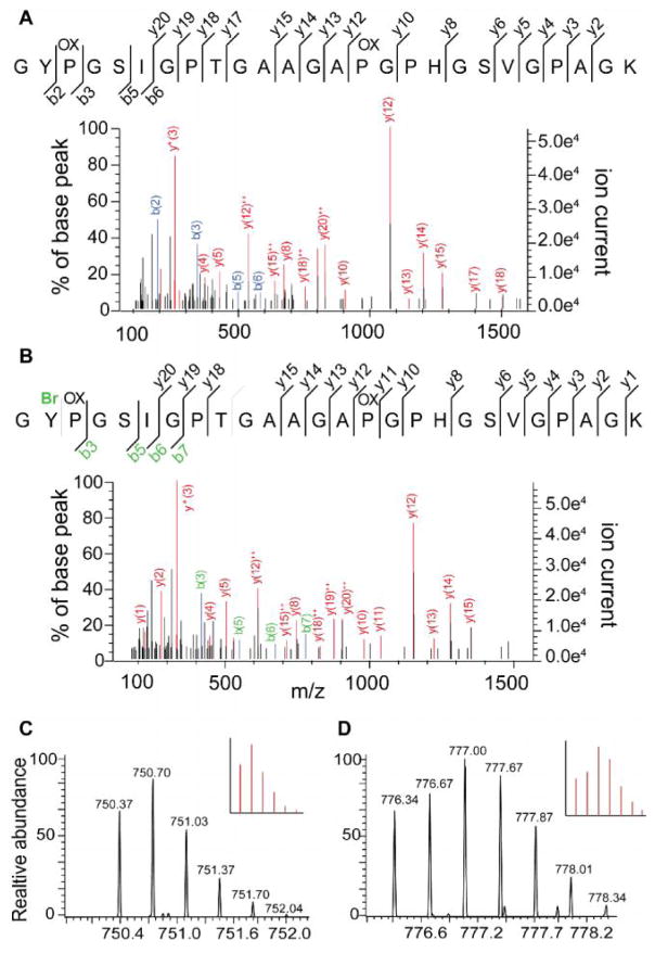
The extracellular matrix (ECM) is readily enriched by decellularizing tissues with nondenaturing detergents to solubilize and deplete the vast majority of cellular components. This approach has been used extensively to generate ECM scaffolds for regenerative medicine technologies and in 3D cell culture to model how the ECM contributes to disease progression. A highly enriched ECM fraction can then be generated using a strong chaotrope buffer that is compatible with downstream bottom-up proteomic analysis or 3D cell culture experiments after extensive dialysis. With most tissues, an insoluble pellet remains after chaotrope extraction that is rich in structural ECM components. Previously, we showed that this understudied fraction represented approximately 80% of total fibrillar collagen from the lung and other ECM fiber components that are known to be covalently cross-linked. Here, we present a hydroxylamine digestion approach for chaotrope-insoluble ECM analysis with comparison to an established CNBr method for matrisome characterization. Because ECM characteristics vary widely among tissues, we chose five tissues that represent unique and diverse ECM abundances, composition, and biomechanical properties. Hydroxylamine digestion is compatible with downstream proteomic workflows, yields high levels of ECM peptides from the insoluble ECM fraction, and reduces analytical variability when compared to CNBr digestion. Data are available via ProteomeXchange with identifier PXD006428.

Keywords: LC−SRM; chemical digestion; collagen; extracellular matrix; insoluble matrix; mass spectrometry; matrisome; proteomics; tissue extraction.


Conflict of interest statement

Conflict of Interest disclosure

The authors declare no competing financial interest.

Figures

Figure 1
Figure 1. Workflow diagram for comparative analysis of CNBr and NH2OH using quantitative QconCAT ECM proteomics
(A) Tissues are sequentially extracted to obtain cellular, soluble ECM (sECM) fractions. The pellet remaining after chaotrope extraction is subjected to either CNBr or NH2OH digestion. QconCATs are spiked into CNBr or NH2OH iECM fractions and samples are then enzymatically digested prior to data acquisition using an LC-SRM approach. (B) Schematic representation of the mature fibrillar collagen fibers of collagen alpha-1(1). CNBr cleavage sites (8 for Col1a1) are denoted by a double black slash while NH2OH cleavage sites (13 for Col1a1) are denoted by a double red (NG) or double orange (NX) slash. Green hexagonal markings denote known sites of cross-linking in the telopeptide region fibrillar collagen. Transparent purple boxes mark sequence regions that match to stable isotope labeled (SIL) QconCAT peptides (Uniprot IDs: Col1a1: P11087 and Col1a2: Q01149). ‡ denotes cleavage sites that were detected by LC-MS.
Figure 2
Figure 2. Characterization of hydroxylamine cleavage specificity and fibrillar collagen abundance iECM fraction
(A) Observed hydroxylamine cleavage specificity across all tissues (1534 peptides total) from data-dependent LC-MS/MS experiments (B) Fibrillar collagen abundance derived from LC-SRM data plotted as percentage of total quantified protein in the iECM fraction.
Figure 3
Figure 3. Comparison of quantitative yield, protein and peptide identifications between CNBr and NH2OH digestion approaches
(A) Protein group abundance plots for cellular proteins, collagens, glycoproteins and proteoglycan protein groups derived from LC-SRM data for tissues prepared with cyanogen bromide (C) or hydroxylamine (H). (B) Venn diagrams of average shared and unique protein and peptide identifications from all tissues derived from data-dependent LC-MS/MS experiments.
Figure 4
Figure 4. CNBr selectively brominates tyrosine containing collagen peptides
(A) Non-brominated form of collagen alpha-2(I) peptide GYPGSIGPTGAAGAPGPHGSVGPAGK. (B) Brominated form of collagen alpha-2(I) peptide GYPGSIGPTGAAGAPGPHGSVGPAGK at position Y2. (C) Isotopic distribution of non-brominated collagen alpha-2(I) peptide, theoretical pattern inset. (D) Isotopic distribution of brominated collagen alpha-2(I) peptide, theoretical pattern inset.

References

    1. Frantz C, Stewart KM, Weaver VM. The extracellular matrix at a glance. Journal of cell science. 2010;123(24):4195–4200. - PMC - PubMed
    1. Mouw JK, Ou G, Weaver VM. Extracellular matrix assembly: a multiscale deconstruction. Nature Reviews Molecular Cell Biology. 2014;15(12):771–785. - PMC - PubMed
    1. DuFort CC, Paszek MJ, Weaver VM. Balancing forces: architectural control of mechanotransduction. Nature reviews Molecular cell biology. 2011;12(5):308–319. - PMC - PubMed
    1. Nelson CM, Bissell MJ. Of extracellular matrix scaffolds and signaling: tissue architecture regulates development homeostasis and cancer. Annual review of cell and developmental biology. 2006;22:287. - PMC - PubMed
    1. Pickup MW, Mouw JK, Weaver VM. The extracellular matrix modulates the hallmarks of cancer. EMBO reports. 2014:e201439246. - PMC - PubMed

Publication types

Substances