. 2018 Jan 1;314(1):H52-H64.
doi: 10.1152/ajpheart.00478.2017. Epub 2017 Sep 29.

TRAF3IP2 mediates high glucose-induced endothelin-1 production as well as endothelin-1-induced inflammation in endothelial cells

Affiliations

TRAF3IP2 mediates high glucose-induced endothelin-1 production as well as endothelin-1-induced inflammation in endothelial cells

Jaume Padilla et al. Am J Physiol Heart Circ Physiol. .

Abstract

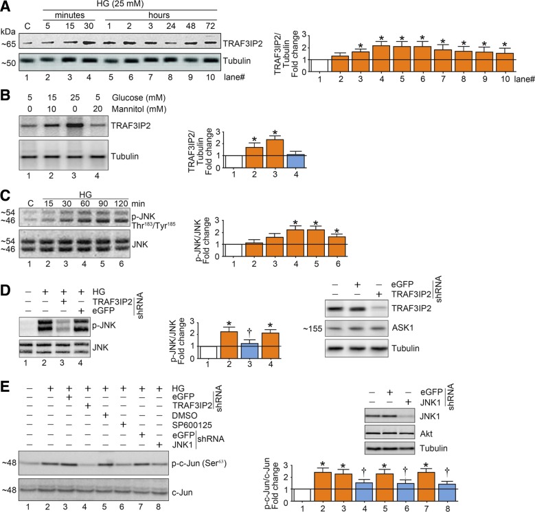
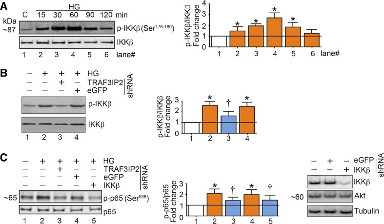
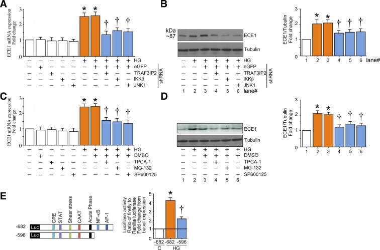
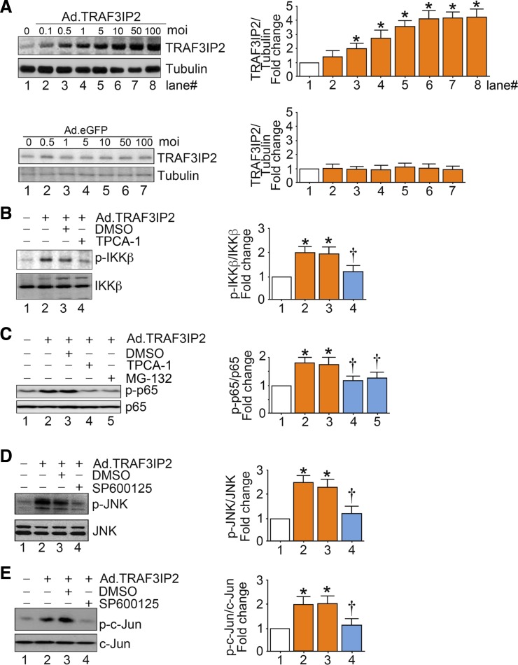
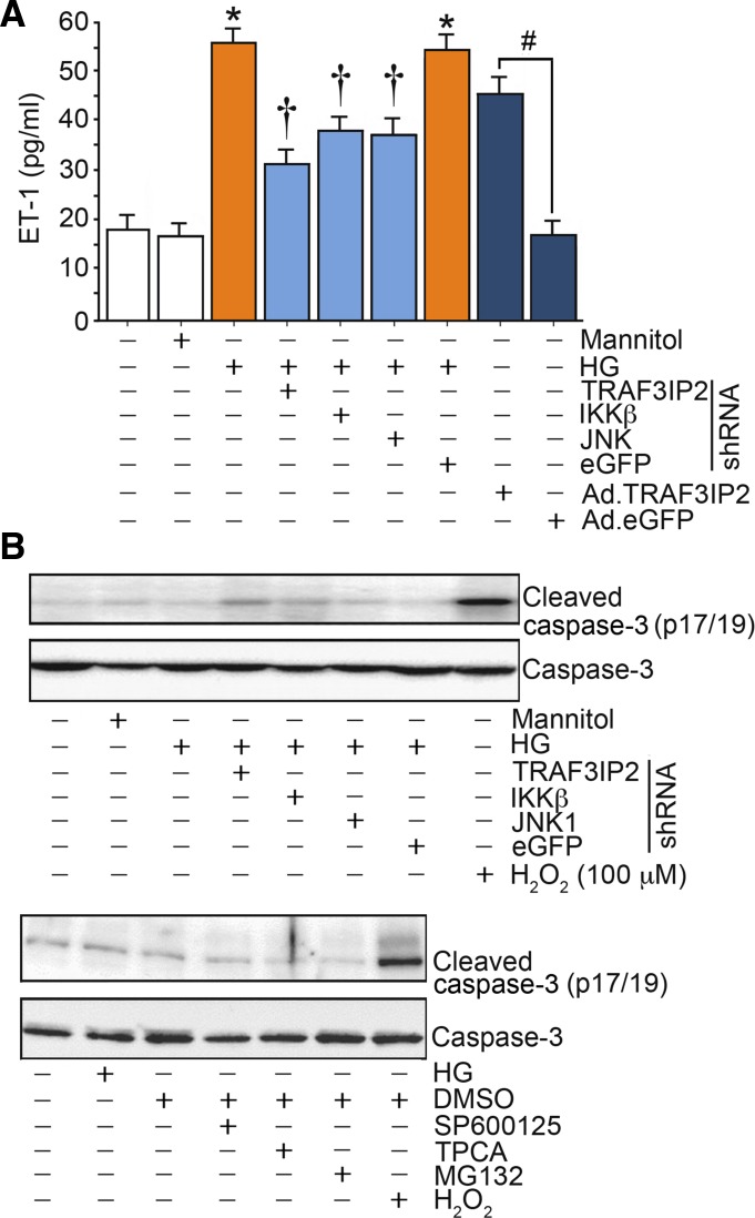
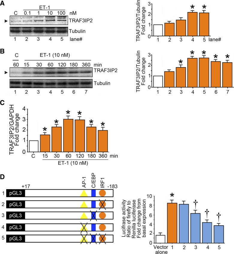
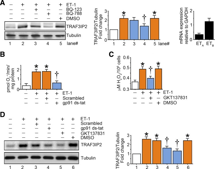
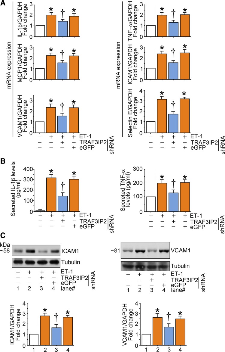
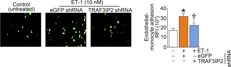
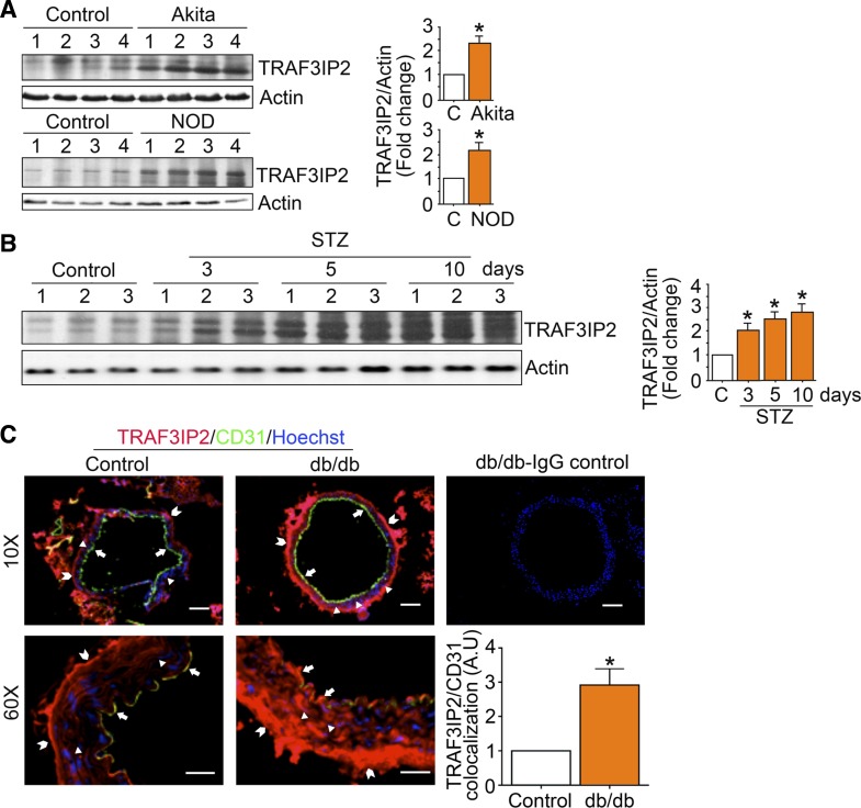
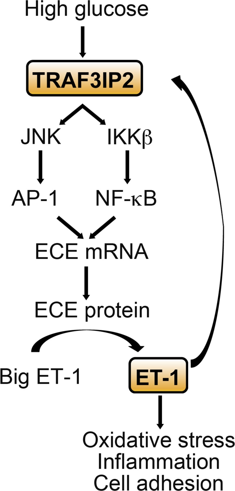
Hyperglycemia-induced production of endothelin (ET)-1 is a hallmark of endothelial dysfunction in diabetes. Although the detrimental vascular effects of increased ET-1 are well known, the molecular mechanisms regulating endothelial synthesis of ET-1 in the setting of diabetes remain largely unidentified. Here, we show that adapter molecule TRAF3 interacting protein 2 (TRAF3IP2) mediates high glucose-induced ET-1 production in endothelial cells and ET-1-mediated endothelial cell inflammation. Specifically, we found that high glucose upregulated TRAF3IP2 in human aortic endothelial cells, which subsequently led to activation of JNK and IKKβ. shRNA-mediated silencing of TRAF3IP2, JNK1, or IKKβ abrogated high-glucose-induced ET-converting enzyme 1 expression and ET-1 production. Likewise, overexpression of TRAF3IP2, in the absence of high glucose, led to activation of JNK and IKKβ as well as increased ET-1 production. Furthermore, ET-1 transcriptionally upregulated TRAF3IP2, and this upregulation was prevented by pharmacological inhibition of ET-1 receptor B using BQ-788, or inhibition of NADPH oxidase-derived reactive oxygen species using gp91ds-tat and GKT137831. Notably, we found that knockdown of TRAF3IP2 abolished ET-1-induced proinflammatory and adhesion molecule (IL-1β, TNF-α, monocyte chemoattractant protein 1, ICAM-1, VCAM-1, and E-selectin) expression and monocyte adhesion to endothelial cells. Finally, we report that TRAF3IP2 is upregulated and colocalized with CD31, an endothelial marker, in the aorta of diabetic mice. Collectively, findings from the present study identify endothelial TRAF3IP2 as a potential new therapeutic target to suppress ET-1 production and associated vascular complications in diabetes. NEW & NOTEWORTHY This study provides the first evidence that the adapter molecule TRAF3 interacting protein 2 mediates high glucose-induced production of endothelin-1 by endothelial cells as well as endothelin-1-mediated endothelial cell inflammation. The findings presented herein suggest that TRAF3 interacting protein 2 may be an important therapeutic target in diabetic vasculopathy characterized by excess endothelin-1 production.

Keywords: TRAF3 interacting protein 2; endothelial dysfunction; endothelin-1; hyperglycemia.


Figures

Fig. 1.
Fig. 1.
High glucose (HG) induces activation of activator protein 1 (AP-1) in part via TRAF3 interacting protein 2 (TRAF3IP2) and JNK in human aortic endothelial cells (HAECs). A: HG (25 mM d-glucose) induces time-dependent TRAF3IP2 expression. At 70% confluency, the complete medium on HAECs was replaced with endothelial basal medium-2 (without supplements) for 2 h, and cells were then incubated with HG for the indicated time periods and analyzed for TRAF3IP2 protein expression by immunoblot analysis. B: HG (15 and 25 mM), but not mannitol, induces TRAF3IP2 expression. HAECs treated as in A with HG or mannitol for 30 min were analyzed for TRAF3IP2 by immunoblot analysis. C: HG (25 mM) induces time-dependent JNK activation. HAECs treated as in A with HG were analyzed for JNK activation using activation-specific antibodies. Total JNK served as a control. D: HG (25 mM) induces JNK activation via TRAF3IP2. At 50% confluency, HAECs were infected with lentiviral TRAF3IP2 or control enhanced green fluorescent protein (eGFP) shRNA (multiplicity of infection 0.5 for 48 h). Cells were then treated with HG for 60 min. Total and activated JNK were analyzed as in C. Knockdown of TRAF3IP2 was confirmed by immunoblot analysis and is shown on the right. ASK1 served as an off-target control. E: HG (25 mM) induced AP-1 activation via TRAF3IP2 and JNK. HAECs infected with TRAF3IP2 or JNK1 shRNA or pretreated with the JNK inhibitor SP600125 (20 µM for 30 min) were incubated with HG for 60 min and analyzed for AP-1 activation by immunoblot analysis using antibodies that specifically detect phosphorylated c-Jun at Ser73. Knockdown of JNK1 is shown in the top right. Bar graphs in AE represent densitometric analyses from 3 independent experiments. *P ≤ 0.05 vs. control (i.e., open bar); †P ≤ 0.05 vs. HG (n = 3).
Fig. 2.
Fig. 2.
High glucose (HG) induces NF-κB activation in part via TRAF3 interacting protein 2 (TRAF3IP2) and IKKβ in aortic endothelial cells (HAECs). A: HG (25 mM) induces time-dependent IKKβ activation. At 70% confluency, the complete medium on HAECs was replaced with endothelial basal medium-2 (without supplements) for 2 h, and cells were then incubated with HG for the indicated time periods and analyzed for IKKβ activation using activation-specific antibodies. Total IKKβ served as a control. B: HG (25 mM) induces IKKβ activation via TRAF3IP2. At 50% confluency, HAECs were infected with lentiviral TRAF3IP2 or control enhanced green fluorescent protein (eGFP) shRNA [multiplicity of infection (MOI) 0.5 for 48 h]. Cells were then incubated with HG for 60 min. Total and activated IKKβ were analyzed by immunoblot analysis. C: HG (25 mM) induces NF-κB activation via TRAF3IP2 and IKKβ. HAECs infected with TRAF3IP2 or IKKβ shRNA (MOI 0.5 for 48 h) were incubated with HG for 60 min and then analyzed for NF-κB activation by immunoblot analysis using antibodies that specifically detect phosphorylated p65 at Ser536. Knockdown of IKKβ was confirmed by immunoblot analysis, as shown on the right. Akt served as an off-target control. Bar graphs in AC represent densitometric analyses from 3 independent experiments. *P ≤ 0.05 vs. control (i.e., open bar); †P ≤ 0.05 vs. HG (n = 3).
Fig. 3.
Fig. 3.
High glucose (HG) induces endothelin converting enzyme 1 (ECE1) expression in part via TRAF3 interacting protein 2 (TRAF3IP2) and its downstream signaling intermediates. A and B: HG (25 mM) induces ECE1 mRNA and protein expression in part via TRAF3IP2, IKKβ, and JNK. At 50% confluency, human aortic endothelial cells (HAECs) infected with lentiviral TRAF3IP2, IKKβ, JNK1, or control eGFP shRNA [multiplicity of infection (MOI) 0.5] for 48 h were treated with HG for 120 min. ECE1 mRNA expression (A) was analyzed by quantitative RT-PCR and protein levels (B) by immunoblot analysis. C and D: pharmacological inhibition of IKKβ, NF-κB, and JNK inhibit HG (25 mM)-induced ECE1 expression. At 70% confluency, the complete medium on HAECs was replaced with endothelial basal medium-2 (without supplements) for 2 h, pretreated with TPCA-1 (5 µM in DMSO), MG-132 (5 µM in DMSO for 1h), SP600125 (20 µM for 30 min), or DMSO vehicle, and then incubated with HG and analyzed for ECE1 mRNA (C) and protein expression (D) after 2 h as in A. E: HG (25 mM) stimulates ECE1 promoter-dependent reporter gene activation via NF-κB and AP-1. HAECs were transfected with a reporter vector containing a 682-bp fragment of the 5′-flanking region of the human ECE1 gene (3 μg for 24 h) with and without the deletions. pGL3-Basic served as a vector control. Cells were cotransfected with the Renilla luciferase vector (100 ng). After transfection, cells were treated with HG for 12 h and harvested for the dual-luciferase assay (n = 6). Firefly luciferase data were normalized to that of corresponding Renilla luciferase activity. Bar graphs in AD represent densitometric analyses from at least 3 independent experiments. AE: *P ≤ 0.01 vs. control (i.e., first open bar); †P ≤ 0.05 vs. HG (n = 3–6).
Fig. 4.
Fig. 4.
Overexpression of TRAF3 interacting protein 2 (TRAF3IP2), by itself, activates IKKβ/NF-κB and JNK/activator protein 1 (AP-1). A: adenoviral transduction of TRAF3IP2. At 70% confluency, human aortic endothelial cells (HAECs) were infected at the indicated multiplicity of infection (MOI) with an adenoviral vector expressing TRAF3IP2. After 24 h, TRAF3IP2 protein levels were analyzed by immunoblot analysis. B: ectopic expression of TRAF3IP2 activates IKKβ. HAECs infected with Ad.TRAF3IP2 (MOI 10 for 24 h) or incubated with TPCA-1 (5 µM in DMSO) were analyzed for IKKβ activation by immunoblot analysis. C: ectopic expression of TRAF3IP2 activates NF-κB via IKKβ and IκB degradation. HAECs treated as in B but with TPCA-1 (5 µM in DMSO) or MG-132 (5 µM in DMSO) were analyzed for NF-κB activation by immunoblot analysis using activation-specific anti-p65 antibodies. D: ectopic expression of TRAF3IP2 activates JNK. HAECs infected with Ad.TRAF3IP2 (MOI 10 for 24 h) and incubated with SP600125 (20 µM in DMSO for 30 min) were analyzed for JNK activation by immunoblot analysis. E: ectopic expression of TRAF3IP2 activates AP-1 via JNK. HAECs treated as in D were analyzed for AP-1 activation by immunoblot analysis using activation-specific anti-c-Jun antibodies. Bar graphs in AE represent densitometric analyses from 3 independent experiments. *P ≤ 0.05 vs. control (i.e., open bar); †P ≤ 0.05 vs. HG (n = 3).
Fig. 5.
Fig. 5.
High glucose (HG) as well as ectopic expression of TRAF3 interacting protein 2 (TRAF3IP2) by itself stimulate endothelin-1 (ET-1) production. A: human aortic endothelial cells (HAECs) infected with lentiviral TRAF3IP2, IKKβ, JNK1, or control eGFP shRNA [multiplicity of infection (MOI) 0.5 for 48 h] or infected with adenoviral vector expressing TRAF3IP2 or control eGFP (MOI 10 for 24 h) were treated with HG (25 mM) for 24 h. ET-1 levels in culture supernatants were analyzed by ELISA. B: demonstration that neither pharmacological inhibitors nor silencing of TRAF3IP2, IKKβ, or JNK1 induced cell death, as evidenced by the low levels of cleaved caspase-3. *P ≤ 0.001 vs. control (i.e., first open bar); †P ≤ 0.05 vs. HG (n = 12); #P < 0.001 vs. Ad.eGFP (n = 12).
Fig. 6.
Fig. 6.
Endothelin-1 (ET-1) induces TRAF3 interacting protein 2 (TRAF3IP2) expression. A and B: ET-1 induces TRAF3IP2 expression in a dose- and time-dependent manner. At 70% confluency, the complete medium on human aortic endothelial cells (HAECs) was replaced with endothelial basal medium-2 (without supplements) for 2 h, and cells were then treated with ET-1 at the indicated concentrations for 1 h (A) or for up to 6 h with ET-1 at 10 nM (B). TRAF3IP2 expression was analyzed by immunoblot analysis. C: ET-1 induces TRAF3IP2 mRNA expression. HAECs were treated with ET-1 (10 nM) for up to 6 h and then analyzed for TRAF3IP2 mRNA expression by quantitative RT-PCR. D: ET-1 stimulates TRAF3IP2 promoter-dependent reporter gene activation in part via CCAAT/enhancer binding protein-β (c/EBPb) and activator protein 1. HAECs were transfected with a reporter vector containing a 200-bp fragment of the 5′-flanking region of the human TRAF3IP2 gene (3 µg for 24 h) with and without mutations (n = 6). pGL3-basic served as a vector control. Cells were cotransfected with the Renilla luciferase vector (100 ng). After transfection, cells were treated with ET-1 (10 nM) for 12 h and harvested for the dual-luciferase assay. Firefly luciferase data were normalized to that of corresponding Renilla luciferase activity. Bar graphs in A and B represent densitometric analyses from 3 independent experiments. AD: *P ≤ 0.01 vs. control (i.e., open bar); †P ≤ 0.05 vs. control or untreated HAECs transfected with the intact TRAF3IP2 promoter reporter construct (n = 3–6).
Fig. 7.
Fig. 7.
Oxidative stress mediates endothelin-1 (ET-1)-induced TRAF3 interacting protein 2 (TRAF3IP2) expression. A: ET-1 induces TRAF3IP2 expression in part via ET-1 receptor B (ETB). At 70% confluency, the complete medium on human aortic endothelial cells (HAECs) was replaced with endothelial basal medium-2 (without supplements) for 2 h, and cells were treated with BQ-123, BQ-788 (1 µM for 1 h), or solvent control DMSO and then with ET-1 (10 nM for 1 h). TRAF3IP2 expression was analyzed by immunoblot analysis (left and middle). ETA and ETB expression at basal conditions was analyzed by quantitative RT-PCR (right). B and C: ET-1 induces oxidative stress. HAECs were treated with the NADPH oxidase (NOX)2 inhibitor gp91ds-tat (1 µM for 1 h) or NOX1/4 dual inhibitor GKT137831 (5 µM in DMSO for 15 min) followed by ET-1 (10 nM) for 15 (B) or 30 (C) min. Superoxide anion (O2−·; B) and H2O2 (C) production were analyzed by cytochrome c assay and Amplex red assay, respectively (n = 6). D: ET-1-induced TRAF3IP2 expression is oxidative stress responsive. HAECs treated as in B and C but for 1 h with ET-1 (10 nM) were analyzed for TRAF3IP2 expression by immunoblot analysis. Bar graphs in A and D represent densitometric analyses from 3 independent experiments. AD: *P ≤ 0.01 vs. control (i.e., open bar); †P ≤ 0.05 vs. ET-1 (n = 3–6).
Fig. 8.
Fig. 8.
Endothelin-1 (ET-1) induces the expression of multiple inflammatory mediators in human aortic endothelial cells (HAECs) in part via TRAF3 interacting protein 2 (TRAF3IP2). AC: ET-1 stimulates proinflammatory cytokine and adhesion molecule expression. At 70% confluency, the complete medium on HAECs was replaced with endothelial basal medium-2 (without supplements) for 2 h, and cells were then incubated with ET-1 (10 nM) for 2 h (A and C) or 24 h (B). mRNA expression was analyzed by quantitative RT-PCR (n = 6), protein expression by immunoblot analysis (n = 3), and secreted cytokine levels by ELISA (data are expressed as absolute values or fold changes; n = 6). Bar graphs in C represent densitometric analyses from 3 independent experiments. AC: *P ≤ 0.01 vs. control (i.e., open bar); †P ≤ 0.05 vs. ET-1 (n = 3–6).
Fig. 9.
Fig. 9.
Endothelin-1 (ET-1) promotes monocyte-endothelial cell adhesion in part via TRAF3 interacting protein 2 (TRAF3IP2). Human aortic endothelial cells (HAECs) at 50% confluency were infected with lentiviral particles expressing TRAF3IP2 or control eGFP shRNA [multiplicity of infection (MOI) 0.5 for 48 h], treated with ET-1 (10 nM) for 12 h, and then incubated with calcein AM-loaded THP-1 monocytic cells for 1 h. Monocyte-endothelial cell adhesion was quantified by measuring fluorescence at excitation and emission wavelengths of 485 and 535 nm, and relative fluorescence intensity was quantified. Wells containing HAECs without THP-1 cells served as blanks. *P ≤ 0.01 vs. control (i.e., open bar); †P ≤ 0.01 vs. ET-1 (n = 6).
Fig. 10.
Fig. 10.
TRAF3 interacting protein 2 (TRAF3IP2) expression is increased in aortic tissues from type 1 and type 2 diabetic mouse models. A and B: TRAF3IP2 expression in aortas from type 1 diabetic Akita and nonobese diabetic (NOD) mice (A) and streptozotocin (STZ)-induced diabetes (B). TRAF3IP2 expression was analyzed by immunoblot analysis. C: colocalization of TRAF3IP2 and CD31, an endothelial cell-specific marker, in aortas from type 2 diabetic db/db mice by immunofluorescence. Positive TRAF3IP2 expression was detected not only in endothelial cells (arrows) but also in the media (arrowheads) and adventitia (block arrows). Magnification: ×10 and ×60. Colocalization of TRAF3IP2 and CD31 demonstrated an ~3-fold increase in TRAF3IP2 expression in CD31-positive endothelial cells (bottom right). A.U., arbitrary units. Scale bars = 10 and 75 µm.
Fig. 11.
Fig. 11.
Summary of findings. Schematic illustrating a vicious cycle resulting from the cross talk between high glucose- and endothelin (ET)-1-mediated TRAF3 interacting protein 2 (TRAF3IP2) induction and TRAF3IP2-dependent ET-converting enzyme 1 (ECE1) transcription, ET-1 production, and ET-1-induced upregulation of proinflammatory and adhesion molecule expression as well as increased monocyte-endothelial cell adhesion. Together, these findings suggest that TRAF3IP2 may be an important therapeutic target in diabetic vasculopathy characterized by excess ET-1 formation. AP-1, activator protein 1.

Comment in

Similar articles

Cited by

References

    1. Adamopoulos C, Piperi C, Gargalionis AN, Dalagiorgou G, Spilioti E, Korkolopoulou P, Diamanti-Kandarakis E, Papavassiliou AG. Advanced glycation end products upregulate lysyl oxidase and endothelin-1 in human aortic endothelial cells via parallel activation of ERK1/2-NF-kappaB and JNK-AP-1 signaling pathways. Cell Mol Life Sci 73: 1685–1698, 2016. doi:10.1007/s00018-015-2091-z. - DOI - PMC - PubMed
    1. Amiri F, Virdis A, Neves MF, Iglarz M, Seidah NG, Touyz RM, Reudelhuber TL, Schiffrin EL. Endothelium-restricted overexpression of human endothelin-1 causes vascular remodeling and endothelial dysfunction. Circulation 110: 2233–2240, 2004. doi:10.1161/01.CIR.0000144462.08345.B9. - DOI - PubMed
    1. Aroor AR, Habibi J, Kandikattu HK, Garro-Kacher M, Barron B, Chen D, Hayden MR, Whaley-Connell A, Bender SB, Klein T, Padilla J, Sowers JR, Chandrasekar B, DeMarco VG. Dipeptidyl peptidase-4 (DPP-4) inhibition with linagliptin reduces western diet-induced myocardial TRAF3IP2 expression, inflammation and fibrosis in female mice. Cardiovasc Diabetol 16: 61, 2017. doi:10.1186/s12933-017-0544-4. - DOI - PMC - PubMed
    1. Babaei S, Picard P, Ravandi A, Monge JC, Lee TC, Cernacek P, Stewart DJ. Blockade of endothelin receptors markedly reduces atherosclerosis in LDL receptor deficient mice: role of endothelin in macrophage foam cell formation. Cardiovasc Res 48: 158–167, 2000. doi:10.1016/S0008-6363(00)00169-3. - DOI - PubMed
    1. Bansilal S, Farkouh ME, Fuster V. Role of insulin resistance and hyperglycemia in the development of atherosclerosis. Am J Cardiol 99: 6B–14B, 2007. doi:10.1016/j.amjcard.2006.11.002. - DOI - PubMed

Publication types

MeSH terms