. 2018 Jan 1;314(1):L144-L149.
doi: 10.1152/ajplung.00379.2017. Epub 2017 Sep 28.

Human lung branching morphogenesis is orchestrated by the spatiotemporal distribution of ACTA2, SOX2, and SOX9

Affiliations

Human lung branching morphogenesis is orchestrated by the spatiotemporal distribution of ACTA2, SOX2, and SOX9

Soula Danopoulos et al. Am J Physiol Lung Cell Mol Physiol. .

Abstract

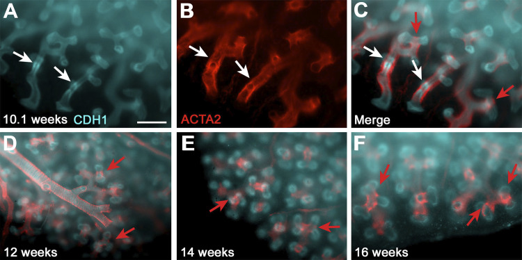
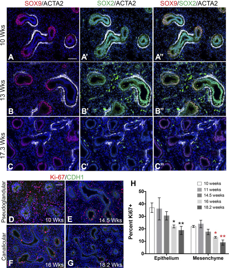
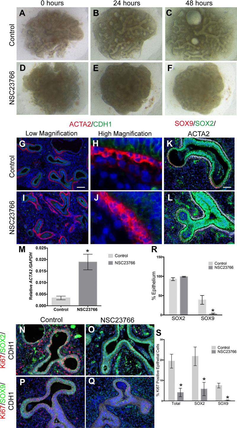
Lung morphogenesis relies on a number of important processes, including proximal-distal patterning, cell proliferation, migration and differentiation, as well as epithelial-mesenchymal interactions. In mouse lung development, SOX2+ cells are localized in the proximal epithelium, whereas SOX9+ cells are present in the distal epithelium. We show that, in human lung, expression of these transcription factors differs, in that during the pseudoglandular stage distal epithelial progenitors at the tips coexpress SOX2 and SOX9. This double-positive population was no longer present by the canalicular stages of development. As in mouse, the human proximal epithelial progenitors express solely SOX2 and are surrounded by smooth muscle cells (SMCs) both in the proximal airways and at the epithelial clefts. Upon Ras-related C3 botulinum toxin substrate 1 inhibition, we noted decreased branching, as well as increased SMC differentiation, attenuated peristalsis, and a reduction in the distal double-positive SOX2/SOX9 progenitor cell population. Thus, the presence of SOX2/SOX9 double-positive progenitor cells in the distal epithelium during the pseudoglandular stage of human lung development appears to be critical to proximal-distal patterning and lung branching. Moreover, SMCs promote a SOX2 proximal phenotype and seem to suppress the SOX9+ population.

Keywords: human lung development; progenitor cells; smooth muscle cells; α-actin 2.


Conflict of interest statement

No conflicts of interest, financial or otherwise, are declared by the authors.

Figures

Fig. 1.
Fig. 1.
Smooth muscle cell (SMC) distribution in the human developing lung. AC: whole mount staining of 10.1 wk human lung for cadherin 1(CDH1, cyan, A) and α-actin 2 (ACTA2, red, B); white arrows show epithelial condensation (A) and SMCs that gathering in a ring-like structure (B). C: merge of A and B; white arrows point to the ring of SMCs around the condensation of epithelial cells, and red arrows show the knot-forming SMCs at the cleft of very distal epithelial branches. DF: CDH1 and ACTA2 staining on intact lung edges of 12 (D)-, 14 (E)-, and 16 (F)-wk lungs. Red arrows point to the knot-forming SMCs in the cleft. Scale: 500 μm.
Fig. 2.
Fig. 2.
Epithelial cells in the branch tips of pseudoglandular stage human lungs coexpress SOX2 and SOX9 and are highly proliferative. AC′′: immunostaining of human fetal lung tissue in the pseudoglandular (10 and 13 wk gestation) and canalicular (17.3 wk gestation) stages of development with SOX2, SOX9, and ACTA2 antibodies. AA′′: 10 wk human fetal lung stained with SOX9/ACTA2 (red/white) (A), SOX2/ACTA2 (green/white) (A′), and the overlay of the triple stain (A′′). BB′′: 13-wk human fetal lung stained with SOX9/ACTA2 (red/white) (B), SOX2/ACTA2 (green/white) (B′), and the overlay of the triple stain (B′′). CC′′: 17.3-wk human fetal lung stained with SOX9/ACTA2 (red/white) (C), SOX2/ACTA2 (green/white) (C′), and the overlay of the triple stain (C′′). DG: immunostaining of human fetal lung tissue in the pseudoglandular (10 and 14.5 wk gestation; D and E) and canalicular (16 and 18.2 wk gestation; F and G) stages of development with Ki-67 and CDH1. H: graph comparing percent of Ki-67+ epithelial cells with the total no. of epithelial cells and percent of Ki-67+ mesenchymal cells with the total no. mesenchymal cells at each gestational time point (n = 3 for each time point). Epithelium: *P = 0.0197 and **P = 0.0227; mesenchyme: *P = 0.0014 and **P = 0.0046. Scale bars: 50 μm.
Fig. 3.
Fig. 3.
SMC function is important for human lung branching. AF: 11.1 wk human fetal lung explants (C and F) in the presence (DF) or absence (AC) of NSC-23766 (25 μM). GJ: immunostaining of NSC-23766 (I and J) and control explants (G and H) with ACTA2 (red) and CDH1 (green) antibodies at low (G and I) and high (H and J) magnifications. Scale bar is 100 μm. M: qRT-PCR for ACTA2 expressed as relative expression compared with GAPDH (n = 5, P = 0.0049). K and L: immunostaining of NSC-23766 (L) and control explants (K) with SOX9 (red), SOX2 (green), and ACTA2 (white). Scale bar is 50 μm. N and O: immunostaining of NSC-23766 (O) and control (N) explants with Ki-67 (red), SOX2 (green), and CDH1 (white). P and Q: immunostaining of NSC-23766 (Q) and control (P) explants with Ki-67 (red), SOX9 (green), and CDH1 (white). R: percentage of epithelial SOX2+ cells and epithelial SOX9+ cells to total epithelial cells in control vs. NSC-23766-treated explants (n = 3, SOX9: P = 0.035). S: percentage of total epithelial Ki-67+ (n = 5, P = 0.0049), Ki-67/SOX2+ (n = 3, P = 0.046), and Ki-67/SOX9+ (n = 3, P = 0.0034) cells in control vs. NSC-23766-treated explants. *Statistically significant difference.
Fig. 4.
Fig. 4.
Schematic distribution of ACTA2, SOX2, and SOX9 in the developing lung. A: SOX2/SOX9 progenitor cells are present in the distal lung epithelial tips in the pseudoglandular stage; their presence is restricted by SMCs. SMCs repress the expression of SOX9 while simultaneously allowing an expansion of SOX2. B: SOX2/SOX9 progenitor cells then disappear in the canalicular stage during which SOX2+ cells are solely localized in the proximal airways, and SOX9+ cells are localized in the distal epithelial tips, coinciding with decreased proliferation. C: in NSC3766-treated explants, an expansion of ACTA2 and SOX2 and a decrease in SOX9 and cell proliferation are observed. D: in mouse lungs, distal tip progenitors contain only SOX9+ cells.

References

    1. Alanis DM, Chang DR, Akiyama H, Krasnow MA, Chen J. Two nested developmental waves demarcate a compartment boundary in the mouse lung. Nat Commun 5: 3923, 2014. doi:10.1038/ncomms4923. - DOI - PMC - PubMed
    1. Badri KR, Zhou Y, Schuger L. Embryological origin of airway smooth muscle. Proc Am Thorac Soc 5: 4–10, 2008. doi:10.1513/pats.200704-049VS. - DOI - PMC - PubMed
    1. Blanc P, Coste K, Pouchin P, Azaïs JM, Blanchon L, Gallot D, Sapin V. A role for mesenchyme dynamics in mouse lung branching morphogenesis. PLoS One 7: e41643, 2012. doi:10.1371/journal.pone.0041643. - DOI - PMC - PubMed
    1. Danopoulos S, Krainock M, Toubat O, Thornton M, Grubbs B, Al Alam D. Rac1 modulates mammalian lung branching morphogenesis in part through canonical Wnt signaling. Am J Physiol Lung Cell Mol Physiol 311: L1036–L1049, 2016. doi:10.1152/ajplung.00274.2016. - DOI - PMC - PubMed
    1. Dekkers BG, Maarsingh H, Meurs H, Gosens R. Airway structural components drive airway smooth muscle remodeling in asthma. Proc Am Thorac Soc 6: 683–692, 2009. doi:10.1513/pats.200907-056DP. - DOI - PubMed

Publication types