. 2018 Jan 1;314(1):L165-L176.
doi: 10.1152/ajplung.00490.2016. Epub 2017 Sep 28.

Activation of the sweet taste receptor, T1R3, by the artificial sweetener sucralose regulates the pulmonary endothelium

Affiliations

Activation of the sweet taste receptor, T1R3, by the artificial sweetener sucralose regulates the pulmonary endothelium

Elizabeth O Harrington et al. Am J Physiol Lung Cell Mol Physiol. .

Abstract

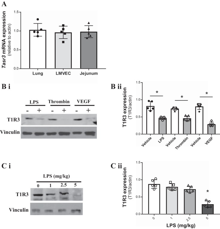
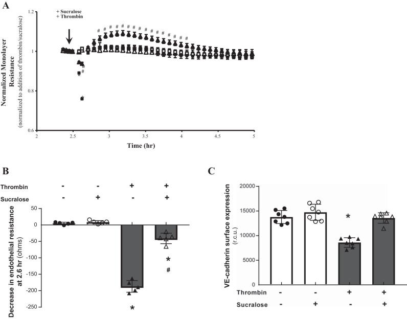
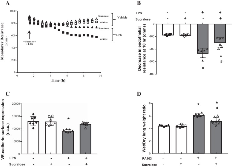
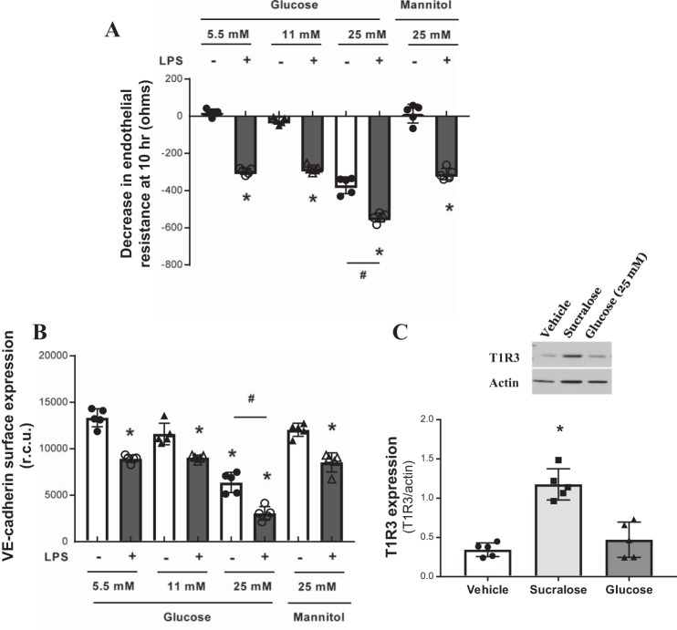
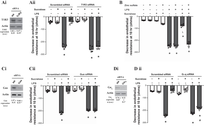
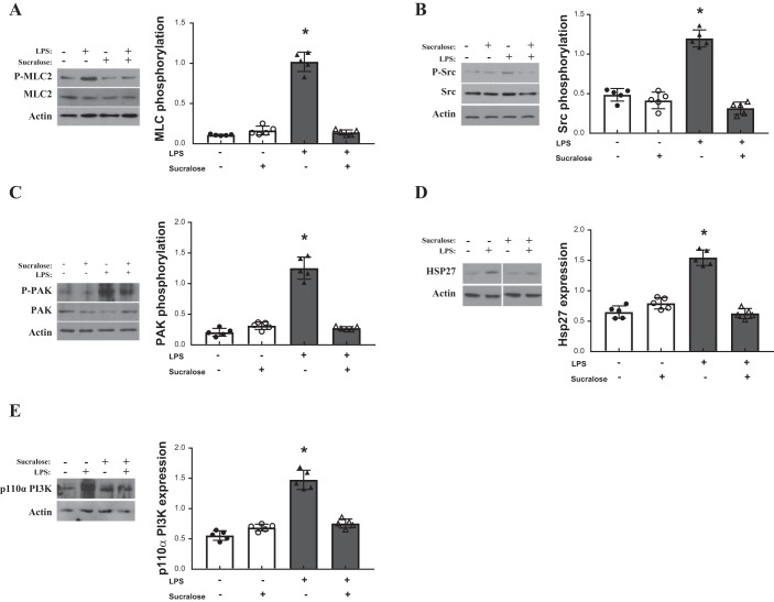
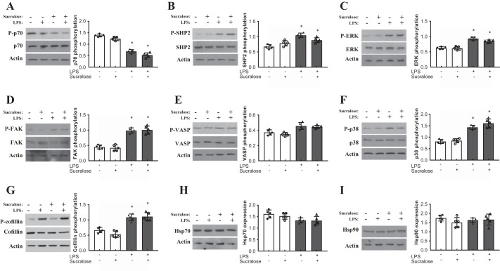
A hallmark of acute respiratory distress syndrome (ARDS) is pulmonary vascular permeability. In these settings, loss of barrier integrity is mediated by cell-contact disassembly and actin remodeling. Studies into molecular mechanisms responsible for improving microvascular barrier function are therefore vital in the development of therapeutic targets for reducing vascular permeability in ARDS. The sweet taste receptor T1R3 is a G protein-coupled receptor, activated following exposure to sweet molecules, to trigger a gustducin-dependent signal cascade. In recent years, extraoral locations for T1R3 have been identified; however, no studies have focused on T1R3 within the vasculature. We hypothesize that activation of T1R3, in the pulmonary vasculature, plays a role in regulating endothelial barrier function in settings of ARDS. Our study demonstrated expression of T1R3 within the pulmonary vasculature, with a drop in expression levels following exposure to barrier-disruptive agents. Exposure of lung microvascular endothelial cells to the intensely sweet molecule sucralose attenuated LPS- and thrombin-induced endothelial barrier dysfunction. Likewise, sucralose exposure attenuated bacteria-induced lung edema formation in vivo. Inhibition of sweet taste signaling, through zinc sulfate, T1R3, or G-protein siRNA, blunted the protective effects of sucralose on the endothelium. Sucralose significantly reduced LPS-induced increased expression or phosphorylation of the key signaling molecules Src, p21-activated kinase (PAK), myosin light chain-2 (MLC2), heat shock protein 27 (HSP27), and p110α phosphatidylinositol 3-kinase (p110αPI3K). Activation of T1R3 by sucralose protects the pulmonary endothelium from edemagenic agent-induced barrier disruption, potentially through abrogation of Src/PAK/p110αPI3K-mediated cell-contact disassembly and Src/MLC2/HSP27-mediated actin remodeling. Identification of sweet taste sensing in the pulmonary vasculature may represent a novel therapeutic target to protect the endothelium in settings of ARDS.

Keywords: T1R3; acute respiratory distress syndrome; artificial sweeteners; pulmonary endothelium; sweet taste.


Figures

Fig. 1.
Fig. 1.
Expression of the sweet taste receptor T1R3 at the pulmonary endothelium is regulated by barrier disruptive agents. A: mRNA expression of the T1R3 gene Tas1r3 in rat lung and jejunum tissue and cultured rat lung microvascular endothelial cells (LMVECs). Gene expression is relative to the housekeeping gene β-actin and normalized to the positive control jejunum tissue; n = 6. B and C: protein expression of T1R3 in cultured rat LMVECs exposed to LPS (1 µg/ml), thrombin (2 U/ml), or VEGF (50 ng/ml) for 24 h (B) and homogenates of lungs from C57/BL6 mice exposed to varying doses of LPS (0–5 mg/kg) (C); n = 5. A representative blot and densitometry relative to the load control (i) and β-actin (ii) are shown. Data are expressed as means ± SD. *P < 0.05 vs. vehicle.
Fig. 2.
Fig. 2.
Stimulation of the sweet taste receptor with the artificial sweetener sucralose attenuates thrombin-induced barrier disruption and VE-cadherin internalization. A and B: changes in rat LMVEC endothelial monolayer resistance were measured using electrical cell impedance sensor in the presence (■, ▲) and absence (□, △) of thrombin (2 U/ml). Monolayers were exposed to sucralose (0.1 mM; ▲, △) or vehicle (H2O; ■, □) at the same time as thrombin. Permeability is shown as an experimental trace, normalized to the addition of thrombin and sucralose (A, arrow) and drop in endothelial resistance (B) measured at 12 min postthrombin and sucralose treatment; n = 5. C: cell surface expression of VE-cadherin was determined, with whole cell indirect ELISA using chemiluminescence, following exposure to thrombin and sucralose as per A; n = 6. Data are expressed as means ± SD. *P < 0.05 vs. vehicle for thrombin; #P < 0.05 vs vehicle for sucralose.
Fig. 3.
Fig. 3.
Stimulation of the sweet taste receptor with the artificial sweetener sucralose attenuates LPS-induced barrier disruption and VE-cadherin internalisation in vitro and bacteria-induced edema formation in vivo. A and B: changes in rat LMVEC endothelial monolayer resistance were measured using electrical cell impedance sensor (ECIS) in the presence (■, ▲) and absence (□, △) of LPS (1 µg/ml). Monolayers were exposed to sucralose (0.1 mM; ▲, △) or vehicle (H2O; ■, □) at the same time as LPS. Permeability is shown as an experimental trace, normalized to the addition of LPS and sucralose (A, arrow) and drop in endothelial resistance (B) measured at 10 h; n = 5. C: cell surface expression of VE-cadherin was determined, with whole cell indirect ELISA using chemiluminescence, following exposure to LPS and sucralose as per A; n = 6. D: lung edema formation was determined by measuring wet-to-dry lung weight ratio in mice following daily gavage of sucralose (1 g/kg) for 1 wk and 4-h exposure to Pseudomonas aeruginosa (PA103); n = 5–8. Data are expressed as means ± SD. *P < 0.05 vs. vehicle for LPS; #P < 0.05 vs vehicle for sucralose.
Fig. 4.
Fig. 4.
High-glucose exposure increases endothelial barrier permeability and VE-cadherin internalisation. A: changes in rat LMVEC endothelial monolayer resistance was measured using ECIS in the presence (closed bars) and absence (open bars) of LPS (1 µg/ml). Monolayers were exposed to different concentrations of glucose (5.5, 11, and 25 mM) or osmotic control mannose (25 mM) at the same time as LPS. Permeability is shown as drop in endothelial resistance measured at 10 h. n = 5. B: cell surface expression of VE-cadherin was determined, with whole cell indirect ELISA using chemiluminescence, following exposure to LPS and glucose as per A. C: protein expression of T1R3 in cultured rat LMVEC exposed to sucralose (0.1 mM), glucose (25 mM), or vehicle for both (H2O) for 24 h. A representative blot (top) and densitometry relative to the load control β-actin (bottom) are shown; n = 5. Data are expressed as means ± SD. *P < 0.05 vs. vehicle for LPS; #P < 0.05 vs. 5.5 mM control.
Fig. 5.
Fig. 5.
Barrier-protective effect of sucralose is mediated through sensing by the sweet taste receptor. A, C, and D: equivalent numbers of rat LMVECs were transiently transfected with scrambled (300 nM, open bars) or T1R3 (300 nM, closed bars) siRNA (Aii), gustducin (Gus, 300 nM, closed bars) siRNA (Cii) or Gαq (300 nM, closed bars) siRNA (Dii). After 48 h, changes in endothelial monolayer resistance were measured using ECIS in the presence and absence of LPS (1 µg/ml) and sucralose (0.1 mM). Permeability is shown as drop in endothelial resistance measured at 10 h (ii). Knockdown of endogenous protein was confirmed by immunoblot analysis of lysates from transiently transfected cells with an antibody specific to T1R3 (Ai), gustducin (Ci), and Gαq (Di). B: monolayer permeability was assessed in the presence and absence of the sweet taste inhibitor zinc sulfate (0.7 mM). Changes in endothelial monolayer resistance were measured using ECIS in the presence and absence of LPS (1 µg/ml) and sucralose (0.1 mM). Permeability is shown as drop in endothelial resistance measured at 10 h; n = 5–6. Data are expressed as means ± SD. *P < 0.05 vs. vehicle for LPS; ʎP < 0.05 vs vehicle for sucralose; #P < 0.05 vs. LPS + vehicle for sucralose.
Fig. 6.
Fig. 6.
Sucralose attenuates LPS-induced elevated heat shock protein 27 (HSP27) and p110α phosphatidylinositol 3-kinase (p110αPI3K) and activation of myosin light chain-2 (MLC2), Src, and p21-activated kinase (PAK). Rat LMVECs were treated in the presence or absence of LPS (1 µg/ml) and sucralose (0.1 mM) for 24 h. Phosphorylation of MLC-2 (A), Src (B), and PAK (C) was assessed in whole cell lysates by immunoblot analysis with an antibody specific to each phosphorylated protein. Blots were stripped and reprobed for total protein expression and actin as a loading control. Total protein expression of HSP27 (D) and p110αPI3K (E) was also assessed in whole cell lysates, followed strip and reprobe of blots for actin as a loading control. Representative blots are shown. Nonessential lanes from the HSP27 representative blot (D) have been removed; n = 6. Data are expressed as means ± SD. *P < 0.05 vs. vehicle for LPS.
Fig. 7.
Fig. 7.
Role of sucralose on LPS-mediated signaling is independent of several key molecules. Rat LMVECs were treated in the presence or absence of LPS (1 µg/ml) and sucralose (0.1 mM) for 24 h. Phosphorylation of HSP70 (A), SHP2 (B), ERK (C), FAK (D), VASP (E), p38 (F), and cofillin (G) was assessed in whole-cell lysates by immunoblot analysis with an antibody specific to each phosphorylated protein. Blots were stripped and reprobed for total protein expression and actin as a loading control. Total protein expression of HSP70 (H) and HSP90 (I) was also assessed in whole-cell lysates, followed strip and reprobe of blots for actin as a loading control. Representative blots are shown. n = 6. Data are expressed as means ± SD. *P < 0.05 vs. vehicle for LPS.

Similar articles

Cited by

References

    1. Abe M, Ono J, Sato Y, Okeda T, Takaki R. Effects of glucose and insulin on cultured human microvascular endothelial cells. Diabetes Res Clin Pract 9: 287–295, 1990. doi: 10.1016/0168-8227(90)90058-2. - DOI - PubMed
    1. Alvarado AG, Thiagarajan PS, Mulkearns-Hubert EE, Silver DJ, Hale JS, Alban TJ, Turaga SM, Jarrar A, Reizes O, Longworth MS, Vogelbaum MA, Lathia JD. Glioblastoma cancer stem cells evade innate immune suppression of self-renewal through reduced TLR4 expression. Cell Stem Cell 20: 450–461.e4, 2017. doi: 10.1016/j.stem.2016.12.001. - DOI - PMC - PubMed
    1. Antonov A, Snead C, Gorshkov B, Antonova GN, Verin AD, Catravas JD. Heat shock protein 90 inhibitors protect and restore pulmonary endothelial barrier function. Am J Respir Cell Mol Biol 39: 551–559, 2008. doi: 10.1165/rcmb.2007-0324OC. - DOI - PMC - PubMed
    1. Aoki T, Tsunekawa K, Araki O, Ogiwara T, Nara M, Sumino H, Kimura T, Murakami M. Type 2 iodothyronine deiodinase activity is required for rapid stimulation of PI3K by thyroxine in human umbilical vein endothelial cells. Endocrinology 156: 4312–4324, 2015. doi: 10.1210/en.2014-1988. - DOI - PMC - PubMed
    1. Barabutis N, Handa V, Dimitropoulou C, Rafikov R, Snead C, Kumar S, Joshi A, Thangjam G, Fulton D, Black SM, Patel V, Catravas JD. LPS induces pp60c-src-mediated tyrosine phosphorylation of Hsp90 in lung vascular endothelial cells and mouse lung. Am J Physiol Lung Cell Mol Physiol 304: L883–L893, 2013. doi: 10.1152/ajplung.00419.2012. - DOI - PMC - PubMed

Publication types

MeSH terms

LinkOut - more resources