. 2017 Nov 1;37(44):10679-10689.
doi: 10.1523/JNEUROSCI.1246-17.2017. Epub 2017 Oct 2.

Submillisecond Optogenetic Control of Neuronal Firing with Two-Photon Holographic Photoactivation of Chronos

Affiliations

Submillisecond Optogenetic Control of Neuronal Firing with Two-Photon Holographic Photoactivation of Chronos

Emiliano Ronzitti et al. J Neurosci. .

Abstract

Optogenetic neuronal network manipulation promises to unravel a long-standing mystery in neuroscience: how does microcircuit activity relate causally to behavioral and pathological states? The challenge to evoke spikes with high spatial and temporal complexity necessitates further joint development of light-delivery approaches and custom opsins. Two-photon (2P) light-targeting strategies demonstrated in-depth generation of action potentials in photosensitive neurons both in vitro and in vivo, but thus far lack the temporal precision necessary to induce precisely timed spiking events. Here, we show that efficient current integration enabled by 2P holographic amplified laser illumination of Chronos, a highly light-sensitive and fast opsin, can evoke spikes with submillisecond precision and repeated firing up to 100 Hz in brain slices from Swiss male mice. These results pave the way for optogenetic manipulation with the spatial and temporal sophistication necessary to mimic natural microcircuit activity.SIGNIFICANCE STATEMENT To reveal causal links between neuronal activity and behavior, it is necessary to develop experimental strategies to induce spatially and temporally sophisticated perturbation of network microcircuits. Two-photon computer generated holography (2P-CGH) recently demonstrated 3D optogenetic control of selected pools of neurons with single-cell accuracy in depth in the brain. Here, we show that exciting the fast opsin Chronos with amplified laser 2P-CGH enables cellular-resolution targeting with unprecedented temporal control, driving spiking up to 100 Hz with submillisecond onset precision using low laser power densities. This system achieves a unique combination of spatial flexibility and temporal precision needed to pattern optogenetically inputs that mimic natural neuronal network activity patterns.

Keywords: holography; optogenetics; two-photon excitation.


Figures

Figure 1.
Figure 1.
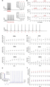
Chronos 2P kinetics and spectra. a, 2P holographic photostimulation and 2P scanning imaging optical setup. PC, polarizer cube; λ/2, half-wave plate; M, mirror; L, lens; CL, cylindrical lens; D, dichroic; LC-VR, liquid crystal variable retarder; GM, galvanometric mirrors; F, filter; OBJ, objective; C, condenser. Inset, IR-transmitted (left) and 2P fluorescence (right) images of Chronos-expressing CHO cells. A patched cell is photostimulated by a holographic illumination pattern matching its surface (red pattern). b, Normalized peak photocurrent (n = 6 cells) over different illumination power densities. Inset, Representative photocurrents generated by irradiating a Chronos-expressing CHO cell with different illumination power densities (p = 0.04, 0.06, 0.09, 0.1, 0.12, 0.14 mW/μm2; Δt = 4 ms; λ = 1030 nm). c, d, On kinetics (c; measured as time to 90% of the peak) and off kinetics over different illumination power densities (d; n = 4 cells; different symbols and colors correspond to different cells). e, Chronos 2P action spectra (data are shown as mean ± SEM; n = 4 cells). Inset, Representative photocurrents generated by irradiating a Chronos-expressing CHO cell with different illumination wavelengths using equal photon flux.
Figure 2.
Figure 2.
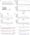
2P activation of Chronos-expressing interneurons. a, 2P image of a portion of an acute cortical brain slice of a virus-injected mouse. A patched/Alexa Fluor 594-filled (white) interneuron of layer 2/3 expressing Chronos-GFP (green) is photostimulated by a 10 μm holographic spot (red spot). Scale bar, 20 μm. b, c, Representative light-evoked photocurrents (b) and membrane potential depolarization (c) over different illumination powers in a Chronos-expressing interneuron. d, Light-evoked currents in spiking (black) and nonspiking (red) neurons for power densities between 0.05 and 0.12 mW/μm2. Each circle corresponds to one cell. e, f, Latencies (e; calculated from the start of the light pulse to the spike depolarization threshold) and jitter (f; calculated as mean deviation of latency) of light-evoked spikes (n = 24 cells) by 10- to 15-μm-diameter spots (data are shown as mean ± SD). Red lines connect data from same cells.
Figure 3.
Figure 3.
Light-driven spiking fidelity across different frequencies in Chronos-expressing interneurons. a, Sample trains of 10 photostimulation pulses of 2 ms duration each over different frequencies: 10 Hz, p = 0.09 mW/μm2 (bottom); 50 Hz, p = 0.09 mW/μm2 (middle); 100 Hz, p = 0.12 mW/μm2 (top). b, Spike probability within the 10 pulses (cells have been photostimulated either with four or 10 light-pulse trains; red shows the number of cells in which the i-th spike has been tested). c, d, Latency (c) and jitter (d) of each light-evoked spike of the train. Illumination power range: 0.05–0.17 mW/μm2, 2–3 ms illumination pulse duration, 10–15 μm diameter spot, λ = 1030 nm. Data are shown as mean ± SD. e, Trains of 10 pulses at 10 Hz triggered every 6 s after 5 min (black line) and after 15 min (blue) preceded by resistance steps. f, Latencies of each light-evoked spike within the train after 0 min (black) and 3.5 min (red; Top, n = 5 cells) and 0 min (black), 3.5 min (red), and 7 min (blue; bottom, n = 3 cells).
Figure 4.
Figure 4.
Light-driven spiking fidelity across different frequencies in CoChR-expressing interneurons. a, Representative light-driven firing induced by a train of 10 illumination pulses at (top) 10 Hz (p = 0.1 mW/μm2; pulse width 2 ms; spot diameter 15 μm) and (bottom) 50 Hz (p = 0.12 mW/μm2; pulse width 1 ms; spot diameter 15 μm). b, Spike probability within the 10 pulses for illumination at 10 Hz (top) and 50 Hz (bottom; n = 4). c, d, Latency (c) and jitter (d) of each light-evoked spike of the train. Illumination power range: 0.05–0.12 mW/μm2, 1–3 ms illumination pulse duration, 15 μm diameter spot, λ = 1030 nm. Data are shown as mean ± SD (n = 4). e, Representative light-driven firing induced by a train of 10 illumination pulses at 100 Hz using different power and pulse width durations. f, Representative normalized photocurrents evoked illuminating with five pulses (2 ms pulse width) at different frequencies Chronos-expressing (black) and a CoChR-expressing (blue) CHO cells (top: 10 Hz, middle: 50 Hz, and bottom: 100 Hz).
Figure 5.
Figure 5.
Scanning imaging effects on Chronos-expressing interneurons. a, 2P image of a patched Chronos-exressing neuron of acute cortical brain slices acquired by raster scanning a 920 nm femtosecond beam (FOV 294 μm; 1.15 μm/line; 2.96 Hz frame rate; p = 14 mW; top); corresponding membrane potential depolarization is shown at bottom. be, Membrane depolarizations obtained on different cells by raster scanning a 920 nm beam over 294 μm (b, d) and 147 μm (c, e) with 14 mW (b, c) and 5 mW (d, e) excitation power for various line-scanning resolutions at 1.32 ms/line.

Similar articles

Cited by

References

    1. Baker CA, Elyada YM, Parra A, Bolton MM (2016) Cellular resolution circuit mapping with temporal-focused excitation of soma-targeted channelrhodopsin. Elife 5. - PMC - PubMed
    1. Baranauskas G. (2015) Can optogenetic tools determine the importance of temporal codes to sensory information processing in the brain? Front Syst Neurosci 9:174. 10.3389/fnsys.2015.00174 - DOI - PMC - PubMed
    1. Bègue A, Papagiakoumou E, Leshem B, Conti R, Enke L, Oron D, Emiliani V (2013) Two-photon excitation in scattering media by spatiotemporally shaped beams and their application in optogenetic stimulation. Biomed Opt Express 4:2869–2879. 10.1364/BOE.4.002869 - DOI - PMC - PubMed
    1. Behrens MM, Ali SS, Dao DN, Lucero J, Shekhtman G, Quick KL, Dugan LL (2007) Ketamine-induced loss of phenotype of fast-spiking interneurons is mediated by NADPH-oxidase. Science 318:1645–1647. 10.1126/science.1148045 - DOI - PubMed
    1. Berndt A, Schoenenberger P, Mattis J, Tye KM, Deisseroth K, Hegemann P, Oertner TG (2011) High-efficiency channelrhodopsins for fast neuronal stimulation at low light levels. Proc Natl Acad Sci U S A 108:7595–7600. 10.1073/pnas.1017210108 - DOI - PMC - PubMed

Publication types

MeSH terms

LinkOut - more resources