. 2017 Nov 1;37(44):10690-10699.
doi: 10.1523/JNEUROSCI.2141-17.2017. Epub 2017 Oct 2.

CaMKII Regulates Synaptic NMDA Receptor Activity of Hypothalamic Presympathetic Neurons and Sympathetic Outflow in Hypertension

Affiliations

CaMKII Regulates Synaptic NMDA Receptor Activity of Hypothalamic Presympathetic Neurons and Sympathetic Outflow in Hypertension

De-Pei Li et al. J Neurosci. .

Abstract

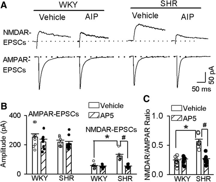
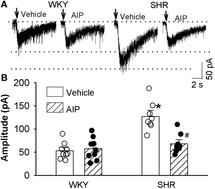
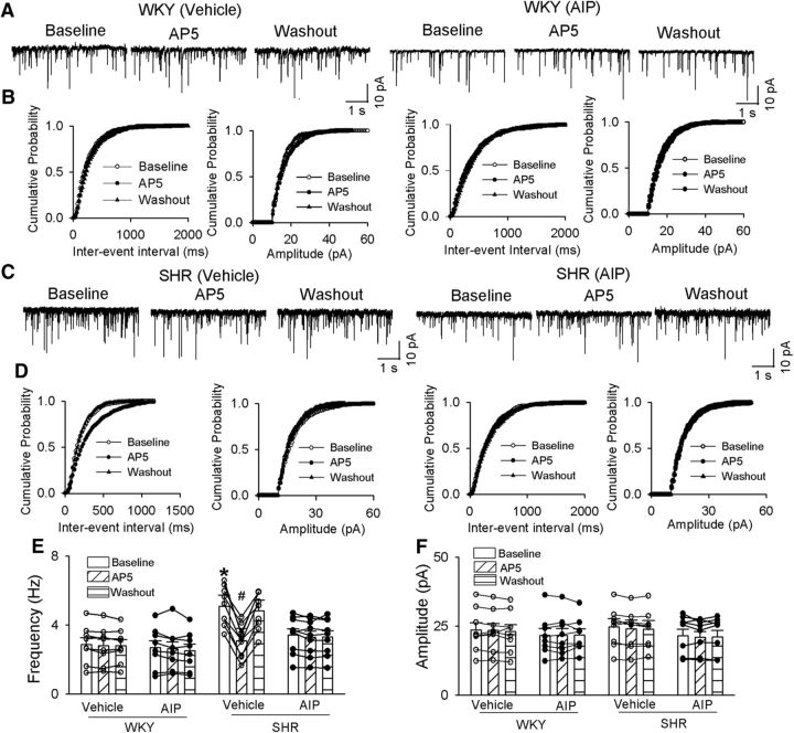
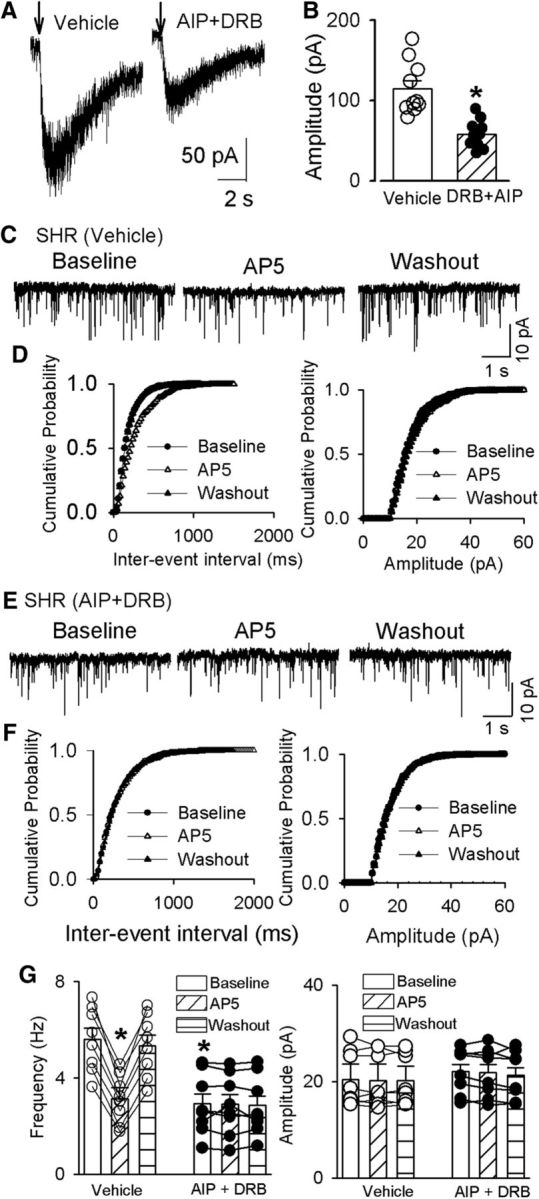
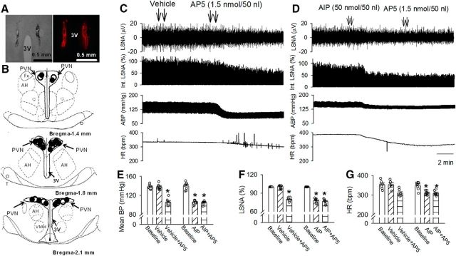
NMDAR activity in the hypothalamic paraventricular nucleus (PVN) is increased and critically involved in heightened sympathetic vasomotor tone in hypertension. Calcium/calmodulin-dependent protein kinase II (CaMKII) binds to and modulates NMDAR activity. In this study, we determined the role of CaMKII in regulating NMDAR activity of PVN presympathetic neurons in male spontaneously hypertensive rats (SHRs). NMDAR-mediated EPSCs and puff NMDA-elicited currents were recorded in spinally projecting PVN neurons in SHRs and male Wistar-Kyoto (WKY) rats. The basal amplitude of evoked NMDAR-EPSCs and puff NMDA currents in retrogradely labeled PVN neurons were significantly higher in SHRs than in WKY rats. The CaMKII inhibitor autocamtide-2-related inhibitory peptide (AIP) normalized the increased amplitude of NMDAR-EPSCs and puff NMDA currents in labeled PVN neurons in SHRs but had no effect in WKY rats. Treatment with AIP also normalized the higher frequency of NMDAR-mediated miniature EPSCs of PVN neurons in SHRs. CaMKII-mediated phosphorylation level of GluN2B serine 1303 (S1303) in the PVN, but not in the hippocampus and frontal cortex, was significantly higher in SHRs than in WKY rats. Lowering blood pressure with celiac ganglionectomy in SHRs did not alter the increased level of phosphorylated GluN2B S1303 in the PVN. In addition, microinjection of AIP into the PVN significantly reduced arterial blood pressure and lumbar sympathetic nerve discharges in SHRs. Our findings suggest that CaMKII activity is increased in the PVN and contributes to potentiated presynaptic and postsynaptic NMDAR activity to elevate sympathetic vasomotor tone in hypertension.SIGNIFICANCE STATEMENT Heightened sympathetic vasomotor tone is a major contributor to the development of hypertension. Although glutamate NMDA receptor (NMDAR)-mediated excitatory drive in the hypothalamus plays a critical role in increased sympathetic output in hypertension, the molecular mechanism involved in potentiated NMDAR activity of hypothalamic presympathetic neurons remains unclear. Here we show that the activity of calcium/calmodulin-dependent protein kinase II (CaMKII) is increased and plays a key role in the potentiated presynaptic and postsynaptic NMDAR activity of hypothalamic presympathetic neurons in hypertension. Also, the inhibition of CaMKII in the hypothalamus reduces elevated blood pressure and sympathetic nerve discharges in hypertension. This new knowledge extends our understanding of the mechanism of synaptic plasticity in the hypothalamus and suggests new strategies to treat neurogenic hypertension.

Keywords: autonomic nervous system; hypertension; hypothalamus; sympathetic nerve activity; synaptic transmission.


Figures

Figure 1.
Figure 1.
CaMKII is involved in increased synaptic NMDAR activity of spinally projecting PVN neurons in SHRs. A, Representative current traces of evoked AMPAR-EPSCs (at a holding potential of −60 mV) and NMDAR-EPSCs (at a holding potential of +40 mV) recorded in labeled PVN neurons from WKY rats and SHRs treated with vehicle or 10 μm AIP. B, Summary data of evoked AMPAR-EPSCs and NMDAR-EPSCs in labeled PVN neurons recorded from WKY rats and SHRs treated with vehicle (n = 7 in the SHR group; n = 9 in the WKY group) or AIP (n = 9 in the SHR group; n = 8 in the WKY group). C, Group data show the ratio of NMDAR-EPSCs to AMPAR-EPSCs in labeled PVN neurons recorded from WKY rats or SHRs treated with vehicle or AIP. Data are presented as the mean ± SEM. One-way ANOVA with Tukey's post hoc test was used to analyze the difference in the NMDAR-EPSCs and the ratio of NMDAR-EPSCs to AMPAR-EPSCs among the four groups. *p < 0.05 compared with the values in the vehicle group in WKY rats. #p < 0.05 compared with the values in the SHR vehicle group.
Figure 2.
Figure 2.
CaMKII contributes to the increased postsynaptic NMDAR activity of spinally projecting PVN neurons in SHRs. A, B, Original current traces (A) and summary data (B) show the effect of 10 μm AIP on puff-NMDA (100 μm)-elicited currents of labeled PVN neurons recorded from WKY rats (vehicle, n = 9; AIP, n = 9) and SHRs (vehicle, n = 8; AIP, n = 9). Data are presented as the mean ± SEM. One-way ANOVA with Tukey's post hoc test was used to analyze the difference in the NMDAR currents among the four groups. *p < 0.05 compared with the respective values in WKY rats; #p < 0.05 compared with the SHR vehicle group.
Figure 3.
Figure 3.
CaMKII increases presynaptic NMDAR activity of spinally projecting PVN neurons in SHRs. A–D, Original traces (A, C) and cumulative probability analysis (B, D) show the effect of bath application of AP-5 (50 μm) on mEPSCs of labeled PVN neurons recorded from WKY rats (A, B) and SHRs (C, D) that were treated with vehicle or 10 μm AIP. E, F, Summary data show the effects of AIP and AP-5 on the frequency and amplitude of mEPSCs in labeled PVN neurons of WKY rats (vehicle, n = 8; AIP, n = 8) and SHRs (vehicle, n = 9; AIP, n = 9). Data are presented as the mean ± SEM. *p < 0.05 compared with the respective value in WKY rats; #p < 0.05 compared with the baseline in the SHR vehicle group.
Figure 4.
Figure 4.
CaMKII and CK2 contributes to enhanced synaptic NMDAR activity of spinally projecting PVN neurons in SHRs. A, B, Original current traces (A) and summary data (B) show the effect of AIP (10 μm) plus DRB (100 μm) on puff NMDA (100 μm)-elicited currents of labeled PVN neurons recorded from SHRs (vehicle, n = 7; AIP plus DRB, n = 8). C–F, Original traces (C, E) and cumulative probability analyses (D, F) show that DRB plus AIP decreased the basal frequency of mEPSCs of labeled PVN neurons recorded from SHRs (C, E) and eliminated the effect of AP-5 (50 μm) on the frequency of mEPSCs (E, F). G, Summary data show the effects of AIP plus DRB and AP-5 on the frequency and amplitude of mEPSCs in labeled PVN neurons of SHRs (vehicle, n = 10; AIP plus DRB, n = 10). Data are presented as the mean ± SEM. *p < 0.05 compared with the baseline in the SHR vehicle group.
Figure 5.
Figure 5.
CaMKII-mediated phosphorylation of NMDARs in PVNs in WKY rats and SHRs. A, B, Representative gel images (A) and summary data (B) show the phosphorylated GluN2B (Ser1303) levels (normalized to GAPDH) in the PVN, hippocampus, and frontal cortex in WKY rats and SHRs (n = 6 rats in each group). C, Original ABP traces and group data show the effect of CGx and sham surgery on the mean ABP in SHRs (n = 6 rats in each group). D, Representative images and summary data show the phosphorylated GluN2B (Ser1303) levels (normalized to GAPDH) in the PVN in SHRs subjected to CGx or sham surgery (n = 6 rats in each group). E, Summary data show the mRNA levels of CaMKII-α and CaMKII-β subunits in PVN tissues from WKY and SHR (n = 7 rats in each group). F, Representative gel images and summary data show that CaMKII-α and CaMKII-β protein levels in the PVN tissue in WKY rats and SHRs (n = 6 rats in each group). Data are presented as the mean ± SEM. *p < 0.05 compared with the WKY group; #p < 0.05 compared with the sham group.
Figure 6.
Figure 6.
CaMKII-dependent NMDAR activity in the PVN maintains elevated sympathetic vasomotor tone in SHRs. A, B, A representative image (A) and schematic drawing (B) show the microinjection sites for vehicle plus AP-5 (●) and AIP plus AP-5 (○) in the PVN in SHRs. C, D, Representative recording traces show the effect of vehicle (C) or AIP (D) followed by AP-5 bilateral injections into the PVNs on ABP, LSNA, and HR in SHRs. E–G, Summary data show changes in mean ABP (E), LSNA (F), and HR (G) in response to AP-5 after microinjection of vehicle (n = 6) or AIP (n = 7) into the PVN in SHRs. Data are presented as the mean ± SEM. *p < 0.05 compared with the respective baseline. 3V, Third ventricle; AH, anterior hypothalamus.

Similar articles

Cited by

References

    1. Allen AM. (2002) Inhibition of the hypothalamic paraventricular nucleus in spontaneously hypertensive rats dramatically reduces sympathetic vasomotor tone. Hypertension 39:275–280. 10.1161/hy0202.104272 - DOI - PubMed
    1. Barcomb K, Hell JW, Benke TA, Bayer KU (2016) The CaMKII/GluN2B protein interaction maintains synaptic strength. J Biol Chem 291:16082–16089. 10.1074/jbc.M116.734822 - DOI - PMC - PubMed
    1. Bard L, Groc L (2011) Glutamate receptor dynamics and protein interaction: lessons from the NMDA receptor. Mol Cell Neurosci 48:298–307. 10.1016/j.mcn.2011.05.009 - DOI - PubMed
    1. Barria A, Muller D, Derkach V, Griffith LC, Soderling TR (1997) Regulatory phosphorylation of AMPA-type glutamate receptors by CaM-KII during long-term potentiation. Science 276:2042–2045. 10.1126/science.276.5321.2042 - DOI - PubMed
    1. Bayer KU, De Koninck P, Leonard AS, Hell JW, Schulman H (2001) Interaction with the NMDA receptor locks CaMKII in an active conformation. Nature 411:801–805. 10.1038/35081080 - DOI - PubMed

Publication types

Substances

LinkOut - more resources