. 2017 Dec 1;292(48):19935-19951.
doi: 10.1074/jbc.M117.812792. Epub 2017 Sep 28.

Photorhabdus luminescens lectin A (PllA): A new probe for detecting α-galactoside-terminating glycoconjugates

Affiliations

Photorhabdus luminescens lectin A (PllA): A new probe for detecting α-galactoside-terminating glycoconjugates

Ghamdan Beshr et al. J Biol Chem. .

Abstract

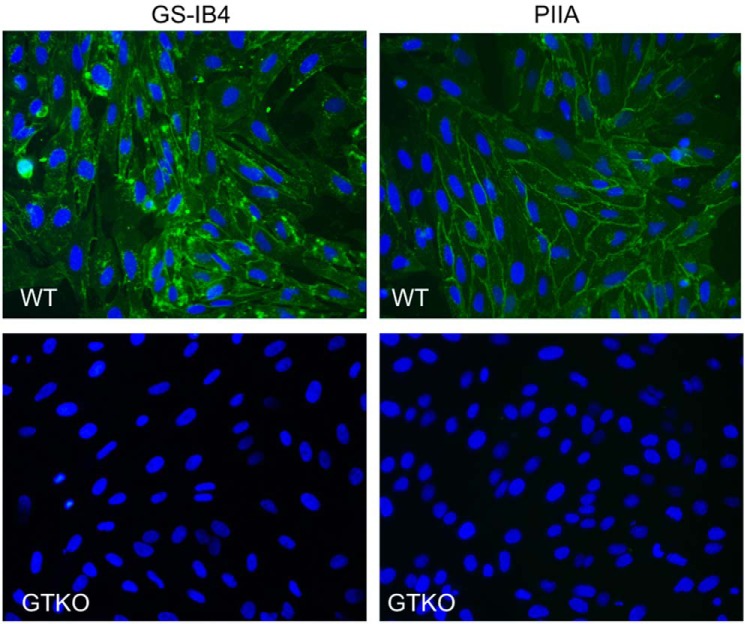
Lectins play important roles in infections by pathogenic bacteria, for example, in host colonization, persistence, and biofilm formation. The Gram-negative entomopathogenic bacterium Photorhabdus luminescens symbiotically lives in insect-infecting Heterorhabditis nematodes and kills the insect host upon invasion by the nematode. The P. luminescens genome harbors the gene plu2096, coding for a novel lectin that we named PllA. We analyzed the binding properties of purified PllA with a glycan array and a binding assay in solution. Both assays revealed a strict specificity of PllA for α-galactoside-terminating glycoconjugates. The crystal structures of apo PllA and complexes with three different ligands revealed the molecular basis for the strict specificity of this lectin. Furthermore, we found that a 90° twist in subunit orientation leads to a peculiar quaternary structure compared with that of its ortholog LecA from Pseudomonas aeruginosa We also investigated the utility of PllA as a probe for detecting α-galactosides. The α-Gal epitope is present on wild-type pig cells and is the main reason for hyperacute organ rejection in pig to primate xenotransplantation. We noted that PllA specifically recognizes this epitope on the glycan array and demonstrated that PllA can be used as a fluorescent probe to detect this epitope on primary porcine cells in vitro In summary, our biochemical and structural analyses of the P. luminescens lectin PllA have disclosed the structural basis for PllA's high specificity for α-galactoside-containing ligands, and we show that PllA can be used to visualize the α-Gal epitope on porcine tissues.

Keywords: carbohydrate; carbohydrate-binding protein; glycobiology; lectin; protein structure; structural biology.


Conflict of interest statement

The authors declare that they have no conflicts of interest with the contents of this article. The content is solely the responsibility of the authors and does not necessarily represent the official views of the National Institutes of Health.

Figures

Figure 1.
Figure 1.
Sequence alignment of LecA from P. aeruginosa with hypothetical LecA-like proteins from Photorhabdus and Xenorhabdus species (one single ortholog per organism selected based on highest identity to LecA). Strictly conserved amino acids are shaded black, and similarly conserved amino acids are shaded gray. Black dot, amino acid of LecA involved in Ca2+ binding; black triangles, amino acids of LecA involved in sugar binding; asterisks, amino acids of LecA involved in both Ca2+ and sugar binding. Amino acid numbering follows the LecA crystal structure where the N-terminal methionine is lacking. The depicted protein sequence of P. luminescens (PllA) is encoded by the plu2096 gene.
Figure 2.
Figure 2.
A, recombinant expression and affinity purification of PllA analyzed by SDS-PAGE (15%). E. coli whole-cell extracts of uninduced (lane 1) and IPTG-induced cultures (lane 3), and purified PllA (lane 4), molecular mass marker in kDa (lane 2) are shown. B, Sepharose size-exclusion chromatogram of PllA; C, DLS analysis of PllA.
Figure 3.
Figure 3.
Carbohydrate specificity of PllA on a glycan array. A, profiling of the glycan-binding specificity of PllA on a glycan array. Glycans containing terminal α-galactoside are colored black; those with terminal β-galactoside are red, and oligosaccharides with neither of these terminal moieties are colored blue. Selected structures showing highest apparent binding are illustrated in CFG notation. B, comparison of glycan-binding specificity between PllA (this work) and LecA (PA-IL (32)) from P. aeruginosa based on normalized CFG glycan array data. Normalization was done by dividing RFU averages of the glycan of interest by the RFU average of the ligand with the highest apparent affinity on the respective array (i.e. glycan 550 for PllA and glycan 31 for LecA). Glycan structures are indicated and depicted with the different spacers of the array for LecA. For the PllA array, identical glycans generally contain β-Sp8 spacers except 105 (Sp0) and 116 (β-Sp0). Numbers on top of columns indicate glycan number of the respective arrays. A and B, error bars correspond to six replicates.
Figure 4.
Figure 4.
Establishing a carbohydrate-binding assay for PllA in solution. A, structure of fluorescent ligands 1–4 based on d-galactose. B, titration of fluorescent ligands 1–4 with PllA. C, dissociation constant for 1 was obtained from a four-parameter fitting procedure to the dose-dependent increase in fluorescence polarization (Kd, 62.7 ± 3.8 μm). D, competitive inhibition of the binding of 1 to PllA with methyl α-d-galactoside (12, IC50 = 0.52 ± 0.07 mm) and raffinose (25, IC50 = 0.11 ± 0.01 mm). One representative titration experiment of triplicates on one plate is shown. Dissociation constant and standard deviations given were obtained from at least three independent replicates of triplicates on three plates each.
Figure 5.
Figure 5.
A, sequence of PllA. Secondary structure elements are shown above the sequence (blue arrows, β-strands; red barrel, α-helix). Residues responsible for sugar binding are highlighted with magenta stars, Ca2+-binding residues with cyan circles, and amino acids coordinating both as yellow triangles. Tail residues unique to PllA and its close homologs are highlighted with a blue box. B, schematic representation of a PllA apo monomer. C, fold diagram for the structure shown in B.
Figure 6.
Figure 6.
Overall structure of PllA and comparison to LecA. A, schematic representation of the PllA tetramer. Two parallel dimers (yellow/magenta and green/cyan) form tail-to-tail dimers with a 90° twist. B, detailed view of the PllA tail-to-tail interface. We observe two hydrogen bonds between the side chains of tail Ser-119 (cyan) and Lys-82 (yellow) and the C terminus of the tail and the backbone nitrogen of Ala-74 (yellow). In addition, tail residue Leu-121 is inserted into a hydrophobic pocket of its binding partner. C, LecA tetramer is planar, formed by tail-to-tail dimerization of two parallel dimers (yellow/magenta and green/cyan). D, much shorter tail of LecA provides several stabilizing hydrogen bonds (dashed lines), but the interactions are not sufficient to cause a twist of the two dimers relative to each other.
Figure 7.
Figure 7.
PllA–carbohydrate complex structures. A and B, PllA bound to methyl α-d-galactoside (12). This interaction is stabilized through 10 hydrogen bonds (dashed lines). Eight of them are between the ligand and the protein, and two are provided by the Ca2+ ion. C and D, PllA bound to raffinose (25). In addition to the hydrogen bonds observed in A, the glucose moiety forms a hydrogen bond with the side chain of Gln-57, whereas terminal fructofuranoside forms two hydrogen bonds with the side chain of Glu-44, which results in the ligand adopting a horseshoe shape. E and F, PllA bound to fluorescent tracer 1. No interactions with the protein are observed beyond the carbohydrate moiety. The fluorescein can only be observed as the result of fortuitous crystal contacts in half of the monomers in the asymmetric unit. PllA is shown as a yellow schematic/surface representation, ligand as gray sticks, oxygen atoms in red, nitrogen atoms in blue, sulfur atoms in yellow, and Ca2+ ions as green spheres. Difference electron density (FoFc) contoured to 3σ with phases calculated from a model that was refined in the absence of metal ions is shown as gray isomesh (B, D, and F).
Figure 8.
Figure 8.
Rationalizing PllA α-galactoside specificity. A, representation of the PllA (yellow)-binding pocket with methyl α-d-galactoside (12, gray sticks). 4-Nitrophenyl β-d-galactoside (salmon sticks and isomesh, taken from PDB 3ZYF) was superposed onto α-d-galactoside. Because of the restricted ligand-binding site of PllA only α-substituted ligands, leading away from the surface, can be accommodated, whereas β-substituted ligands clash. B, superposition of the binding site amino acid residues of PllA (blue) with LecA (magenta), oxygen atoms, red; nitrogen atoms, blue. Residue numbers correspond to PllA.
Figure 9.
Figure 9.
Staining of primary porcine kidney cells from wild-type pigs (WT) and GGTA1 KO (GTKO) animals unable to produce the α-Gal antigen. Fluorescein-tagged PllA or GS-IB4 were used as probes and detected the α-Gal antigen in WT cells. Lectin concentration: PllA, 50 μg/ml; GS-IB4, 500 μg/ml, 400× magnification.
Figure 10.
Figure 10.
Hemagglutination of porcine red blood cells by PllA. A, wild-type pig RBCs. B, GTKO pig RBCs. C, inhibition of PllA-mediated agglutination of wild-type pig RBCs with raffinose.

Similar articles

Cited by

References

    1. Poinar G. O., Thomas G. M., and Hess R. (1977) Characteristics of the specific bacterium associated with Heterorhabditis bacteriophora (Heterorhabditidae: Rhabditida). Nematologica 23, 97–102
    1. Thomas G. M., and Poinar J. R. (1979) Xenorhabdus gen. nov., a genus of entomopathogenic, nematophilic bacteria of the family Enterobacteriaceae. Int. J. Syst. Evol. Microbiol. 29, 352–360
    1. Boemare N., Akhurst R., and Mourant R. (1993) DNA relatedness between Xenorhabdus spp. (Enterobacteriaceae), symbiotic bacteria of entomopathogenic nematodes, and a proposal to transfer Xenorhabdus luminescens to a new genus, Photorhabdus gen. nov. Int. J. Syst. Evol. Microbiol. 43, 249–255
    1. Forst S., and Nealson K. (1996) Molecular biology of the symbiotic-pathogenic bacteria Xenorhabdus spp., and Photorhabdus spp. Microbiol. Rev. 60, 21–43 - PMC - PubMed
    1. Fischer-Le Saux M., Viallard V., Brunel B., Normand P., and Boemare N. E. (1999) Polyphasic classification of the genus Photorhabdus and proposal of new taxa: P. luminescens subsp. luminescens subsp. nov., P. luminescens subsp. akhurstii subsp. nov., P. luminescens subsp. laumondii subsp. nov., P. temperata sp. nov., P. temperata subsp. temperata subsp. nov., and P. asymbiotica sp. nov. Int. J. Syst. Bacteriol. 49, 1645–1656 - PubMed

Publication types

LinkOut - more resources