. 2017 Nov 24;292(47):19356-19365.
doi: 10.1074/jbc.M117.795187. Epub 2017 Sep 28.

Interphotoreceptor retinoid-binding protein removes all- trans-retinol and retinal from rod outer segments, preventing lipofuscin precursor formation

Affiliations

Interphotoreceptor retinoid-binding protein removes all- trans-retinol and retinal from rod outer segments, preventing lipofuscin precursor formation

Chunhe Chen et al. J Biol Chem. .

Abstract

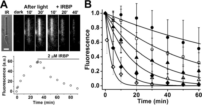
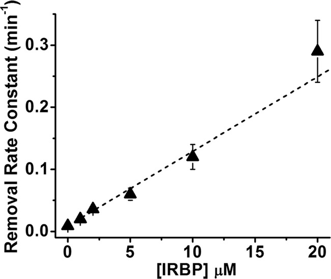
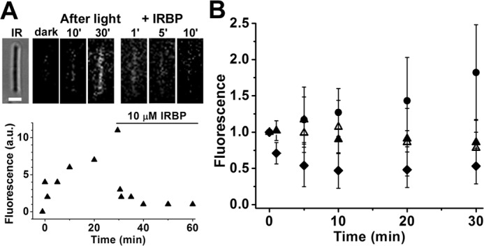
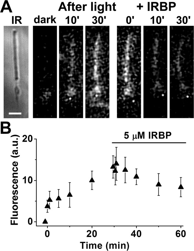
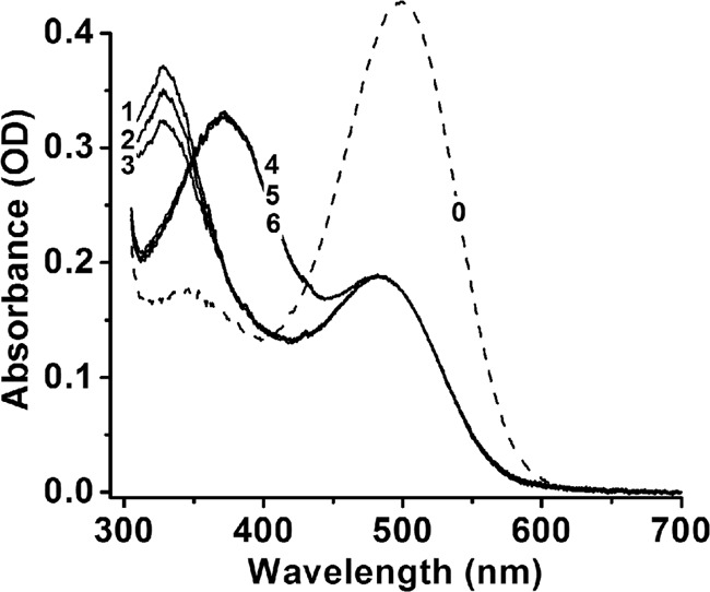
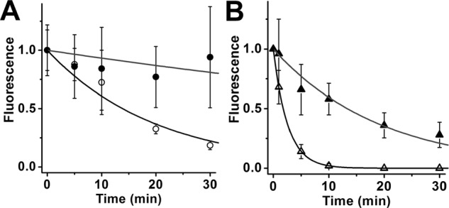
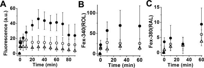
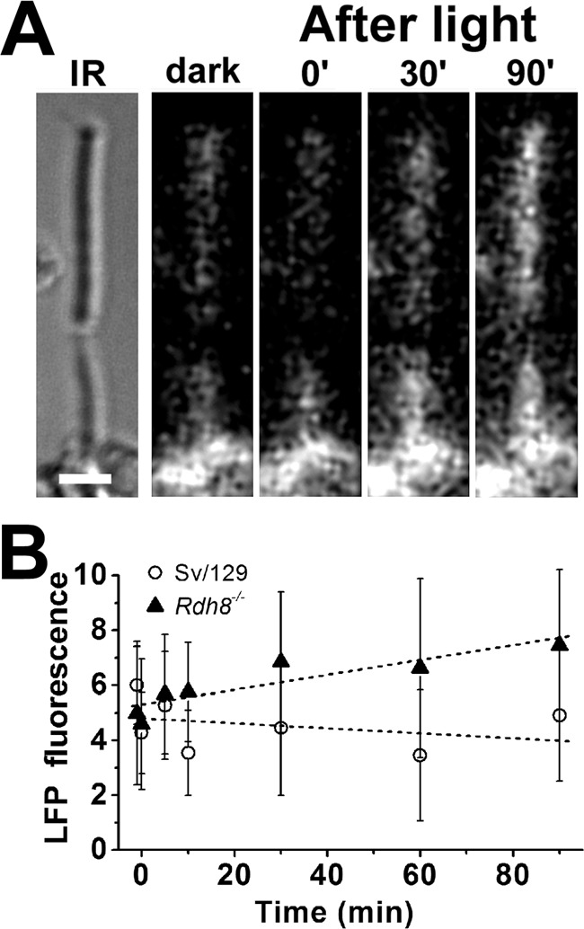
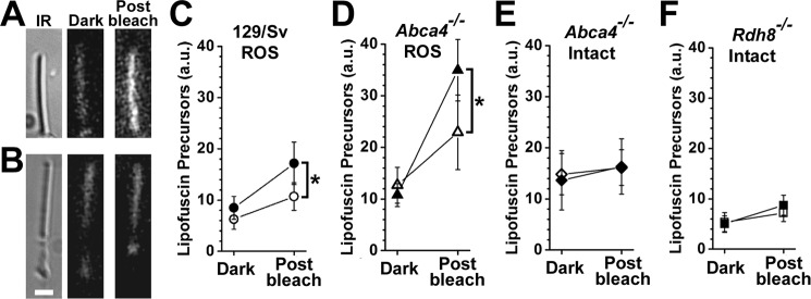
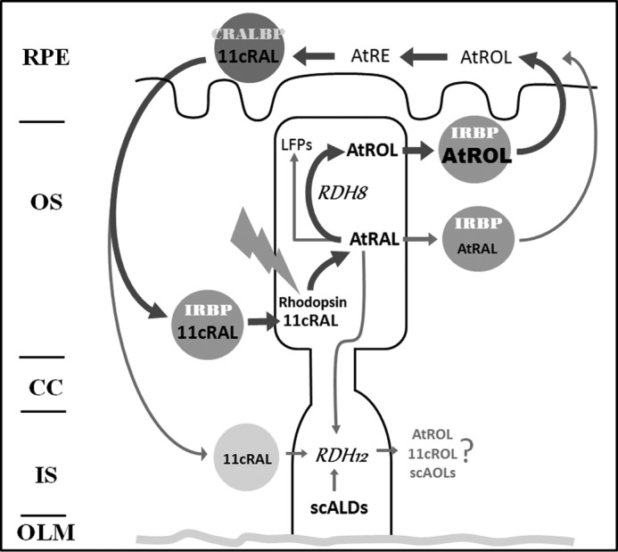
Interphotoreceptor retinoid-binding protein (IRBP) is a specialized lipophilic carrier that binds the all-trans and 11-cis isomers of retinal and retinol, and this facilitates their transport between photoreceptors and cells in the retina. One of these retinoids, all-trans-retinal, is released in the rod outer segment by photoactivated rhodopsin after light excitation. Following its release, all-trans-retinal is reduced by the retinol dehydrogenase RDH8 to all-trans-retinol in an NADPH-dependent reaction. However, all-trans-retinal can also react with outer segment components, sometimes forming lipofuscin precursors, which after conversion to lipofuscin accumulate in the lysosomes of the retinal pigment epithelium and display cytotoxic effects. Here, we have imaged the fluorescence of all-trans-retinol, all-trans-retinal, and lipofuscin precursors in real time in single isolated mouse rod photoreceptors. We found that IRBP removes all-trans-retinol from individual rod photoreceptors in a concentration-dependent manner. The rate constant for retinol removal increased linearly with IRBP concentration with a slope of 0.012 min-1 μm-1 IRBP also removed all-trans-retinal, but with much less efficacy, indicating that the reduction of retinal to retinol promotes faster clearance of the photoisomerized rhodopsin chromophore. The presence of physiological IRBP concentrations in the extracellular medium resulted in lower levels of all-trans-retinal and retinol in rod outer segments following light exposure. It also prevented light-induced lipofuscin precursor formation, but it did not remove precursors that were already present. These findings reveal an important and previously unappreciated role of IRBP in protecting the photoreceptor cells against the cytotoxic effects of accumulated all-trans-retinal.

Keywords: fluorescence; photoreceptor; retina; retinoid-binding protein; retinol.


Conflict of interest statement

The authors declare that they have no conflicts of interest with the contents of this article

Figures

Figure 1.
Figure 1.
Irbp-deficient mice exhibit early and progressive photoreceptor cell loss. Eyes from wild-type (C57BL/6) and Irbp-deficient (Rbp3−/−) mice were fixed in 2% paraformaldehyde and 2% glutaraldehyde, embedded in JB-4 plastic, and Lee's stained sections of retina/RPE/choroid (5 μm) were imaged on a Nikon Eclipse E800 microscope with DMX1200 digital camera. Measurements of outer nuclear layer (ONL) thickness obtained using Image ProPlus 5.0 software were plotted versus distance from the optic nerve head (ONH) ± S.D. and are shown alongside representative micrographs of the superior retina about 500 μm from the optic nerve. Top, C57BL/6 mice: 5 weeks (■, n = 3); 3 months (●, n = 3); 1 year (▴, n = 5). Bottom, Irbp-deficient mice: 6 weeks (□, n = 3); 3 months (○, n = 3); 1 year (▵, n = 4).
Figure 2.
Figure 2.
Measurement of all-trans-retinol removal by different concentrations of IRBP in isolated metabolically intact rod photoreceptors from wild-type mice. A, removal of all-trans-retinol formed after bleaching by 2 μm IRBP. IR, infrared image of a single rod photoreceptor showing its straight outer segment over the rounded tapered inner segment; bar, 5 μm. Bleaching was carried out between t = −1 min and t = 0; IRBP was added 30 min after bleaching. Fluorescence images of the cell were acquired with 360 nm excitation and >420 nm emission. B, removal of all-trans-retinol by different concentrations of IRBP added at t = 0, 30 min after the bleaching of rhodopsin. Retinol outer segment fluorescence intensities have been normalized over the value at t = 0. The lines are simple exponentials, ek·t, decaying to 0 with unitary amplitude at t = 0 min; they have been drawn with rate constants k determined from the experimental data points. Without addition of IRBP, retinol fluorescence decreased with a rate constant 0.009 ± 0.001 min−1 (●, n = 8). IRBP concentrations and removal rate constants are, in μm and min−1, respectively: 1, 0.020 ± 0.001 (○, n = 6); 2, 0.036 ± 0.003 (▴, n = 7); 5, 0.06 ± 0.01 (▵, n = 7); 10, 0.12 ± 0.02 (♦, n = 8); 20, 0.29 ± 0.05 (♢, n = 8). Error bars represent standard deviations. The errors for the removal rate constants were obtained from the curve fits.
Figure 3.
Figure 3.
Linear dependence of the rate constant of retinol removal on the extracellular IRBP concentration (R = 0.99). The slope of the line is 0.012 ± 0.001 min−1 μm−1. Rate constant data are from the experiments shown in Fig. 2.
Figure 4.
Figure 4.
Measurement of all-trans-retinal removal by different concentrations of IRBP in isolated metabolically compromised bROS from wild-type mice. A, removal of all-trans-retinal released after bleaching by 10 μm IRBP. IR, infrared image of the cell; bar, 5 μm. Bleaching was carried out between t = −1 min and t = 0. IRBP was added 30 min after bleaching. Fluorescence images of the cell were acquired with 360 nm excitation and >420 nm emission. B, removal of all-trans-retinal by different concentrations of IRBP added at t = 0, 30 min after the bleaching of rhodopsin. Without addition of IRBP (●, n = 10), retinal fluorescence kept increasing. IRBP concentrations are in μm: 2 (▴, n = 6); 5 (▵, n = 7); 10 (♦, n = 7). Retinal outer segment fluorescence intensities have been normalized over the value at t = 0. Error bars represent standard deviations.
Figure 5.
Figure 5.
Removal of all-trans-retinal by IRBP in isolated metabolically intact rod photoreceptors from Rdh8−/− mice, outer segments that lack Rdh8 cannot reduce the all-trans-retinal released after bleaching. A, IR, infrared image of a metabolically intact rod photoreceptor cell isolated from an Rdh8−/− mouse; bar, 5 μm. Bleaching was carried out between t = −1 min and t = 0; 5 μm IRBP was added 30 min after bleaching. Fluorescence images were acquired with 360 nm excitation and >420 nm emission. B, rod outer segment fluorescence intensity after bleaching between t = −1 min and t = 0, and following the addition of 5 μm IRBP (▴, n = 8). Error bars represent standard deviations.
Figure 6.
Figure 6.
Removal of all-trans-retinol by IRBP in purified bovine ROS membranes. Rhodopsin (spectrum 0, concentration 10 μm) in purified ROS membranes was bleached for 5 min with >495 nm light in the presence of different concentrations of IRBP (0, 2, and 5 μm) and with or without NADPH (100 μm). After 30 min at 37 °C, in the presence of NADPH the released all-trans-retinal had been quantitatively converted to all-trans-retinol (spectra 1–3), whereas in the absence of NADPH it remained unconverted (spectra 4–6). In the absence of IRBP, both all-trans-retinol and all-trans-retinal remained in the membranes (spectra 1 and 4). 2 μm (spectrum 2) and 5 μm (spectrum 3) IRBP removed all-trans-retinol but had no effect on all-trans-retinal (spectra 5 and 6). To obtain the spectra, ROS membranes were solubilized in 1% Ammonyx LO at the end of incubation.
Figure 7.
Figure 7.
Removal of exogenous all-trans-retinal and retinol from rod outer segments. bROS from Rpe65−/− mice, containing negligible amounts of endogenous retinoids, were loaded with 50 μm retinal or retinol for 10 min, supplied with 1% BSA as carrier. Supply of exogenous retinoid was terminated at t = 0. Retinoid outer segment fluorescence intensities have been normalized over the value at t = 0. The lines are simple exponentials, ek·t, decaying to 0 with unitary amplitude at t = 0 min, and have been drawn with rate constants k determined from the experimental data points. A, removal of exogenous all-trans-retinal. In the absence of IRBP, retinal fluorescence declined with rate constant 0.007 ± 0.003 min−1 (●, n = 9); in the presence of 5 μm IRBP, fluorescence decreased with rate constant 0.05 ± 0.01 min−1 (○, n = 4). B, removal of exogenous all-trans-retinol. In the absence of IRBP, retinol fluorescence declined with rate constant 0.052 ± 0.005 min−1 (▴, n = 8); in the presence of 5 μm IRBP, fluorescence decreased with rate constant 0.390 ± 0.002 min−1 (▵, n = 5). Error bars represent standard deviations. The errors for the removal rate constants were obtained from the curve fits.
Figure 8.
Figure 8.
Bleaching rod photoreceptors in the presence of IRBP results in strong reduction of outer segment fluorescence due to all-trans-retinol and all-trans-retinal. Metabolically intact rod photoreceptors from wild-type mice were bleached in the absence and in the presence of different concentrations of IRBP. Bleaching took place between t = −1 and 0 min. A, outer segment fluorescence (excitation, 360 nm; emission, >420 nm) due to both all-trans-retinol and all-trans-retinal at different times after bleaching in the absence (●, n = 8) of IRBP and in the presence of 2 μm (○, n = 10) or 5 μm (▵, n = 13) IRBP. B and C, outer segment fluorescence due to all-trans-retinol (B) and all-trans-retinal (C) at different times after bleaching in the absence (●, n = 7) of IRBP and in the presence of 2 μm (○, n = 6) or 5 μm (▵, n = 5) IRBP. B and C, fluorescence signals Fex-340(ROL) and Fex-380(RAL) due, respectively, to all-trans-retinol and all-trans-retinal were measured simultaneously in the same cells. Error bars represent standard deviations.
Figure 9.
Figure 9.
Formation of LFP in metabolically intact rod photoreceptors of Rdh8−/− mice. A, IR, infrared image of a rod photoreceptor cell isolated from an Rdh8−/− mouse; bar, 5 μm. Bleaching was carried out between t = −1 min and t = 0. Fluorescence images were acquired with 490 nm excitation and >515 nm emission. B, outer segment LFP fluorescence (excitation, 490 nm; emission, >515 nm) after bleaching in rod photoreceptors isolated from Rdh8−/− mice (▴, n = 12). Data from wild-type mice (○, n = 11) are re-plotted from Ref. for comparison. Error bars represent standard deviations.
Figure 10.
Figure 10.
IRBP prevents the formation of lipofuscin precursors but does not remove precursors already formed. LFP levels were measured from rod outer segment fluorescence (excitation, 490 nm; emission, >515 nm) before bleaching (dark) and 60 min after bleaching (post-bleach). A, bROS from a 129/Sv mouse without added IRBP. B, metabolically intact rod photoreceptors from an Abca4−/− mouse without added IRBP. Bar, 5 μm. C, bROS from 129/Sv wild-type mice without added IRBP (●, n = 6) and in the presence of 5 μm IRBP (○, n = 10). D, bROS from Abca4−/− mice without added IRBP (▴, n = 6), and in the presence of 5 μm IRBP (▵, n = 8). E, metabolically intact rod photoreceptors from Abca4−/− mice without added IRBP (♦, n = 7) and in the presence of 5 μm IRBP (♢, n = 7). F, metabolically intact rod photoreceptors from Rdh8−/− mice without added IRBP (■, n = 11) and in the presence of 5 μm IRBP (□, n = 9). Asterisks denote statistical significance. Error bars represent standard deviations.
Figure 11.
Figure 11.
Schematic of the functional coupling of IRBP and RDH8 that facilitates efficient removal of all-trans-retinol (AtROL) from photoreceptor outer segments following rhodopsin bleaching, which is necessary to prevent formation of LFPs. Also shown are lesser pathways of all-trans-retinal (AtRAL) removal, involving direct interaction with IRBP, as well as leakage into the photoreceptor inner segment and reduction by RDH12, a broad specificity reductase for multiple short-chain aldehydes. CRALBP, cellular retinaldehyde-binding protein; 11cRAL, 11-cis-retinal; 11cROL, 11-cis-retinol; scALDs, short-chain aldehydes; scAOLs, short-chain alcohols; RPE, retinal pigment epithelium; OS, outer segment; CC, connecting cilium; IS, inner segment; OLM, outer limiting membrane.

Similar articles

Cited by

References

    1. Ebrey T., and Koutalos Y. (2001) Vertebrate photoreceptors. Prog. Retin. Eye Res. 20, 49–94 - PubMed
    1. Wald G. (1968) Molecular basis of visual excitation. Science 162, 230–239 - PubMed
    1. Chen C., Thompson D. A., and Koutalos Y. (2012) Reduction of all-trans-retinal in vertebrate rod photoreceptors requires the combined action of RDH8 and RDH12. J. Biol. Chem. 287, 24662–24670 - PMC - PubMed
    1. Maeda A., Maeda T., Imanishi Y., Kuksa V., Alekseev A., Bronson J. D., Zhang H., Zhu L., Sun W., Saperstein D. A., Rieke F., Baehr W., and Palczewski K. (2005) Role of photoreceptor-specific retinol dehydrogenase in the retinoid cycle in vivo. J. Biol. Chem. 280, 18822–18832 - PMC - PubMed
    1. Rattner A., Smallwood P. M., and Nathans J. (2000) Identification and characterization of all-trans-retinol dehydrogenase from photoreceptor outer segments, the visual cycle enzyme that reduces all-trans-retinal to all-trans-retinol. J. Biol. Chem. 275, 11034–11043 - PubMed

Publication types