. 2017 Nov 24;292(47):19250-19265.
doi: 10.1074/jbc.M117.797092. Epub 2017 Sep 29.

Pharmacochaperoning in a Drosophila model system rescues human dopamine transporter variants associated with infantile/juvenile parkinsonism

Affiliations

Pharmacochaperoning in a Drosophila model system rescues human dopamine transporter variants associated with infantile/juvenile parkinsonism

H M Mazhar Asjad et al. J Biol Chem. .

Abstract

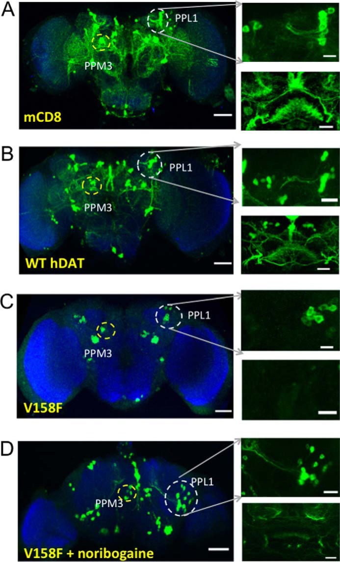
Point mutations in the gene encoding the human dopamine transporter (hDAT, SLC6A3) cause a syndrome of infantile/juvenile dystonia and parkinsonism. To unravel the molecular mechanism underlying these disorders and investigate possible pharmacological therapies, here we examined 13 disease-causing DAT mutants that were retained in the endoplasmic reticulum when heterologously expressed in HEK293 cells. In three of these mutants, i.e. hDAT-V158F, hDAT-G327R, and hDAT-L368Q, the folding deficit was remedied with the pharmacochaperone noribogaine or the heat shock protein 70 (HSP70) inhibitor pifithrin-μ such that endoplasmic reticulum export of and radioligand binding and substrate uptake by these DAT mutants were restored. In Drosophila melanogaster, DAT deficiency results in reduced sleep. We therefore exploited the power of targeted transgene expression of mutant hDAT in Drosophila to explore whether these hDAT mutants could also be pharmacologically rescued in an intact organism. Noribogaine or pifithrin-μ treatment supported hDAT delivery to the presynaptic terminals of dopaminergic neurons and restored sleep to normal length in DAT-deficient (fumin) Drosophila lines expressing hDAT-V158F or hDAT-G327R. In contrast, expression of hDAT-L368Q in the Drosophila DAT mutant background caused developmental lethality, indicating a toxic action not remedied by pharmacochaperoning. Our observations identified those mutations most likely amenable to pharmacological rescue in the affected children. In addition, our findings also highlight the challenges of translating insights from pharmacochaperoning in cell culture to the clinical situation. Because of the evolutionary conservation in dopaminergic neurotransmission between Drosophila and people, pharmacochaperoning of DAT in D. melanogaster may allow us to bridge that gap.

Keywords: chaperone; dopamine; dopamine transporter; endoplasmic reticulum (ER); neurotransmitter transport.


Conflict of interest statement

The authors declare that they have no conflicts of interest with the contents of this article

Figures

Figure 1.
Figure 1.
Human DAT mutants associated with infantile dystonia/parkinsonism generate ER-retained non-functional proteins. A, specific uptake of [3H]dopamine was determined as described under “Experimental procedures” in HEK293 cells transiently transfected with plasmids driving the expression of YFP–tagged WT hDAT as a reference and 13 hDAT mutants responsible for infantile dystonia/parkinsonism. B, confocal imaging of YFP–tagged WT hDAT and the indicated DAT mutants in transiently transfected HEK293 cells. The transfected cells were seeded onto poly-d-lysine–coated ibidi® glass-bottomed chambers 24 h after transfection. The cells were stained with trypan blue (0.04% in PBS, shown in red) to visualize the plasma membrane. The images were captured on a Zeiss LSM710 microscope after an additional 24 h. Overlay images were produced to show co-localization between the different signals. Scale bars represent 10 μm.
Figure 2.
Figure 2.
Screening of hDAT mutants for their response to pharmacochaperoning by noribogaine and the HSP70 inhibitor pifithrin-μ. A and B, HEK293 cells were transiently transfected with plasmids driving the expression of YFP–tagged wild-type hDAT and 13 hDAT mutants responsible for infantile dystonia/parkinsonism. After 24 h, the cells were seeded onto 48-well plates for 24 h and treated with 17-DMAG (2 μm), noribogaine (30 μm), or pifithrin-μ (5 μm) for another 24 h. The cells were repeatedly washed to remove the compounds, and uptake of [3H]dopamine was subsequently measured as outlined under “Experimental procedures.” Nonspecific uptake was defined as the radioactivity accumulated in the presence of 30 μm mazindole and subtracted. The values for control uptake, i.e. uptake in the absence of any pretreatment, are the same as those shown in Fig. 1. The data are from three independent experiments, done in triplicate; the error bars indicate S.E.
Figure 3.
Figure 3.
Kinetics of [3H]dopamine uptake by hDAT-V158F, -G327R, and -L368Q before and after pharmacological rescue. HEK293 cells were transiently transfected with plasmids driving the expression of YFP–tagged wild-type hDAT (A), hDAT-V158F (B), hDAT-G327R (C), and hDAT-L368Q (D) were seeded onto 48-well plates. After 24 h, the cells were seeded onto 48-well plates 24 h and incubated in the absence (squares) and presence of 30 μm noribogaine (circles) and 5 μm pifithrin-μ (triangles) for another 24 h. Thereafter, the medium was exchanged, the cells repeatedly washed, and specific [3H]dopamine uptake was determined as outlined under “Experimental procedures.” Nonspecific uptake was defined as the radioactivity accumulated in the presence of 30 μm mazindole and subtracted. The data were obtained in four to five independent experiments, done in triplicate; the error bars indicate S.D.
Figure 4.
Figure 4.
Deglycosylation of wild-type DAT (A) and hDAT-L368Q (B) in the presence of endoglycosidase H and PNGase F. Detergent lysates were prepared from HEK293 cells transiently expressing YFP–tagged WT hDAT (A) and hDAT-L368Q (B); aliquots thereof (15 μg) were diluted in the appropriate buffer and incubated in the absence (second and fourth lane) and presence of PNGase F (1000 units, lane 3) and of endoglycosidase H (1500 units, Endo H, lane 5) for 2 h at 37 °C. Thereafter, the proteins were subjected to denaturing gel electrophoresis and transferred to nitrocellulose. Immunoreactivity for DAT was visualized by immunoblotting with an antibody directed against the YFP tag. The endoglycosidase H-sensitive and -resistant bands are designate C (core-glycosylated) and M (mature, complex glycosylated), respectively; D denotes the deglycosylated protein.
Figure 5.
Figure 5.
Increase in mature, complex glycosylated species of hDAT-V158F, -G327R, and -L368Q but not of hDAT-Y470S (A) and of [3H]CFT binding to hDAT-V158F, -G327R, and -L368Q after cellular preincubation in the presence of noribogaine and pifithrin-μ (B). A, after cellular preincubation in the absence (control lane) and presence of noribogaine (Nor, 30 μm), pifithrin-μ (Pif, 5 μm), and the combination thereof for 24 h, detergent lysates were prepared from HEK293 cells transiently expressing YFP–tagged wild-type DAT (WT, left-hand panel), hDAT-V158F, -G327R, -L368Q, and -Y470S as indicated. After electrophoretic separation of proteins and their transfer to nitrocellulose membranes, immunoreactivity of hDAT and of the other mutants was detected via their N-terminal tag with an antibody directed against GFP. M and C indicate the position of the mature and core glycosylated (ER resident) forms of the proteins, respectively. Lysates were also blotted for the G protein β-subunits (Gβ), HSP70-1A (HSP70), and calnexin (CNX) as a loading control for plasma membranes, cytosole, and ER membranes, respectively. The diagrams at the bottom show a quantitative assessment from three independent experiments: the immunoreactivity of the mature (M) and core glycosylated band (C) was quantified by densitometry, and the ratio M/C was plotted. The box plots show the median and the interquartile range; whiskers indicate the 5–95% confidence interval. In all instances but hDAT-Y460S, the ratio was significantly higher in cells pretreated with noribogaine, pifithrin-μ, or the combination thereof than in the corresponding untreated control cells (p < 0.05, Friedmann test followed by a post hoc comparison according to the Holm–Sidak method). B, binding of [3H]CFT to intact HEK293 cells transiently expressing wild-type hDAT and hDAT mutants, which had been treated with noribogaine (30 μm) or pifithrin-μ (5 μm) for 24 h prior to the binding assays. Shown are the individual values from three to six independent experiments carried out in triplicate for wild-type hDAT and hDAT-V158F, -G327R, and -L368Q and a box plot with the median and the interquartile range; whiskers indicate the 5–95% confidence interval. Binding after preincubation with noribogaine or prifithrin-μ differed in a statistically significant manner from the corresponding control binding (p < 0.05, Friedmann test followed by a post hoc comparison according to the Holm–Sidak method).
Figure 6.
Figure 6.
Pharmacochaperoning of hDAT-V158F, -G327R, and -L368Q with noribogaine and pifithrin-μ reduces the amount of associated HSP70-1A. HEK293 cells transiently expressing YFP–tagged hDAT or hDAT-V158F, -G327R, and -L368Q were incubated in the absence (first lane in each blot) and presence of noribogaine (Nor, 30 μm), pifithrin-μ (Pif, 5 μm), and the combination thereof for 24 h. Detergent lysates were prepared with and the transporters was immunoprecipitated with an anti-GFP antibody and immunoreactive bands were detected with appropriate antibodies directed against GFP (for DAT), and HSP70-1A as described under “Experimental procedures.” The diagrams at the bottom show a quantitative assessment from three independent experiments; the immunoreactivity of HSP70-1A was quantified by densitometry and related to the immunoreactivity of DAT. The value determined in the absence of any treatment was set 1 to normalize for interassay variations. Shown are the individual values from three independent experiments and a box plot with the median and the interquartile range. The difference between untreated cells and all other treatments was statistically significant (p < 0.05, Friedman test followed by a post hoc comparison according to the Holm–Sidak method).
Figure 7.
Figure 7.
Additive effect of pifithrin-μ on noribogaine-induced rescue of folding-deficient hDAT-V158F (A), hDAT-G327R (B), and hDAT-L368Q (C). HEK293 cells transiently transfected with YFP–tagged hDAT-V158F, hDAT-G327R, and hDAT-L368Q were seeded onto 48-well plates and incubated in the absence (control, closed squares) and presence of pifithrin-μ (5 μm, open circles) with increasing concentrations of noribogaine for 24 h. Transporter function was quantified by measuring specific [3H]dopamine uptake. The data are from three independent experiments done in triplicate; error bars indicate S.D. The curves were generated by fitting the data points to the Hill equation. The Emax values (pmol·min−1·10−6 cells) in the absence and presence of pifithrin-μ, respectively, are for hDAT-V158F 0.33 (95% confidence interval: 0.28–0.39) and 0.64 (0.56–0.71), for hDAT-G327R 0.44 (0.34–0.53) and 0.96 (0.82–1.09), and for hDAT-L368Q 0.47 (0.36–0.59) and 0.92 (0.78–1.07), respectively. A calculation of the Z scores showed that the pifithrin-μ–induced increase in dopamine uptake was statistically significant in all three instances (p < 0.001). In contrast, pifithrin-μ did not affect the EC50 values for noribogaine; these EC50 values were for V158F 7.1 (5.1–9.9 μm) and 6.4 (5.0–8.2 μm), for hDAT-G327R 8.9 (5.4–14.7 μm) and 8.3 (6.2–10.9 μm), and for hDAT-L368Q 6.6 (3.8.0–11.6 μm) and 6.0 (3.8–11.6 μm) in the absence and presence of pifithrin-μ, respectively.
Figure 8.
Figure 8.
Effect of pharmacochaperoning on the trafficking of hDAT-V158F in dopaminergic neurons of adult fly brain. Confocal images of the posterior half of mounted adult fly brains, respectively. Neurons were visualized by staining with an antibody against neuronal cadherin (blue background). TH-GAL4–driven expression in neurons of mCD8-GFP (A), YFP-hDAT-WT (B), YFP-hDAT-V158F in brains from untreated (C), and noribogaine-treated flies (D) was visualized by staining with an antibody against GFP; the left-hand panel gives an overview, where clusters of PPL1 (dorsolateral posterior protocerebral) neurons and PPM3 (dorsomedial posterior protocerebral) neurons are marked. The right top and bottom panels show a magnified view of dorsolateral posterior protocerebral neuronal cluster (PPL1) and of the FB, respectively. Note that the wild-type hDAT enters the neurites of PPL1 cluster neurons (B), whereas hDAT-V158F mutant fails to enter the neuronal processes (C). In hDAT-V158F flies, which received 30 μm noribogaine through feeding, the mutant protein entered into neurites and was delivered to the presynaptic terminals in the FB (D). Each image is representative of at least 10 additional images per condition. Scale bars represent 50 μm for the left panels and 20 μm for the right panels.
Figure 9.
Figure 9.
Locomotor activity of w1118, dDAT-deficient fumin flies (A), hDAT-V158F (B), and hDAT-G327R flies (C). Locomotor activity of individual flies was measured using a Drosophila activity monitoring system (TriKinetics). Beam crossings were recorded over 1-min time intervals for 7 days and combined into 60-min bins. The graph shows data from days 2–6 (on day 1 flies were allowed to recover from CO2 anesthesia; data from day 7 were omitted because the circadian rhythm became progressively desynchronized). Flies received the indicated concentrations of drugs in the food pellet, i.e. 30 μm noribogaine and 30 μm pifithrin-μ denoted by blue and light green symbols and lines for w1118 and dark green and purple for fumin (fmn) flies, respectively (A), and red, blue, and green symbols/lines for 10, 30, and 100 μm noribogaine (B and C), respectively. The data are means ± S.D. from three independent experiments, which were carried out in parallel with 10 flies/condition.
Figure 10.
Figure 10.
Restoring normal sleep duration by treating flies harboring transgenic hDAT-V158F and hDAT-G327R with noribogaine and pifithrin-μ. 3–5-day-old male flies expressing UAS-hDAT-V158F (A and B) and UAS-hDAT-G327R (C and D) in the fumin background received food lacking or containing the indicated concentrations of noribogaine (A and C) or of pifithrin-μ (B and D); fumin (E and F) and w1118 (G and H) flies were used as controls. Total amount of sleep was calculated from activity recordings similar to those shown in Fig. 9 using pySolo as outlined under “Experimental procedures” for every condition. Empty circles shown in A–H represent recordings of individual flies, which were collected in five independent experiments using 8–15 flies/condition. The means ± S.E. are indicated. Statistical significance of the observed differences was determined by analysis of variance followed by Dunn's post hoc test (**, p < 0.01; ***, p < 0.001, significantly different from control).
Figure 11.
Figure 11.
Noribogaine treatment alters grooming behavior of hDAT-V158F and -G327R flies. Grooming was recorded and quantified as described under “Experimental procedures.” Briefly, flies harboring hDAT-V158F and hDAT-G327R and control (w1118 and fumin) flies were transferred individually into a chamber, where their behavior was video recorded for a period of 5 min. Videos were converted into seven images/s; 2100 images/fly were studied for grooming of front legs, head (including eyes), abdomen, wings, and back legs. Total grooming (A) and head grooming (B) were calculated for untreated and noribogaine (Nor)-treated flies. Box plots show median and interquartile range, and whiskers indicate the minimum and maximum range. Statistical analysis was performed by a Kruskal–Wallis test followed by Dunn's post hoc test (n = 20 per genotype; ***, p < 0.001, significantly different from control).

References

    1. Rudnick G., Krämer R., Blakely R. D., Murphy D. L., and Verrey F. (2014) The SLC6 transporters: perspectives on structure, functions, regulation, and models for transporter dysfunction. Pflügers Arch. 466, 25–42 - PMC - PubMed
    1. Bröer S., and Gether U. (2012) The solute carrier 6 family of transporters. Br. J. Pharmacol. 167, 256–278 - PMC - PubMed
    1. Carta E., Chung S. K., James V. M., Robinson A., Gill J. L., Remy N., Vanbellinghen J. F., Drew C. J., Cagdas S., Cameron D., Cowan F. M., Del Toro M., Graham G. E., Manzur A. Y., Masri A., et al. (2012) Mutations in the GlyT2 gene (SLC6A5) are a second major cause of startle disease. J. Biol. Chem. 287, 28975–28985 - PMC - PubMed
    1. Rosenberg E. H., Almeida L. S., Kleefstra T., deGrauw R. S., Yntema H. G., Bahi N., Moraine C., Ropers H. H., Fryns J. P., deGrauw T. J., Jakobs C., and Salomons G. S. (2004) High prevalence of SLC6A8 deficiency in X-linked mental retardation. Am. J. Hum. Genet. 75, 97–105 - PMC - PubMed
    1. Kurian M. A., Zhen J., Cheng S. Y., Li Y., Mordekar S. R., Jardine P., Morgan N. V., Meyer E., Tee L., Pasha S., Wassmer E., Heales S. J., Gissen P., Reith M. E., and Maher E. R. (2009) Homozygous loss-of-function mutations in the gene encoding the dopamine transporter are associated with infantile parkinsonism-dystonia. J. Clin. Invest. 119, 1595–1603 - PMC - PubMed

Publication types

MeSH terms

LinkOut - more resources