. 2017 Nov 24;292(47):19209-19225.
doi: 10.1074/jbc.M117.815126. Epub 2017 Sep 26.

An inhibitor of the proteasomal deubiquitinating enzyme USP14 induces tau elimination in cultured neurons

Affiliations

An inhibitor of the proteasomal deubiquitinating enzyme USP14 induces tau elimination in cultured neurons

Monica Boselli et al. J Biol Chem. .

Abstract

The ubiquitin-proteasome system (UPS) is responsible for most selective protein degradation in eukaryotes and regulates numerous cellular processes, including cell cycle control and protein quality control. A component of this system, the deubiquitinating enzyme USP14, associates with the proteasome where it can rescue substrates from degradation by removal of the ubiquitin tag. We previously found that a small-molecule inhibitor of USP14, known as IU1, can increase the rate of degradation of a subset of proteasome substrates. We report here the synthesis and characterization of 87 variants of IU1, which resulted in the identification of a 10-fold more potent USP14 inhibitor that retains specificity for USP14. The capacity of this compound, IU1-47, to enhance protein degradation in cells was tested using as a reporter the microtubule-associated protein tau, which has been implicated in many neurodegenerative diseases. Using primary neuronal cultures, IU1-47 was found to accelerate the rate of degradation of wild-type tau, the pathological tau mutants P301L and P301S, and the A152T tau variant. We also report that a specific residue in tau, lysine 174, is critical for the IU1-47-mediated tau degradation by the proteasome. Finally, we show that IU1-47 stimulates autophagic flux in primary neurons. In summary, these findings provide a powerful research tool for investigating the complex biology of USP14.

Keywords: IU1; IU1-47; neurodegenerative disease; proteasome; small molecule; tauopathy; ubiquitin.


Conflict of interest statement

Patents 8933087 and 9201073 are held on IU1, IU1-47, and USP14 inhibition. A patent application on this work has been filed by Harvard University on behalf of the authors

Figures

Figure 1.
Figure 1.
IU1-47, a specific inhibitor of USP14 with improved potency. A, summary of structure–activity relationship analysis of IU1 derivatives (see also supplemental Tables 1 and 2). B, chemical structures of IU1-47 and related compounds. IU1-47 combines structural features of IU1-2 and IU1-33. C, dose-response curves for small-molecule inhibition of the Ub-AMC hydrolysis activity of proteasome-bound USP14 (top left) and IsoT/USP5 (bottom left), a closely related deubiquitinating enzyme (curves were fitted using nonlinear regression). Values correspond to the average of two duplicates. A representative example of n > 3 independent experiments is shown. A table of IC50 values of IU1 and its more potent derivatives is given at right. D, effect of IU1-47 (17 μm) on Ub-AMC hydrolysis activity of recombinant USP14 (2 μm) in the absence of proteasome. Error bars represent S.D.
Figure 2.
Figure 2.
IU1-47 antagonizes USP14 deubiquitinating activity and stimulates substrate degradation in vitro. A, USP14-mediated deconjugation of polyubiquitinated T7-Sic1PY by human proteasome (Ptsm) (4 nm) purified in the presence of ADP (Ptsm (ADP)) and with the addition of recombinant wild-type USP14 (80 nm). In these assays, ADP was further supplemented to 5 mm to suppress substrate degradation. Samples to which IU1-47 (17 μm) was added are indicated; control samples received the vehicle DMSO. Ubn-Sic1PY conjugates in this experiment were prepared using K63R ubiquitin. B, degradation of polyubiquitinated T7-Sic1PY by USP14-free human proteasome (4 nm) and with the addition of recombinant wild-type USP14 (80 nm) in the presence or absence of IU1-47 (25 μm). This experiment was carried out in the presence of ATP (5 mm). Ubn-Sic1PY adducts were substantially rescued from degradation by the proteasome when USP14 was added. This effect is counteracted by IU1-47. A representative example of two independent experiments is shown.
Figure 3.
Figure 3.
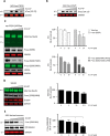
IU1-47 treatment reduces the level of endogenous wild-type tau in murine primary neurons. A, rat primary cortical neurons were infected with lentiviral vector expressing human wild-type tau for 4 days. Cultures were then treated with either IU1-47 (25 μm) alone or in combination with MG-132 (10 μm) for 48 h. Lysates were prepared, and proteins were resolved by SDS-PAGE followed by immunoblot analysis at left to detect total tau (tau5 antibody). GAPDH was used as a loading control. The proteins' molecular masses are indicated. Right, quantification of IU1-47–mediated tau reduction (n = 4; treatment from two independent experiments). Error bars represent S.E. Asterisks denote p < 0.01 (one-way ANOVA, Tukey–Kramer post hoc analyses). ns, not significant. B, murine hippocampal primary neurons (DIV4) were incubated with graded doses of IU1-47 for 48 h. Lysates were prepared, resolved by SDS-PAGE, and immunoblotted with antibodies against endogenous total tau (tau5), unphosphorylated (unP) tau (tau1), and multiple forms of phosphorylated (P) tau (pSer-396/404, pSer-396, and pSer-202). GAPDH was used as a loading control. Proteins of interest were visualized with IRDye-conjugated secondary antibodies using an Odyssey imaging system (left). The proteins' molecular masses are indicated. The 37-kDa band is shown as it recognizes monomeric tau species detected with the tau5 antibody. Quantification of total tau (as detected by tau5 antibody) and of the phosphotau species pSer-396/404 (as detected by PHF1 antibody) and pSer-202 is shown for three independent experiments. Error bars represent S.D. Asterisks denote p values <0.05; double asterisks denote p values <0.01 of differences from appropriate DMSO controls (right panel). C, quantification by AQUA analysis of total tau level from whole-cell lysates of murine primary neurons treated with graded amounts of IU1-47. Elution profiles of heavy and light peptides resolved by LC-MS are shown with their retention time and mass errors (ppm). Peak heights are given in arbitrary units; intensity values have been divided by 106 for simplicity of presentation (left). Quantification was derived from the area under the curves, and values are given as a percentage of tau present as compared with lysate from DMSO-treated cells (right). D, quantitative PCR showing the level of tau transcript normalized to 18S rRNA and relative to DMSO-treated control after 48-h treatment with increasing concentrations of IU1-47 in mouse cortical primary neurons. Values correspond to the average of four replicates. Error bars represent S.D. E, viability of murine cortical primary neurons (DIV6) after 48-h treatment with IU1-47 was assayed using Toxilight bioassay (Lonza), which measures adenylate kinase activity released into the medium upon cell death. No treatment with medium alone and treatment with 1 μm staurosporine are indicated as M and S, respectively. Staurosporine, a nonselective kinase inhibitor that induces rapid programmed cell death in neurons, was used as a positive control for dead cells (84, 85). The graph shows neuronal viability upon IU1-47 treatment from three independent experiments. Values shown are averages, and error bars correspond to S.D. Measurements for medium alone and controls for dead cells were done in two of the three independent experiments. F, murine cortical primary neurons were treated with 12.5 μm IU1-C, a structural analog of IU1-47 that does not inhibit USP14, for 48 h. Lysates were prepared, resolved by SDS-PAGE, probed with antibodies against total tau (tau5) and GAPDH (loading control), and then visualized as above (left). Quantification of total tau indicates that there is no decrease in tau levels during the course of IU1-C treatment (n = 3; right). Error bars represent S.D.
Figure 4.
Figure 4.
IU1-47 treatment reduces the level of pathological tau in primary neurons. A, rat cortical primary neurons were infected with AAV-tau-P301S, which expresses the human tau mutant P301S. After 5 days, IU1-47 was added at the indicated concentrations. Following an additional 2 days of culture, lysates were prepared, and proteins were resolved as described above. Human tau was visualized by immunoblotting using an antibody specific for human tau (CP-27), and GAPDH was probed as a loading control. The proteins' molecular masses are indicated. B, rat cortical primary neurons were infected with AAV-tau-A152T, which expresses human tau variant A152T, for 5 days prior to IU1-47 treatment for 48 h at the indicated concentrations. Lysates were prepared and resolved as described above. Human tau was visualized by immunoblotting using an antibody specific for human tau (CP-27), and GAPDH was probed as a loading control. C, murine cortical primary neurons (DIV5) isolated from APPSwe/P301L transgenic animals were incubated with the indicated doses of IU1-47 for 48 h, then harvested, and processed as above. Immunoblots developed with IRDye-conjugated secondary antibodies show the level of total tau and of several specific phospho (P)-tau species upon IU1-47 treatment. Phosphoepitopes are indicated in parentheses. The top three blots are from one gel; the bottom four blots are from another gel (left). The USP14 blot shows that no change in USP14 protein level was observed upon IU1-47 treatment. Values in the graphs represent an average of two biological replicates representing cortical neurons from littermates. Error bars represent S.E. (right). Monoclonal antibodies are listed below the matching epitope in the bar graph. D, murine cortical primary neurons (DIV6) isolated from 5XFAD mice were incubated with the indicated concentrations of IU1-47 for 48 h. Cells were harvested, and lysates were prepared and resolved as described above. A representative immunoblot shows the levels of total tau (as detected by tau5 antibody) in cortical neurons derived from one of two independently processed embryos upon treatment with IU1-47 (left). Secondary antibodies were IRDye-conjugated. The decrease in phosphorylated tau pSer-396/404 was also visualized with IRDye-conjugated secondary antibodies upon IU1-47 treatment. Quantification of total tau from independent samples is shown (right). E, NPC terminally differentiated into neurons for 6 weeks was treated with increasing concentrations of IU1-47. Lysates were prepared, resolved by SDS-PAGE, and immunoblotted with antibodies against phosphorylated tau (pSer-396/404). GAPDH was used as a loading control. βΙΙΙ-tubulin levels were also assayed as a marker for differentiation and to assess cytoskeletal integrity. Proteins of interest were visualized with IRDye-conjugated secondary antibodies using an Odyssey imaging system. Similar results were obtained using an antibody against tau pSer-202 (not shown). Quantification of phosphotau (pSer-396/404) as detected by PHF1 (right) is shown for three independent experiments for the indicated time points or for two independent experiments. Error bars represent S.D. for n = 3 (DMSO and 10 and 30 μm IU1-47) and S.E. for n = 2 (3 μm IU1-47). Asterisks denote p values <0.05.
Figure 5.
Figure 5.
Lysine 174 of tau is required for IU1-47–mediated degradation. A, primary neurons were infected with AAV vectors that express either wild-type human tau or htau-K174Q. Four days after infection, the cultures were treated with either 25 μm IU1-47 or DMSO for 48 h. Cells were harvested, and lysates were prepared and resolved by SDS-PAGE. Total human tau was visualized by immunoblotting using the tau5 antibody. GAPDH was probed as a loading control. Right, quantification of IU1-47–mediated tau reduction is shown (htau wild type treated with DMSO (n = 6), htau wild type treated with IU1-47 (n = 7), htau-K174Q treated with DMSO (n = 6), and htau-K174Q treated with IU1-47 (n = 7) from four independent experiments). Asterisks denote p < 0.01 (Mann–Whitney nonparametric test). Error bars represent S.E. B, HEK293 cells were transfected with vectors expressing either FLAG-tagged human wild-type or mutant tau (K174Q or K274Q) and HA-ubiquitin. Cells were then treated with IU1-47 (10 μm) for 20 h and subsequently treated with MG-132 (20 μm) for 4 h. Cells were lysed with ubiquitination buffer, and supernatant proteins were immunoprecipitated with FLAG M2-agarose beads for 3 h. Reactions were resolved by SDS-PAGE and immunoblotted with anti-HA antibody. Tau5 antibody was used to detect total tau at the (bottom left). The right panel shows the quantification of polyubiquitinated tau normalized to total tau (wild-type tau (n = 7) and tau-K174Q (n = 7) from five independent experiments and tau-K274Q (n = 5) from two independent experiments). Error bars represent S.E. Asterisks denote p < 0.01 (one-way ANOVA, Tukey–Kramer post hoc analyses).
Figure 6.
Figure 6.
USP14 inhibition elicits an increase in autophagic flux in primary neurons. A, rat cortical primary neurons (DIV4) were incubated with graded doses of IU1-47 for 48 h. Cells were harvested, and lysates were prepared and resolved by SDS-PAGE and immunoblotted with antibodies against the LC3, endogenous total tau (tau5), multiple forms of phosphorylated (P) tau (pSer-396/404 and pSer-202/pThr-205), and caspase-3 (full-length and cleaved forms). βIII-tubulin was used as a marker for structural integrity. GAPDH was used as a loading control. Proteins of interest were visualized with IRDye-conjugated secondary antibodies using an Odyssey imaging system. A representative sample of three independent experiments is shown. The right panel shows the quantification of LC3-II expression. Each data point represents the average from two independent samples. Error bars represent S.D. from n = 3 independent experiments. Asterisks denote p values <0.05; double asterisks denote p values <0.01 of differences from appropriate DMSO controls (one-way ANOVA, Tukey–Kramer post hoc analyses). The images were produced from three independent gels to avoid stripping the membrane. B, rat cortical primary neurons (DIV3) were treated with 12.5 μm IU1-47 for 48 h in combination with the calpain inhibitor calpeptin (2 and 10 μm). Lysates were prepared, and proteins were resolved by SDS-PAGE followed by immunoblot analysis using monoclonal tau antibody (amino acids 210–241; clone tau5) to detect full-length tau (tau-FL) and, if present, the 17-kDa tau fragment resulting from calpain-mediated tau cleavage (one asterisk denotes an unspecific band; two asterisks denotes the expected 17-kDa fragment position in the blot above). GAPDH was used as a loading control. The blot above is representative of two independent experiments.
Scheme 1
Scheme 1

Similar articles

Cited by

References

    1. Budenholzer L., Cheng C. L., Li Y., and Hochstrasser M. (2017) Proteasome structure and assembly. J. Mol. Biol. 427, 3500–3524 - PMC - PubMed
    1. Finley D. (2009) Recognition and processing of ubiquitin-protein conjugates by the proteasome. Annu. Rev. Biochem. 78, 477–513 - PMC - PubMed
    1. Collins G. A., and Goldberg A. L. (2017) The logic of the 26S proteasome. Cell 169, 792–806 - PMC - PubMed
    1. Inobe T., and Matouschek A. (2014) Paradigms of protein degradation by the proteasome. Curr. Opin. Struct. Biol. 24, 156–164 - PMC - PubMed
    1. Goldberg A. L. (2007) Functions of the proteasome: from protein degradation and immune surveillance to cancer therapy. Biochem. Soc. Trans. 35, 12–17 - PubMed

MeSH terms