

Structural insights into GDP-mediated regulation of a bacterial acyl-CoA thioesterase
- PMID: 28972175
- PMCID: PMC5733585
- DOI: 10.1074/jbc.M117.800227
Structural insights into GDP-mediated regulation of a bacterial acyl-CoA thioesterase
Abstract
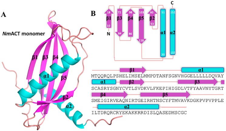
Thioesterases catalyze the cleavage of thioester bonds within many activated fatty acids and acyl-CoA substrates. They are expressed ubiquitously in both prokaryotes and eukaryotes and are subdivided into 25 thioesterase families according to their catalytic active site, protein oligomerization, and substrate specificity. Although many of these enzyme families are well-characterized in terms of function and substrate specificity, regulation across most thioesterase families is poorly understood. Here, we characterized a TE6 thioesterase from the bacterium Neisseria meningitidis Structural analysis with X-ray crystallographic diffraction data to 2.0-Å revealed that each protein subunit harbors a hot dog-fold and that the TE6 enzyme forms a hexamer with D3 symmetry. An assessment of thioesterase activity against a range of acyl-CoA substrates revealed the greatest activity against acetyl-CoA, and structure-guided mutagenesis of putative active site residues identified Asn24 and Asp39 as being essential for activity. Our structural analysis revealed that six GDP nucleotides bound the enzyme in close proximity to an intersubunit disulfide bond interactions that covalently link thioesterase domains in a double hot dog dimer. Structure-guided mutagenesis of residues within the GDP-binding pocket identified Arg93 as playing a key role in the nucleotide interaction and revealed that GDP is required for activity. All mutations were confirmed to be specific and not to have resulted from structural perturbations by X-ray crystallography. This is the first report of a bacterial GDP-regulated thioesterase and of covalent linkage of thioesterase domains through a disulfide bond, revealing structural similarities with ADP regulation in the human ACOT12 thioesterase.
Keywords: Neisseria meningitidis; acetyl coenzyme A (acetyl-CoA); coenzyme A (CoA); enzyme kinetics; enzyme regulation; enzyme structure; hotdog-fold; hydrolase; thioesterase.
© 2017 by The American Society for Biochemistry and Molecular Biology, Inc.
Conflict of interest statement
The authors declare that they have no conflicts of interest with the contents of this article.
Figures


Similar articles
-
Structural and Functional Characterization of the PaaI Thioesterase from Streptococcus pneumoniae Reveals a Dual Specificity for Phenylacetyl-CoA and Medium-chain Fatty Acyl-CoAs and a Novel CoA-induced Fit Mechanism.J Biol Chem. 2016 Jan 22;291(4):1866-1876. doi: 10.1074/jbc.M115.677484. Epub 2015 Nov 4. J Biol Chem. 2016. PMID: 26538563 Free PMC article.
-
Structure of YciA from Haemophilus influenzae (HI0827), a hexameric broad specificity acyl-coenzyme A thioesterase.Biochemistry. 2008 Mar 4;47(9):2797-805. doi: 10.1021/bi702336d. Epub 2008 Feb 9. Biochemistry. 2008. PMID: 18260643
-
Structural basis for nucleotide-independent regulation of acyl-CoA thioesterase from Bacillus cereus ATCC 14579.Int J Biol Macromol. 2021 Feb 15;170:390-396. doi: 10.1016/j.ijbiomac.2020.12.174. Epub 2020 Dec 28. Int J Biol Macromol. 2021. PMID: 33383082
-
Structure, function, and regulation of thioesterases.Prog Lipid Res. 2020 Jul;79:101036. doi: 10.1016/j.plipres.2020.101036. Epub 2020 May 19. Prog Lipid Res. 2020. PMID: 32416211 Review.
-
Functional and structural properties of mammalian acyl-coenzyme A thioesterases.Prog Lipid Res. 2010 Oct;49(4):366-77. doi: 10.1016/j.plipres.2010.04.001. Epub 2010 May 12. Prog Lipid Res. 2010. PMID: 20470824 Review.
Cited by
-
Cloning, heterologous expression, and characterization of a novel thioesterase from natural sample.Heliyon. 2021 Mar 24;7(3):e06542. doi: 10.1016/j.heliyon.2021.e06542. eCollection 2021 Mar. Heliyon. 2021. PMID: 33851045 Free PMC article.
-
Thioesterase enzyme families: Functions, structures, and mechanisms.Protein Sci. 2022 Mar;31(3):652-676. doi: 10.1002/pro.4263. Epub 2022 Jan 4. Protein Sci. 2022. PMID: 34921469 Free PMC article.
-
Fatty acid metabolism and the oxidative stress response support bacterial predation.Proc Natl Acad Sci U S A. 2025 Feb 4;122(5):e2420875122. doi: 10.1073/pnas.2420875122. Epub 2025 Jan 27. Proc Natl Acad Sci U S A. 2025. PMID: 39869799 Free PMC article.
-
Transcriptome analysis reveals candidate genes of the synthesis of branched-chain fatty acids related to mutton flavor in the lamb liver using Allium mongolicum Regel extract.J Anim Sci. 2022 Sep 1;100(9):skac256. doi: 10.1093/jas/skac256. J Anim Sci. 2022. PMID: 35946924 Free PMC article.
References
-
- Kirkby B., Roman N., Kobe B., Kellie S., and Forwood J. K. (2010) Functional and structural properties of mammalian acyl-coenzyme A thioesterases. Prog. Lipid Res. 49, 366–377 - PubMed
-
- Rodríguez-Rodríguez M. F., Salas J. J., Garcés R., and Martínez-Force E. (2014) Acyl-ACP thioesterases from Camelina sativa: cloning, enzymatic characterization and implication in seed oil fatty acid composition. Phytochemistry 107, 7–15 - PubMed
-
- Swarbrick C. M., Roman N., Cowieson N., Patterson E. I., Nanson J., Siponen M. I., Berglund H., Lehtiö L., and Forwood J. K. (2014) Structural basis for regulation of the human acetyl-CoA thioesterase 12 and interactions with the steroidogenic acute regulatory protein-related lipid transfer (START) domain. J. Biol. Chem. 289, 24263–24274 - PMC - PubMed
-
- Park H., Graef G., Xu Y., Tenopir P., and Clemente T. E. (2014) Stacking of a stearoyl-ACP thioesterase with a dual-silenced palmitoyl-ACP thioesterase and 12 fatty acid desaturase in transgenic soybean. Plant Biotechnol. J. 12, 1035–1043 - PubMed
MeSH terms
Substances
Associated data
- Actions
- Actions
- Actions
- Actions
- Actions
- Actions
- Actions
LinkOut - more resources
Full Text Sources
Other Literature Sources
