. 2017 Nov 24;292(47):19238-19249.
doi: 10.1074/jbc.M117.782300. Epub 2017 Sep 27.

Post-translational modifications clustering within proteolytic domains decrease mutant huntingtin toxicity

Affiliations

Post-translational modifications clustering within proteolytic domains decrease mutant huntingtin toxicity

Nicolas Arbez et al. J Biol Chem. .

Abstract

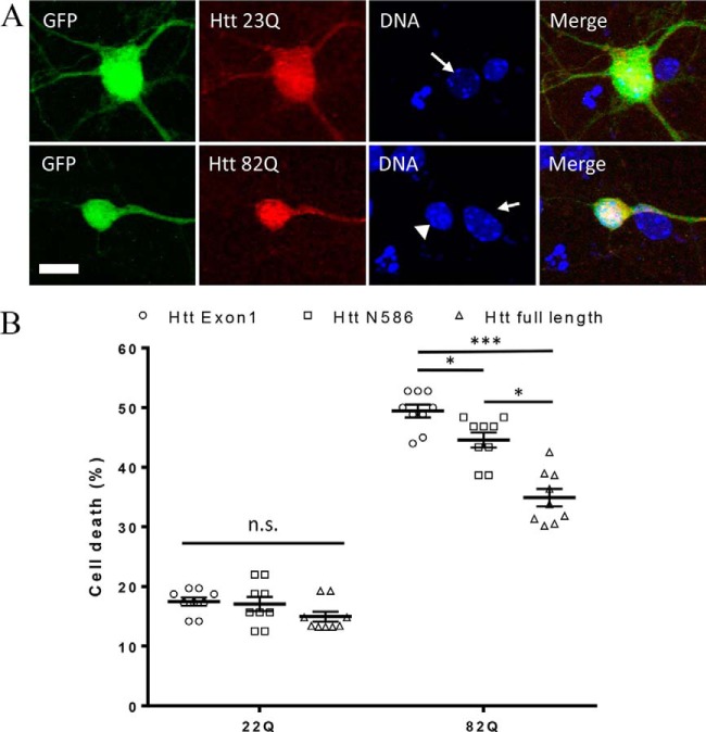
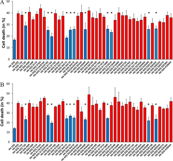
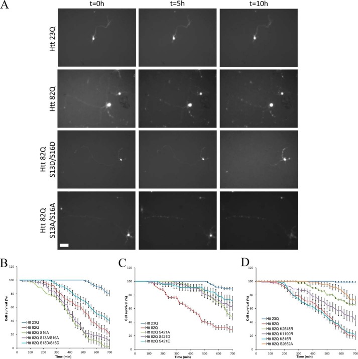
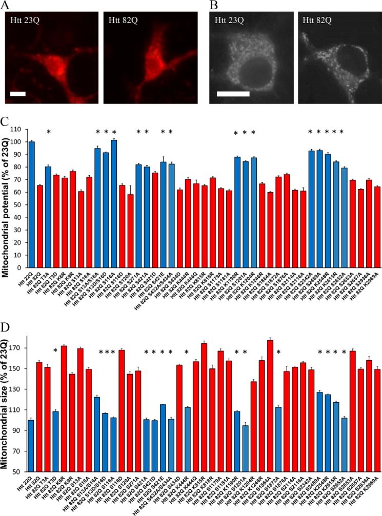
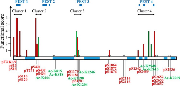
Huntington's disease (HD) is caused in large part by a polyglutamine expansion within the huntingtin (Htt) protein. Post-translational modifications (PTMs) control and regulate many protein functions and cellular pathways, and PTMs of mutant Htt are likely important modulators of HD pathogenesis. Alterations of selected numbers of PTMs of Htt fragments have been shown to modulate Htt cellular localization and toxicity. In this study, we systematically introduced site-directed alterations in individual phosphorylation and acetylation sites in full-length Htt constructs. The effects of each of these PTM alteration constructs were tested on cell toxicity using our nuclear condensation assay and on mitochondrial viability by measuring mitochondrial potential and size. Using these functional assays in primary neurons, we identified several PTMs whose alteration can block neuronal toxicity and prevent potential loss and swelling of the mitochondria caused by mutant Htt. These PTMs included previously described sites such as serine 116 and newly found sites such as serine 2652 throughout the protein. We found that these functionally relevant sites are clustered in protease-sensitive domains throughout full-length Htt. These findings advance our understanding of the Htt PTM code and its role in HD pathogenesis. Because PTMs are catalyzed by enzymes, the toxicity-modulating Htt PTMs identified here may be promising therapeutic targets for managing HD.

Keywords: Huntington disease; neurodegeneration; neuron; phosphorylation; toxicity.


Conflict of interest statement

The authors declare that they have no conflicts of interest with the contents of this article

Figures

Figure 1.
Figure 1.
Neuronal toxicity of expanded full-length Htt. A, example images of primary neurons transfected with full-length Htt containing either 23Q or 82Q and co-transfected with GFP. Transfected Htt was detected by immunocytochemistry using mAb 2166, and DNA is stained with Hoechst. Condensed nuclei (arrowhead) are brighter and smaller than healthy nuclei (arrow). B, quantification of cell death by nuclear condensation assay. Primary cortical neurons were transfected at DIV5 with indicated plasmid and with GFP. The cells were fixed after 48 h, and the nuclei were stained with Hoechst. Automated picture acquisition was performed on Zeiss Axiovert 200 microscope, and automated quantification of the nuclear intensity of transfected cells was performed using Volocity. *, p ≤ 0.05; ***, p ≤ 0.001 (n = 9 independent neuronal preparations).
Figure 2.
Figure 2.
Neuronal toxicity of expanded full-length Htt is modulated by PTMs. Quantification of cell death by nuclear condensation assay is shown. Primary cortical (A) and striatal (B) neurons were transfected at DIV5 with indicated plasmid and with GFP. The cells were fixed after 48 h, and the nuclei were stained with Hoechst. Automated picture acquisition was performed on Zeiss Axiovert 200 microscope, and automated quantification of the nuclear intensity of transfected cells was performed using Volocity. The results are expressed as means ± S.E. of the percentage of dead cells. *, p < 0.05 versus Htt-82Q (n = 4 independent neuronal preparations).
Figure 3.
Figure 3.
Time-lapse imaging quantification of neuronal survival. Primary cortical neurons were co-transfected at DIV5 with indicated full-length constructs and GFP. After 24 h, GFP positive neurons were image every 20 min for 10 h. A, examples of neuronal morphology over time for selected Htt constructs. Scale bar, 100 μm. B–D, morphology of every neuron was quantified as described under “Experimental procedures” for the Ser13/Ser16 (B), Ser421 (C), and downstream new sites (D) series of constructs. The results are expressed as means ± S.E. (n = 200 cells analyzed over five independent neuronal preparations).
Figure 4.
Figure 4.
Mitochondrial abnormalities caused by expanded Htt are modulated by PTMs. A, example images of primary neurons transfected with full-length Htt containing either 23Q or 82Q and stained with the potential sensitive mitochondrial dye TMRM. Scale bar, 10 μm. B, example images of primary neurons transfected with full-length Htt containing either 23Q or 82Q and stained with MitoTracker. C, cells were then imaged at 24 h post-transfection, and individual cell mitochondrial potential was measured using Volocity. *, p ≤ 0.05 versus Htt-82Q (n = 3 independent neuronal preparations). D, mitochondrial swelling assay. Primary cortical neurons were transfected at DIV5 with indicated full-length constructs and GFP. After 24 h, the cells were loaded with MitoTracker Green for 45 min at 37 °C. The cells were then fixed and imaged. Individual mitochondria size was measured using Volocity. *, p ≤ 0.05 versus Htt-82Q (n = 3 independent neuronal preparations).
Figure 5.
Figure 5.
Functional assessment of PTMs on Htt. PTMs clustering within proteolytic domains modulate mutant Htt toxicity. The functional score has been calculated for each PTM, based on the outcome of all the assays (toxicity in cortex, toxicity in striatum, mitochondrial potential, and mitochondrial size). Each PTM was given a score of 1 per assay if significant changes were observed and 0 if there was no difference. For multiple alterations of the same amino acid, a combined score was recorded. Ser13 and Ser16 are scored based on the single and combined alterations results. Based on this scoring, we found four clusters of PTMs that largely modulate expanded Htt functional or toxic properties. These clusters are located within protease-sensitive (PEST) regions (predicted by epestfind-EMBOSS Explorer) as indicated. Phosphorylation sites are shown in red, acetylation sites are in green, and sites that did not score in any of the assays are indicated by short white bars.

References

    1. The Huntington's Disease Collaborative Research Group (1993) A novel gene containing a trinucleotide repeat that is expanded and unstable on Huntington's disease chromosomes. Cell 72, 971–983 - PubMed
    1. Iqbal K., Liu F., Gong C. X., and Grundke-Iqbal I. (2010) Tau in Alzheimer disease and related tauopathies. Curr. Alzheimer Res. 7, 656–664 - PMC - PubMed
    1. Xu Y., Deng Y., and Qing H. (2015) The phosphorylation of α-synuclein: development and implication for the mechanism and therapy of the Parkinson's disease. J. Neurochem. 135, 4–18 - PubMed
    1. Hasegawa M., Nonaka T., Tsuji H., Tamaoka A., Yamashita M., Kametani F., Yoshida M., Arai T., and Akiyama H. (2011) Molecular dissection of tdp-43 proteinopathies. J. Mol. Neurosci. 45, 480–485 - PubMed
    1. Aronin N., and DiFiglia M. (2014) Huntingtin-lowering strategies in Huntington's disease: antisense oligonucleotides, small RNAs, and gene editing. Mov. Disord 29, 1455–1461 - PubMed

Publication types

Substances

LinkOut - more resources