. 2017 Dec 8;292(49):20010-20031.
doi: 10.1074/jbc.M117.807925. Epub 2017 Sep 25.

Calmodulin regulates Cav3 T-type channels at their gating brake

Affiliations

Calmodulin regulates Cav3 T-type channels at their gating brake

Jean Chemin et al. J Biol Chem. .

Abstract

Calcium (Cav1 and Cav2) and sodium channels possess homologous CaM-binding motifs, known as IQ motifs in their C termini, which associate with calmodulin (CaM), a universal calcium sensor. Cav3 T-type channels, which serve as pacemakers of the mammalian brain and heart, lack a C-terminal IQ motif. We illustrate that T-type channels associate with CaM using co-immunoprecipitation experiments and single particle cryo-electron microscopy. We demonstrate that protostome invertebrate (LCav3) and human Cav3.1, Cav3.2, and Cav3.3 T-type channels specifically associate with CaM at helix 2 of the gating brake in the I-II linker of the channels. Isothermal titration calorimetry results revealed that the gating brake and CaM bind each other with high-nanomolar affinity. We show that the gating brake assumes a helical conformation upon binding CaM, with associated conformational changes to both CaM lobes as indicated by amide chemical shifts of the amino acids of CaM in 1H-15N HSQC NMR spectra. Intact Ca2+-binding sites on CaM and an intact gating brake sequence (first 39 amino acids of the I-II linker) were required in Cav3.2 channels to prevent the runaway gating phenotype, a hyperpolarizing shift in voltage sensitivities and faster gating kinetics. We conclude that the presence of high-nanomolar affinity binding sites for CaM at its universal gating brake and its unique form of regulation via the tuning of the voltage range of activity could influence the participation of Cav3 T-type channels in heart and brain rhythms. Our findings may have implications for arrhythmia disorders arising from mutations in the gating brake or CaM.

Keywords: calcium channel; circular dichroism (CD); cryo-electron microscopy; gating; isothermal titration calorimetry (ITC); nuclear magnetic resonance (NMR); patch clamp; short hairpin RNA (shRNA).


Conflict of interest statement

The authors declare that they have no conflicts of interest with the contents of this article

Figures

Figure 1.
Figure 1.
CaM associates with full-length Cav3 T-type channels illustrated by nano-particle cryoelectron microscopy (A) and co-immunoprecipitation (B). A, field of negatively stained (2% w/v uranyl acetate) full-length Cav3.1 in complex with CaM in the presence of 1.5 mm Ca2+. Scale bar is 50 nm. Left panel, CaM has a biotin label that binds streptavidin gold (5 nm). Protein appears white, and the gold that is electron-dense is black. Right panel, top row, montage of purified Cav3.1 particles (white density) presenting different views of the channel. Asterisk indicates “tail” domain that we have previously determined corresponds to the C terminus (38). Middle row, montage of Cav3.1 particles with CaM–biotin bond indicated by the presence of streptavidin gold (black sphere). Bottom row, control images of CaM–biotin–streptavidin gold particle without Cav3.1. Observed relative differences in Cav3.1 and streptavidin gold particle sizes relate to the orientation and contrast of each Cav3.1 channel particle adhered to the EM support film and the size variation in the streptavidin gold particles (3–6 nm with nominal size of 5 nm, according to the manufacturer). B, CaM–GFP bound to anti-GFP magnetic beads associates with hemagglutinin (HA)-tagged Cav3.2 channel (top panel, middle lane) as illustrated by HA antibody labeling (259-kDa band) of the Cav3.2 channel co-immunoprecipitant (IP) bound to beads. 259-kDa HA-tagged Cav3.2 channel band does not appear as a co-immunoprecipitant in the Western blot without co-expression of pCDNA3.1 plasmid inserts containing HA-tagged Cav3.2 channel (top panel, left lane) or without co-expression of CaM–GFP (top panel, right lane) in HEK-293T cells. Middle panel illustrates anti-HA antibody staining of the 259-kDa HA-tagged Cav3.2 channel of a replicate experiment of input proteins for the Western blot shown in the top panel without co-immunoprecipitation. Bottom panel illustrates anti-GFP antibody staining of the 44.2-kDa GFP-tagged CaM or GFP alone (27 kDa), in a replicate experiment of input proteins for the Western blot shown in the top panel without co-immunoprecipitation. C, CaM–GFP (left two lanes, 44.2 kDa) and GFP alone (right lane, 27 kDa) bound to anti-GFP magnetic beads as illustrated by anti-GFP antibody labeling (259-kDa band) of the Cav3.2 channel co-immunoprecipitant bound to beads. GFP alone generated two bands on the Western blot, which may result from differing post-translational modifications. Co-immunoprecipitation experiments were carried out in 33.3 μm CaCl2, pH 7.4. Vector for HEK-293T cell expressed inserts for Western blotting (Cav3.2–HA, EGFP, CaM–pGFP) were contained in pcDNA3.1. Membranes were stained with Ponceau red following protein transfer to evaluate the protein content in each lane. Co-immunoprecipitation experiments were carried out in 33.3 μm CaCl2, pH 7.4. Vector for HEK-293T cell expressed inserts for Western blotting (Cav3.2–HA, EGFP, CaM–pGFP) were contained in pcDNA3.1. Membranes were stained with Ponceau red following protein transfer to evaluate the protein content in each lane.
Figure 2.
Figure 2.
CaM is predicted to associate with Cav3 T-type channels in helix-2 of the gating brake in the proximal I–II linker. A, illustration of the four domain (DI, DII, DIII, and DIV) × 6 transmembrane helices structure common to sodium channels, calcium channels, and NALCN. Cav1, Cav2, and Nav channels contain a canonical proximal C-terminal IQ motif that possess high-affinity CaM-binding site. A proposed equivalence of the CaM-binding C-terminal IQ motif in Cav3 T-type channels is a helix–loop–helix gating brake motif in the proximal I–II linker that is in the analogous position of β-subunit binding to Cav1 and Cav2 calcium channels. B, sequence alignment of T-type channels illustrates a predicted CaM-binding site in helix-2 of the gating brake (illustrated by red color) in representative species from cnidarians to the three human genes. CaM-binding site prediction from CaM Target Database (39). Predictions suggested the putative CaMB peptide sequence is cytoplasmic, and helical wheel analysis indicates its amphipathic nature. In the most primitive metazoan with a T-type channel, Trichoplax (placozoan), the most basal extant T-type channel in multicellular organisms known possesses a gating brake motif resembling the C-terminal IQ motif shared with other calcium (Cav1 and Cav2) and sodium (Nav2 and Nav1) channels (illustrated by blue color). Yellow outlined residues are the sequences for the synthetic CaMB peptides. 39 and 111 amino acid sequence deleted in gating brake deletion mutant, Cav3.2(D453–491) and Cav3.2(D429–539) are indicated. C and D, protein similarities among CaMB peptide sequences in the I–II linker, illustrating the high similarity among invertebrate (snail) LCav3 and the human Cav3.x homologs (C) and the greater similarity of Trichoplax Cav3 CaMB peptide sequences to the C-terminal IQ motif of Cav1.2 channels (D), rather than other gating brake sequences (C).
Figure 3.
Figure 3.
CaMB peptides from snail LCav3 and the three human Cav3.x T-type channels associate with Ca2+–CaM. Gel-mobility shifts with CaM are evident in the presence of 0.1 mm CaCl2-containing solution and increasing molar ratios (0.5–4×) of snail LCav3 or human Cav3.1, Cav3.2, and Cav3.3 channel CaMB peptides but not evident in the presence of mutated control Cav3.2 CaMB peptide (Cav3.2mut). 1st lane in each gel is Ca2+–CaM-only control. The rank order of weakest to strongest apparent interaction of Ca2+–CaM for peptides: Cav3.3 < Cav3.2 < Cav3.1 < LCav3 correlates with the rank order of calculated binding affinities using ITC (see Fig. 5). Note that Cav3.1 and Cav3.2 peptides both completely displace Ca2+–CaM at 1.5× peptide to CaM molar ratio, but Cav3.1 appears to possess a higher affinity based on sub-saturation levels of peptide. Urea attenuates the weaker interacting human Cav3.x channels but does not attenuate the higher affinity LCav3 CaMB peptide.
Figure 4.
Figure 4.
CaMB peptides from snail Cav3 and human Cav3.x T-type channels assume an α-helical structure when bound to Ca2+–CaM. A, differential circular dichroism (CD) spectra of CaMB peptides co-incubated in the presence of helix-stabilizing agent, TFE (gray dashed line), 20 μm CaM (red line), or without CaM (blue line). 26-mer snail LCav3, human Cav3.2, and Cav3.3 CaMB peptides (sequence, bottom right) assume a more helical secondary structure upon addition of CaM and TFE. TFE has a much greater effect on the highest binding affinity peptide, snail LCav3, than the others. Cav3.1 peptide is the only isoform that is α-helical when free in solution, as evidenced by the characteristic negative peaks at 208 and 222 nm. Cav3.2-Gbmut peptide was incapable of adopting an α-helix even at 50% TFE, nor did it appear to interact with CaM. CaM-alone curve was subtracted from each spectra, converted to mean residue ellipticity (θ), and smoothed. For TFE experiments, 50 μm peptide in PBS was used, whereas the baseline was corrected against PBS as background. B, gating brake peptide sequences highlighting the alanine-rich Cav3.1 sequence in blue outline, and the “PGPGPG” substitution in red outline that serves as the Cav3.2 gating brake mutant. The 24 residues of the 26-mer peptides with a yellow-colored background are predicted to contain α-helices according to PSIPRED version 3.3 (Bioinformatics Group at University College London) (91).
Figure 5.
Figure 5.
Thermodynamic basis for CaM-binding peptide interactions with wild-type and mutant CaM. Raw data traces, isothermal titration calorimetry (above), and integrated heats of the measured interaction were fitted with a One Sites model using Malvern MicroCal (ITC200) add-on within Origin software (below). A, representative interactions with snail LCav3, and human Cav3.1 and Cav3.2 CaMB peptides and wild-type CaM used in the calculation for their 12.4, 42.7, and 187.1 average nanomolar affinities, respectively. B, sample interactions with human Cav3.2 and Cav3.1 CaMB peptides and mutant calmodulins, including N-lobe of CaM, and CaM1234 used in the calculation for their 31.1 and 543.3 average nanomolar affinities, respectively. Full table of ITC parameters (mean ± S.E., n = 3) is illustrated in Fig. 6A.
Figure 6.
Figure 6.
ITC and NMR analyses indicate high-affinity, nanomolar binding, and associated conformation changes, respectively, upon Cav3 T-type channel CaM-binding peptides to N- and C-terminal lobes of CaM. A, tabulated data of ITC analysis indicates a 1:1 stoichiometry of CaM binding with a 10, 36, 196, and 383 nanomolar affinity for CaM-binding peptides for snail LCav3 and human Cav3.1, Cav3.2, and Cav3.3 channels respectively. All peptide and CaM interactions are endothermic, except Cav3.2 CaMB peptide, which is exothermic. LCav3 CaMB peptide in particular has a complex ITC curve and will bind to CaM with mutated N-terminal Ca2+-binding sites (CaM12), but not to CaM with mutated C-terminal Ca2+-binding sites (CaM34). N-CaM and C-CaM are 74-aa constructs (exactly half of the CaM molecule) representing each individual lobe. Both N- and C-CaM associate with Cav3.2 CaMB peptide, albeit a higher affinity for the N-lobe of CaM. CaM with all four Ca2+-binding sites mutated in its lobes (apo-CaM, CaM1234) still associates with CaM-binding peptides. Table headings: Binding stoichiometry of the interaction between CaM and CaM-binding peptide in solution (N), Binding affinity (Ka), enthalpy changes (ΔH), entropy changes (ΔS). ITC curves were fitted to a one-set-of-sites model with a high degree of fit for N, Ka, and ΔH values shown for representative experiments in Fig. 5A. The parameters for the all peptides were calculated from three replicate experiments. B, T-type channel GB peptides promote a conformational change involving both CaM lobes confirmed by amide chemical shifts of CaM's amino acids in 1H-15N HSQC NMR spectra. Overlay of 1H-15N HSQC spectra of CaM alone (red color) and CaM bound to gating brake peptides from Cav3.2 (green color), or snail LCav3 (cyan color). C, chemical shift differences between CaM and the CaM–Cav3.2 complex and CaM and the CaM–LCav3 complex. B and C illustrate representative data from one of three replicate experiments.
Figure 7.
Figure 7.
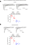
Intracellular dialysis of 5 μm Cav3 CaMB peptide but not mutated (CaMBmut) peptide causes a hyperpolarization shift and faster channel kinetics in Cav3.2 channels. Transfected Cav3.2 channels in HEK-293T cells were evaluated by whole-cell patch-clamp electrophysiology for activation (A) and inactivation (B) at start of patch recording (time 0) and after 20 min of intracellular dialysis of 26-mer Cav3 CaM-binding (CaMB) peptide or mutated CaM-binding (CaMBmut) peptide (peptide sequences in Figs. 2B and 4B). Representative current traces after 20-min dialyses are shown. Calcium currents were measured for their current-voltage relationships and τ mono-exponential fits for the kinetics of activation (A) and inactivation (B). Activation and steady-state inactivation curves were created with peak currents generated from a step depolarization from −110 to −80 to −10 mV and to −30 mV from holding potentials ranging from −140 to −40 mV, respectively. Statistical significance (p < 0.05) using a non-parametric Wilcoxon matched-pairs signed rank test for the kinetic data is shown by * measured before and after equilibration of the intracellular dialysis of Cav3 CaMB peptide or mutated CaM binding (CaMBmut) peptide. n values for current-voltage relationship Cav3 CaMB peptide (n = 7) and CaMBmut (n = 6). n values for inactivation Cav3 CaMB peptide (n = 5) and CaMBmut (n = 6).
Figure 8.
Figure 8.
Intracellular dialysis of 5 μm Cav3 CaMB peptides cause a hyperpolarizing shift in voltage dependence but only with Cav3.2 channels containing an intact gating brake. Scatterplot (left) with mean ± S.E. (right) values of the voltages of 50% activation (A) and steady-state inactivation (B) taken from Boltzmann fits of activation and steady-state inactivation curves for wild-type Cav3.2 channels and Cav3.2 channels with a deleted gating brake (deleted sequence from 429 to 539, see Fig. 2A). Representative current traces at time 0 min and after 20-min dialysis of Cav3.2 CaMB peptide are shown. Dialysis of CaMB peptide (after 20 min) generates a hyperpolarizing shift in voltage dependence of wild-type Cav3.2 channels, but it does not promote additional hyperpolarizing shifts in voltage dependence on the Cav3.2 channels that are highly hyperpolarizing shifted after gating brake deletion. The statistical significance after dialyses of CaMB peptide in Cav3.2 channels containing an intact gating brake is p = 0.0156 (*) and p = 0.0313 (*) for V50% activation and V50% inactivation, respectively, in a Wilcoxon matched-pairs signed rank test.
Figure 9.
Figure 9.
Overexpression of apo-CaM/CaM1234 (Ca2+-binding deficient CaM) causes a hyperpolarizing shift in voltage sensitivities and faster channel kinetics in Cav3.2 channels only in the presence of an intact gating brake in Cav3.2. A and B, Cav3.2 channels with and without co-expressed CaM1234 were measured for their current-voltage relationships and τ for mono-exponential fits of the kinetics of activation (A) and inactivation (B). Representative current traces are shown. Results are presented as mean ± S.E., and n = the number of cells. Statistical analyses were first performed with the Student's t test or with one-way ANOVA combined with a Tukey post-test for multiple comparisons (*, p < 0.05; **, p < 0.01; ***, p < 0.001). C, scatter plot (left) with mean ± S.E. (right) values illustrate the 50% activation and inactivation values taken from Boltzmann fits of activation and steady-state inactivation curves of Cav3.2 channels alone (−), and Cav3.2 channels with co-expressed apo-calmodulin (CaM1234) or calmodulin (CaM). Voltage responses to co-expressed apo-calmodulin (CaM1234) are also illustrated for Cav3.2 deletion mutants spanning sequences 429–539 or 453–491, lacking the 111 or 39 amino acids, respectively, flanking the gating brake in the I–II linker. The region of deletion mutants spanning the gating brake of Cav3.2 channels are illustrated in Fig. 2B. The statistical significance of CaM1234 overexpression compared with wild-type conditions was p = 0.0028 (***) and p = 0.0308 (*) for V50% activation and V50% inactivation, respectively, using a Kruskal-Wallis test followed by a Dunn's multiple comparisons test. The effect of CaM1234 overexpression compared with wild-type conditions for Cav3.2 deletion mutants (D429–539 or D453–491) were non-significant using a Mann-Whitney test.

Similar articles

Cited by

References

    1. Ben-Johny M., Dick I. E., Sang L., Limpitikul W. B., Kang P. W., Niu J., Banerjee R., Yang W., Babich J. S., Issa J. B., Lee S. R., Namkung H., Li J., Zhang M., Yang P. S., et al. (2015) Towards a unified theory of calmodulin regulation (calmodulation) of voltage-gated calcium and sodium channels. Curr. Mol. Pharmacol. 8, 188–205 - PMC - PubMed
    1. Ben-Johny M., Yang P. S., Niu J., Yang W., Joshi-Mukherjee R., and Yue D. T. (2014) Conservation of Ca2+/calmodulin regulation across Na and Ca2+ channels. Cell 157, 1657–1670 - PMC - PubMed
    1. Budde T., Meuth S., and Pape H. C. (2002) Calcium-dependent inactivation of neuronal calcium channels. Nat. Rev. Neurosci. 3, 873–883 - PubMed
    1. Halling D. B., Aracena-Parks P., and Hamilton S. L. (2006) Regulation of voltage-gated Ca2+ channels by calmodulin. Sci. STKE 2006, er1. - PubMed
    1. Minor D. L. Jr., and Findeisen F. (2010) Progress in the structural understanding of voltage-gated calcium channel (CaV) function and modulation. Channels 4, 459–474 - PMC - PubMed

Publication types