. 2017 Nov 17;292(46):19024-19033.
doi: 10.1074/jbc.M117.811265. Epub 2017 Sep 25.

Intramolecular autoinhibition of checkpoint kinase 1 is mediated by conserved basic motifs of the C-terminal kinase-associated 1 domain

Affiliations

Intramolecular autoinhibition of checkpoint kinase 1 is mediated by conserved basic motifs of the C-terminal kinase-associated 1 domain

Ryan P Emptage et al. J Biol Chem. .

Abstract

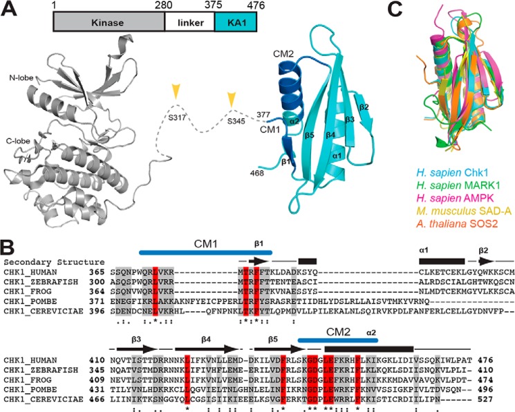
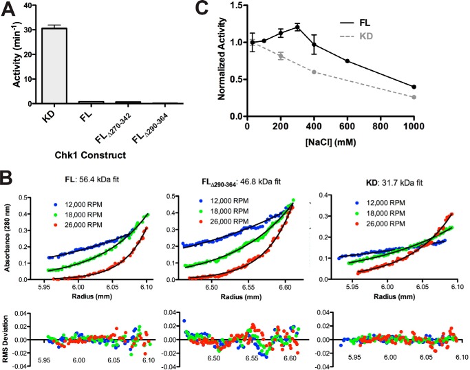
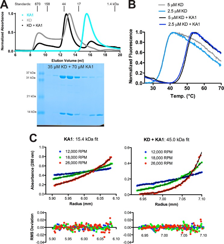
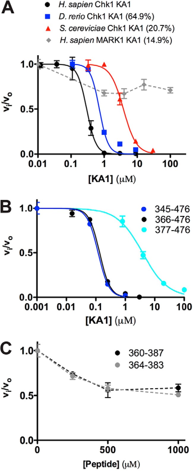
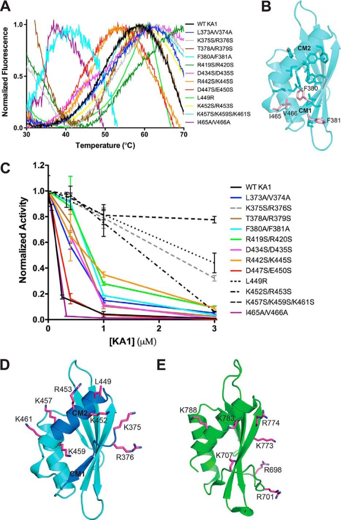
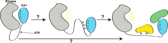
Precise control of the cell cycle allows for timely repair of genetic material prior to replication. One factor intimately involved in this process is checkpoint kinase 1 (Chk1), a DNA damage repair inducing Ser/Thr protein kinase that contains an N-terminal kinase domain and a C-terminal regulatory region consisting of a ∼100-residue linker followed by a putative kinase-associated 1 (KA1) domain. We report the crystal structure of the human Chk1 KA1 domain, demonstrating striking structural homology with other sequentially diverse KA1 domains. Separately purified Chk1 kinase and KA1 domains are intimately associated in solution, which results in inhibition of Chk1 kinase activity. Using truncation mutants and site-directed mutagenesis, we define the inhibitory face of the KA1 domain as a series of basic residues residing on two conserved regions of the primary structure. These findings point to KA1-mediated intramolecular autoinhibition as a key regulatory mechanism of human Chk1, and provide new therapeutic possibilities with which to attack this validated oncology target with small molecules.

Keywords: DNA damage response; cell cycle; checkpoint control; crystal structure; enzyme inactivation; enzyme mechanism; protein domain; serine/threonine protein kinase; structural biology.


Conflict of interest statement

The authors declare that they have no conflicts of interest with the contents of this article

Figures

Figure 1.
Figure 1.
Domain architecture and structure of Chk1. A, domain architecture (top) and crystal structures (bottom) of Chk1. Chk1 consists of a protein kinase domain (gray) (PDB code 1NVR) and a KA1 (cyan) domain (this work) connected by a ∼100-residue linker. Phosphorylation of the linker region (yellow arrows) by ATR kinase activates Chk1. The crystal structure of the KA1 domain reported here highlights the location of conserved motifs CM1 and CM2 (blue). B, multiple sequence alignment of KA1 domains from Chk1 orthologs with secondary structure (arrows for β-strands, bars for α-helices) of human Chk1 indicated. Identical (red, asterisk) and similar (gray, strongly similar: colon, or weakly similar, dot) positions are noted. Locations of CM1 and CM2 are indicated in blue. C, structural alignment of the KA1 domains of Homo sapiens Chk1 (this work, residues 377–468), MARK1 (PDB code 3OSE, 696–795, 18.7% sequence identity), AMPK (PDB code 4CFE, 404–473, 532–551, 18.1%), Mus musculus SAD-A (PDB code 4YOM, 533–636, 15.7%), and A. thaliana SOS2 (PDB code 2EHB, 337–430, 20.4%).
Figure 2.
Figure 2.
Evidence for a KA1-mediated intramolecular autoinhibitory mechanism for Chk1. A, activity of recombinant Chk1 kinase domain (KD, 1–277), full-length Chk1 (FL, 1–476), and linker region deletion mutants (FL Δ270–342, FL Δ290–364). All constructs were assayed at 1 μm except for KD, which was assayed at 0.05 μm. Values plotted are mean ± S.D. of three replicates. B, sedimentation equilibrium analytical ultracentrifugation of 4 μm FL, FL Δ290–364, or KD Chk1 at the indicated speeds. The black lines represent global fits of the three indicated speeds at two concentrations (4 and 8 μm for FL/KD, 2 and 4 μm for FL Δ290–364). Data are representative of two independent protein preparations. C, FL at 0.5 μm and KD at 0.05 μm were assayed in the presence of 30 to 1000 mm NaCl. Values plotted are mean ± S.D. of three replicates with lines connecting the data points for clarity.
Figure 3.
Figure 3.
Separately purified Chk1 kinase and KA1 domains interact in solution. A, size-exclusion chromatography profiles of recombinant Chk1 kinase domain (KD, 1–277) (gray), KA1 domain (366–476) (cyan), or a mixture of KD and KA1 domains (black) at a 1:2 molar ratio. Peaks were normalized to maximum absorbance at 280 nm. Peak positions and molecular weights of protein standards are indicated above the plot. An SDS-PAGE gel of fractions from the black trace is displayed below the chromatogram aligned with the curve. B, representative melting curves of KD alone (light blue and gray) or a mixture of KD and KA1 (blue and black) at 2.5 or 5 μm based on SYPRO Orange fluorescence. Mean ± S.D. melting temperature of six replicates were 36.6 ± 0.3 °C for kinase alone and 49.1 ± 0.4 °C for the mixture. Chk1 KA1 domain alone displayed a melting temperature of ∼50 °C (Fig. 5A). C, sedimentation equilibrium analytical ultracentrifugation of a 24 μm Chk1 KA1 domain or an equimolar mixture of 4 μm KD and KA1 at the indicated speeds. The black lines represent global fits of the three indicated speeds at two concentrations (8 and 24 μm for KA1, 4 and 8 μm for the mixture). Data are representative of two independent protein preparations.
Figure 4.
Figure 4.
KA1 domains inhibit the Chk1 kinase domain in trans. A and B, human Chk1 kinase domain (1–277) at 0.05 μm was assayed in the presence of increasing concentration of KA1 domain constructs. Where applicable, plots of fractional velocity versus concentration (mean ± S.D. of three replicates) were fit (solid lines) to calculate Ki for non-competitive inhibition and a Hill coefficient (n, see “Experimental procedures”). A, fits of KA1 domains from H. sapiens (366–476, black), D. rerio (301–410, blue), and S. cerevisiae (412–527, red) resulted in Ki values of 0.29 ± 0.01, 0.72 ± 0.08, and 3.82 ± 0.24 μm with Hill coefficients of 3.08 ± 0.14, 3.27 ± 0.92, and 2.20 ± 0.28, respectively. The MARK1 KA1 domain (683–795, gray) did not display strong enough inhibition to fit, indicated by a dashed line. Sequence identities compared with the human Chk1 KA1 domain construct are indicated. B, fits of human Chk1 KA1 constructs consisting of 345–476 (blue), 366–476 (black), or 377–476 (cyan) resulted in Ki values of 0.12 ± 0.01, 0.13 ± 0.01, and 3.86 ± 0.39 μm with Hill coefficients of 2.38 ± 0.09, 2.43 ± 0.24, and 1.01 ± 0.10, respectively. C, two peptides made up of CM1 only plus flanking sequence consisting of 360–387 (black) or 364–383 (gray) did not display enough inhibitory activity to be fit. Mean ± S.D. of three replicates are plotted, with dashed lines connecting the data points for clarity.
Figure 5.
Figure 5.
The autoinhibitory face of Chk1 KA1 domains includes CM1 and CM2. A, melting curves of Chk1 KA1 domain mutants at 25 to 50 μm based on SYPRO Orange fluorescence. All melting temperatures cluster from ∼45 to ∼55 °C except for F380A/F381A and I465A/V466V, which show reduced melting temperatures of ∼35 °C. The K365S/R376S, T378A/R379S, and R419S/R420S mutants likely show high fluorescence readings at 30 °C due to SYPRO Orange binding to exposed hydrophobic protein patches prior to temperature-induced protein unfolding. B, residues contributing to the hydrophobic core of the Chk1 KA1 domain (sticks) include Phe-380, Ile-465, and Val-466 (salmon sticks). C, Chk1 kinase domain (1–277) at 0.05 μm was assayed in the presence of up to 3 μm of various surface-mutated KA1 domain (366–476) constructs compared with WT (black line) with mean ± S.D. of three replicates plotted. Lines connecting the data points are included for clarity. Mutants that most significantly abrogated inhibitory activity (dashed lines) were located on or near CM1 (gray) or CM2 (black). D, residues most critical for KA1-mediated inhibition (magenta sticks) are mapped onto the Chk1 KA1 domain structure (cyan schematic) with the location of CM1 and CM2 indicated (blue). Because these were not part of the crystal construct, Lys-375 and Arg-376 were modeled in COOT (39) as was the side chain of Lys-457, which was disordered in the crystal structure. E, residues (magenta sticks) of the MARK1 KA1 domain (green schematic) previously implicated in autoinhibition (16).
Figure 6.
Figure 6.
A proposed model for the role of linker phosphorylation in Chk1 activation. It is not known how phosphorylation of the linker region between kinase (gray) and KA1 (cyan) domains of Chk1 by ATR results in release of autoinhibition and full activation of Chk1 (yellow lines), potentially assisted by additional binding partners (lower arrow, green and yellow proteins). Due to the reliance of KA1-mediated autoinhibition of Chk1 on basic residues of CM1 and CM2, we propose that phosphorylation of the linker region by ATR and subsequent autophosphorylation may interrupt the charge–charge interaction between KA1 and kinase domain by binding to CM1 and CM2 with negatively charged phosphates (middle panel). This conformation could activate Chk1 on its own, or act as an intermediate between the autoinhibited state and sustained activation through binding other partners at sites of Chk1 phosphorylation or the KA1 domain itself.

References

    1. Walworth N., Davey S., and Beach D. (1993) Fission yeast Chk1 protein kinase links the rad checkpoint pathway to cdc2. Nature 363, 368–371 - PubMed
    1. Sanchez Y., Wong C., Thoma R. S., Richman R., Wu Z., Piwnica-Worms H., and Elledge S. J. (1997) Conservation of the Chk1 checkpoint pathway in mammals: linkage of DNA damage to Cdk regulation through Cdc25. Science 277, 1497–1501 - PubMed
    1. Smits V. A., and Gillespie D. A. (2015) DNA damage control: regulation and functions of checkpoint kinase 1. FEBS J. 282, 3681–3692 - PubMed
    1. Zhang Y., and Hunter T. (2014) Roles of Chk1 in cell biology and cancer therapy. Int. J. Cancer 134, 1013–1023 - PMC - PubMed
    1. Lee J., Kumagai A., and Dunphy W. G. (2001) Positive regulation of Wee1 by Chk1 and 14-3-3 proteins. Mol. Biol. Cell 12, 551–563 - PMC - PubMed

Publication types

LinkOut - more resources