. 2017 Oct 3;7(10):e1246.
doi: 10.1038/tp.2017.209.

Genetically driven brain serotonin deficiency facilitates panic-like escape behavior in mice

Affiliations

Genetically driven brain serotonin deficiency facilitates panic-like escape behavior in mice

J Waider et al. Transl Psychiatry. .

Abstract

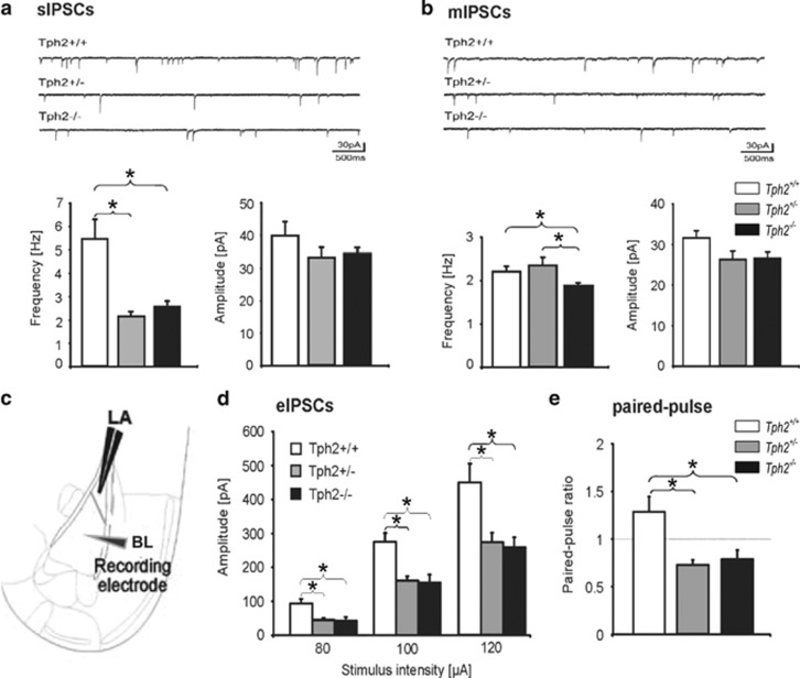
Multiple lines of evidence implicate brain serotonin (5-hydroxytryptamine; 5-HT) system dysfunction in the pathophysiology of stressor-related and anxiety disorders. Here we investigate the influence of constitutively deficient 5-HT synthesis on stressor-related anxiety-like behaviors using Tryptophan hydroxylase 2 (Tph2) mutant mice. Functional assessment of c-Fos after associated foot shock, electrophysiological recordings of GABAergic synaptic transmission, differential expression of the Slc6a4 gene in serotonergic neurons were combined with locomotor and anxiety-like measurements in different contextual settings. Our findings indicate that constitutive Tph2 inactivation and consequential lack of 5-HT synthesis in Tph2 null mutant mice (Tph2-/-) results in increased freezing to associated foot shock and a differential c-Fos activity pattern in the basolateral complex of the amygdala. This is accompanied by altered GABAergic transmission as observed by recordings of inhibitory postsynaptic currents on principal neurons in the basolateral nucleus, which may explain increased fear associated with hyperlocomotion and escape-like responses in aversive inescapable contexts. In contrast, lifelong 5-HT deficiency as observed in Tph2 heterozygous mice (Tph+/-) is able to be compensated through reduced GABAergic transmission in the basolateral nucleus of the amygdala based on Slc6a4 mRNA upregulation in subdivisions of dorsal raphe neurons. This results in increased activity of the basolateral nucleus of the amygdala due to associated foot shock. In conclusion, our results reflect characteristic syndromal dimensions of panic disorder and agoraphobia. Thus, constitutive lack of 5-HT synthesis influence the risk for anxiety- and stressor-related disorders including panic disorder and comorbid agoraphobia through the absence of GABAergic-dependent compensatory mechanisms in the basolateral nucleus of the amygdala.


Conflict of interest statement

The authors declare no conflict of interest.

Figures

Figure 1
Figure 1
Increased freezing and shock reactivity in Tph2-deficient mice. Paired foot shock (FS) protocol (a). Mice were exposed to a novel context for 120 s. After 120 s, a tone was paired with a FS and repeated two times with a 60 s pause. Mice were returned to the home cage 120 s after the last FS and killed 2 h later for c-Fos analysis. Freezing during fear conditioning was compared between FS+ (n=8/genotype) and FS− (n=3–4/genotype) mice (b). Shock reactivity in the FS+ and FS− groups was analyzed (c) and correlated with post-shock freezing (d). Data are shown as means±s.e.m. (b) or +s.e.m. (c). #0.1>P>0.05, **P<0.01 and ***P<0.001 compared to respective controls.
Figure 2
Figure 2
Foot shock differentially activates the basolateral complex of amygdala in Tph2-deficient mice. Anti-parvalbumin (PV) (green) and c-Fos (red) immunofluorescent staining with DAPI (blue) of Tph2+/+, Tph2+/− and Tph2−/− mice in the lateral (LA) and basolateral (BL) nucleus of the basolateral amygdala (a). c-Fos immunostaining was analyzed in Tph2+/+, Tph2+/ and Tph2/− mice under home cage control conditions (Ctrl) (n=5–7), mice that were placed in the conditioning context but did not receive foot shocks (FS−, n=3-4) and after the foot shock presentation (FS+, n=5–7/condition) (b). c-Fos-immunoreactive cell densities were analyzed between Ctrl, FS− and FS+ cohorts in the BL (c) and LA (d). Arrows in a indicate c-Fos ir cells in the anterior BL. Data are shown as means+s.e.m. #0.1>P>0.05, *P<0.05 and compared to respective controls. Scale bar, 100 μm (a).
Figure 3
Figure 3
Inhibitory currents in the basolateral nucleus of the amygdala of Tph2-deficient mice. Electrophysiological investigation of GABAergic synapse function in vitro of naive Tph2−/−, Tph2+/− and Tph2+/+ mice. (a) Upper panel: mean frequency (left) and amplitude (right) of spontaneous inhibitory postsynaptic currents (sIPSCs) in Tph2−/− (n=21/3), Tph2+/ (n=24/4) and Tph2+/+ (n=19/3) mice with example traces shown in the lower panel. (b) Upper panel: mean frequency and amplitude of miniature inhibitory postsynaptic currents (mIPSCs) in Tph2−/− (n=18/4), Tph2+/− (n=18/3) and Tph2+/+ (n=16/3) mice with example traces shown in the lower panel. (c) Scheme of recording indicating the stimulating electrode in the lateral nucleus (LA) and the patch-clamp pipette in the basolateral nucleus of the amygdala (BL). (d) Mean amplitude of evoked inhibitory postsynaptic currents (eIPSCs) recorded at different stimulus intensities in pyramidal neurons from Tph2−/−, Tph2+/ and Tph2+/+ mice. Note significant reduction of eIPSCs at all tested stimulation intensities in Tph2−/− (n=14/4) and Tph2+/− (n=13/3) compared to wild types (n=12/3) as well as paired-pulse ratio (e). Data are shown as means+s.e.m. *P<0.05, **P<0.01 and ***P<0.001.
Figure 4
Figure 4
Expression of Slc6a4 mRNA across the rostrocaudal extent of the dorsal raphe nucleus. (a) Photomicrographs illustrating Slc6a4 (solute carrier family 6, member 4; 5-Htt) mRNA expression in Tph2+/+ mice (left), Tph2+/− mice (middle) and Tph2−/− mice (right) at −4.364 mm bregma. (be) Mean expression of Slc6a4 mRNA across the rostrocaudal extent of (b) the entire dorsal raphe nucleus (DR) and (c) MnR, (d) rDRD and (e) cDRD as well as (f) rDRV and (g) cDRV of Tph2+/+, Tph2+/− and Tph2−/− mice (n=6–8 per genotype). DR, dorsal raphe nucleus; DRD, dorsal raphe nucleus, dorsal part; DRV, dorsal raphe nucleus, ventral part; MnR, median raphe nucleus. n=6–8/genotype; *P<0.01. Scale bar, 200 μm (a).
Figure 5
Figure 5
5-HT deficiency induces hyperlocomotion and escape behaviors in different contexts. Locomotor activity of Tph2+/+, Tph2+/− and Tph2−/− mice was measured over 30 min in the home cage (n=10–11/genotype) and in the open field (n=11/genotype) (a) as well as in a two-trial social interaction test (b) (n=7/genotype). Escape responses were detected as wall jumps in the open field (c). Further anxiety-like measures (n=11/genotype) included mean open-field center time (d) as well as latency to enter (e) and time in the light compartment of the light–dark box (f). Novelty-suppressed feeding (n=10–11/genotype) included latency to feed (g) as well as total feeding time (h). Social behaviors were evaluated using measures of sociability and preference for social novelty (i). Data are shown as means+s.e.m. #P<0.1, *P<0.05 and **P<0.01 compared to respective controls.

References

    1. Wittchen HU, Jacobi F, Rehm J, Gustavsson A, Svensson M, Jonsson B et al. The size and burden of mental disorders and other disorders of the brain in Europe 2010. Eur Neuropsychopharmacol 2011; 21: 655–679. - PubMed
    1. Shekhar A, Sajdyk TS, Keim SR, Yoder KK, Sanders SK. Role of the basolateral amygdala in panic disorder. Ann N Y Acad Sci 1999; 877: 747–750. - PubMed
    1. Rauch SL, Shin LM, Wright CI. Neuroimaging studies of amygdala function in anxiety disorders. Ann NY Acad Sci 2003; 985: 389–410. - PubMed
    1. Naughton M, Mulrooney JB, Leonard BE. A review of the role of serotonin receptors in psychiatric disorders. Hum Psychopharmacol 2000; 15: 397–415. - PubMed
    1. Graeff FG, Guimaraes FS, De Andrade TG, Deakin JF. Role of 5-HT in stress, anxiety, and depression. Pharmacol Biochem Behav 1996; 54: 129–141. - PubMed

Publication types