. 2017 Nov 20;30(11):2023-2032.
doi: 10.1021/acs.chemrestox.7b00227. Epub 2017 Oct 23.

Mechanism of Error-Free DNA Replication Past Lucidin-Derived DNA Damage by Human DNA Polymerase κ

Affiliations

Mechanism of Error-Free DNA Replication Past Lucidin-Derived DNA Damage by Human DNA Polymerase κ

Oliver P Yockey et al. Chem Res Toxicol. .

Abstract

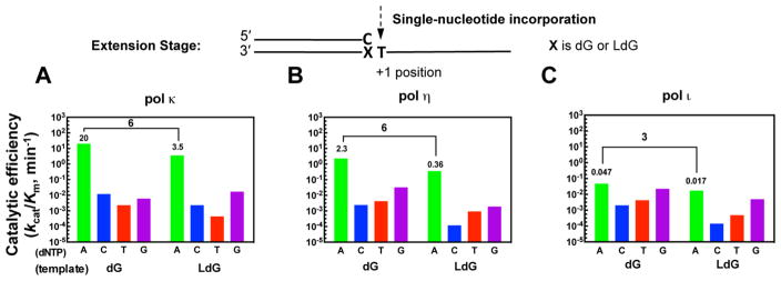
DNA damage impinges on genetic information flow and has significant implications in human disease and aging. Lucidin-3-O-primeveroside (LuP) is an anthraquinone derivative present in madder root, which has been used as a coloring agent and food additive. LuP can be metabolically converted to genotoxic compound lucidin, which subsequently forms lucidin-specific N2-2'-deoxyguanosine (N2-dG) and N6-2'-deoxyadenosine (N6-dA) DNA adducts. Lucidin is mutagenic and carcinogenic in rodents but has low carcinogenic risks in humans. To understand the molecular mechanism of low carcinogenicity of lucidin in humans, we performed DNA replication assays using site-specifically modified oligodeoxynucleotides containing a structural analogue (LdG) of lucidin-N2-dG DNA adduct and determined the crystal structures of DNA polymerase (pol) κ in complex with LdG-bearing DNA and an incoming nucleotide. We examined four human pols (pol η, pol ι, pol κ, and Rev1) in their efficiency and accuracy during DNA replication with LdG; these pols are key players in translesion DNA synthesis. Our results demonstrate that pol κ efficiently and accurately replicates past the LdG adduct, whereas DNA replication by pol η, pol ι is compromised to different extents. Rev1 retains its ability to incorporate dCTP opposite the lesion albeit with decreased efficiency. Two ternary crystal structures of pol κ illustrate that the LdG adduct is accommodated by pol κ at the enzyme active site during insertion and postlesion-extension steps. The unique open active site of pol κ allows the adducted DNA to adopt a standard B-form for accurate DNA replication. Collectively, these biochemical and structural data provide mechanistic insights into the low carcinogenic risk of lucidin in humans.


Conflict of interest statement

Notes

The authors declare no competing financial interest.

Figures

Figure 1
Figure 1
Metabolic activation of lucidin-O-primeveroside and lucidin, and formation of ludicin-derived DNA adducts. The structure of synthetic analogue (LdG) of lucidin-N2-2′-deoxyguanosine adduct is shown.
Figure 2
Figure 2
Translesion DNA synthesis by human DNA polymerases κ, η, ι, or Rev1 with unmodified or LdG-containing DNA substrate. Reaction conditions are described in the Materials and Methods section. (A) Primer extension reactions in the presence of all four dNTPs at their physiological concentrations (i.e., 10 μM for dGTP and 40 μM for dATP, dCTP, and dTTP). (B) Catalytic efficiencies of individual dNTPs opposite unmodified dG or LdG adduct. Enzyme kinetic parameters are shown in the Supporting Information Table S2. Changes in catalytic efficiency relative to a native base pair were calculated from (kcat/Km,dCTP)unmodified/(kcat/Km,dCTP)LdG and indicated as x-fold decrease.
Figure 3
Figure 3
Postlesion extension by human DNA polymerases κ, η, or ι with unmodified or LdG-containing DNA. Catalytic efficiencies of nucleotide incorporations were determined by steady-state kinetic analysis with detailed kinetic parameters shown in the Supporting Information Table S3. Changes in catalytic efficiency relative to a native base pair were calculated from (kcat/Km,dATP)unmodified/(kcat/Km,dATP)LdG and indicated as x-fold decrease.
Figure 4
Figure 4
Crystal structures of pol κ: LdG-DNA:dNTP* ternary complexes at the (A) insertion and (B) extension stages. The overall structures of pol κ:LdG-DNA:dNTP* ternary complexes (central panels) are in the back views with the minor groove DNA facing the viewer. dNTP* is dCMPNPP (dCTP*) or dAMPNPP (dATP*). Template is in light pink, primer in gray, LdG in orange, and Mg2+ ions in green spheres. The protein is depicted in three colors: core domains (palm, finger and thumb) are in cyan, little finger (LF) is in light purple, and N-clasp is in beige. Orthogonal views of the replicating base pairs (bottom panels) at the active site with 2FoFc map (blue mesh) contoured at 1σ. Top panels are zoomed-in views of the detailed stabilization of the bulky LdG ring (orange) by surrounding amino acid residues, solvent molecules, polyethene glycol (PEG) and glycerol (Gol), and backbone of the template strand (pink).
Figure 5
Figure 5
Structures of LdG-harboring DNA within pol κ-bound complexes at the (A) insertion and (B) extension stages. The color scheme of the left and middle panels is the same as described in Figure 4. Incoming dNTP* is in yellow, where dNTP* is dCMPNPP (dCTP*) or dAMPNPP (dATP*). The double arrowed lines illustrate a distance of 4.3–4.5 Å between the 3′–OH of the primer and the α phosphate of dNTP*. In both insertion and extension complexes, the lucidin ring is oriented toward the 3′-end of the template strand. The right panels illustrate the superposition of LdG-containing DNA with unmodified DNA in a pol κ complex (PDB codes: 2OHO). The unmodified DNA is in gray and the adducted DNA (LdG adduct in orange) is in cyan (insertion stage) and blue (extension stage).

References

    1. Poirier MC, Santella RM, Weston A. Carcinogen macromolecular adducts and their measurement. Carcinogenesis. 2000;21:353–359. - PubMed
    1. Hecht SS. Tobacco carcinogens, their biomarkers and tobacco-induced cancer. Nat Rev Cancer. 2003;3:733–744. - PubMed
    1. Poirier MC. Chemical-induced DNA damage and human cancer risk. Nat Rev Cancer. 2004;4:630–637. - PubMed
    1. Balbo S, Turesky RJ, Villalta PW. DNA adductomics. Chem Res Toxicol. 2014;27:356–366. - PMC - PubMed
    1. Hoeijmakers JHJ. Genome maintenance mechanisms for preventing cancer. Nature. 2001;411:366–374. - PubMed

Publication types

LinkOut - more resources