

Optic Nerve Infiltration in Primary Central Nervous System Lymphoma
- PMID: 28973119
- PMCID: PMC5710580
- DOI: 10.1001/jamaneurol.2017.2545
Optic Nerve Infiltration in Primary Central Nervous System Lymphoma
Abstract
Importance: Visual impairment in primary central nervous system lymphoma (PCNSL) is caused mostly by intraocular lymphomatous involvement (vitritis and retinal infiltration), whereas optic nerve infiltration (ONI) is a rare condition.
Objective: To describe the clinical presentation of ONI, its imaging characteristics, and outcome.
Design, setting and participants: A total of 752 patients diagnosed with PCNSL were retrospectively identified from the databases of 3 French hospitals from January 1, 1998, through December 31, 2014. Of these, 7 patients had documented ONI. Exclusion criteria were intraocular involvement, orbital lymphoma, or other systemic lymphoma. Clinical presentation, neuroimaging, biological features, treatment, and outcomes were assessed.
Main outcomes and measures: Treatment response was evaluated clinically and radiologically on follow-up magnetic resonance imaging (MRI) according to the International PCNSL Collaborative Group response criteria.
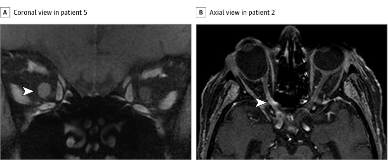
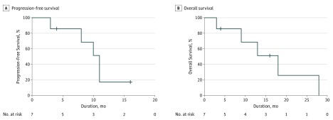
Results: The 7 patients included 5 women and 2 men. Median age at diagnosis was 65 years (range, 49-78 years). Two patients had initial ONI at diagnosis, and 5 had ONI at relapse. Clinical presentation was marked by rapidly progressive and severe visual impairment for all patients. The MRI findings showed optic nerve enlargement in 3 patients and contrast enhancement of the optic nerve in all patients. Additional CNS lesions were seen in 4 patients. Examination of cerebrospinal fluid samples detected lymphomatous meningitis in 2 patients. Clinical outcome was poor and marked by partial recovery for 2 patients and persistent severe low visual acuity or blindness for 5 patients. Median progression-free survival after optic nerve infiltration was 11 months (95% CI, 9-13 months), and median overall survival was 18 months (95% CI, 9-27 months).
Conclusions and relevance: Optic nerve infiltration is an atypical and challenging presentation of PCNSL. Its visual and systemic prognosis is particularly poor compared with vitreoretinal lymphomas even in response to chemotherapy. Although intraocular involvement is frequent in PCNSL and clinically marked by slowly progressive visual deterioration, lymphomatous ONI is rare and characterized by rapidly progressive severe visual impairment.
Conflict of interest statement
Figures
References
-
- Baryla J, Allen LH, Kwan K, Ong M, Sheidow T. Choroidal lymphoma with orbital and optic nerve extension: case and review of literature. Can J Ophthalmol. 2012;47(1):79-81. - PubMed
-
- Zelefsky JR, Revercomb CH, Lantos G, Warren FA. Isolated lymphoma of the anterior visual pathway diagnosed by optic nerve biopsy. J Neuroophthalmol. 2008;28(1):36-40. - PubMed
-
- Gill MK, Jampol LM. Variations in the presentation of primary intraocular lymphoma: case reports and a review. Surv Ophthalmol. 2001;45(6):463-471. - PubMed
MeSH terms
LinkOut - more resources
Full Text Sources
Other Literature Sources
Medical
