. 2017 Nov 1;26(21):4153-4167.
doi: 10.1093/hmg/ddx304.

Homeostatic plasticity can be induced and expressed to restore synaptic strength at neuromuscular junctions undergoing ALS-related degeneration

Affiliations

Homeostatic plasticity can be induced and expressed to restore synaptic strength at neuromuscular junctions undergoing ALS-related degeneration

Sarah Perry et al. Hum Mol Genet. .

Abstract

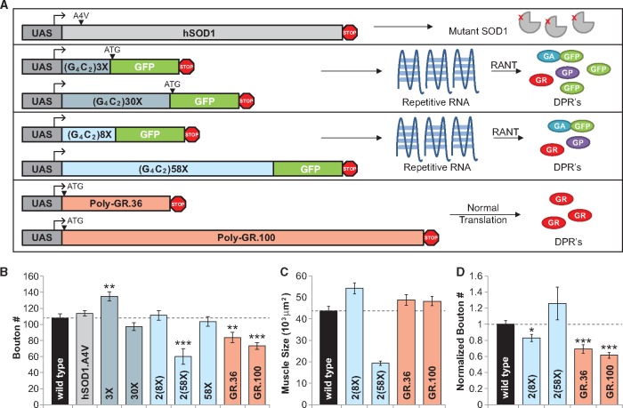
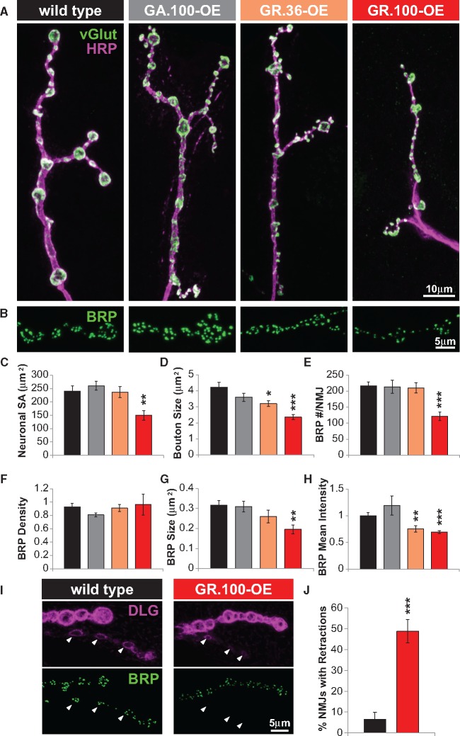
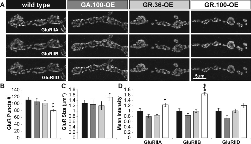
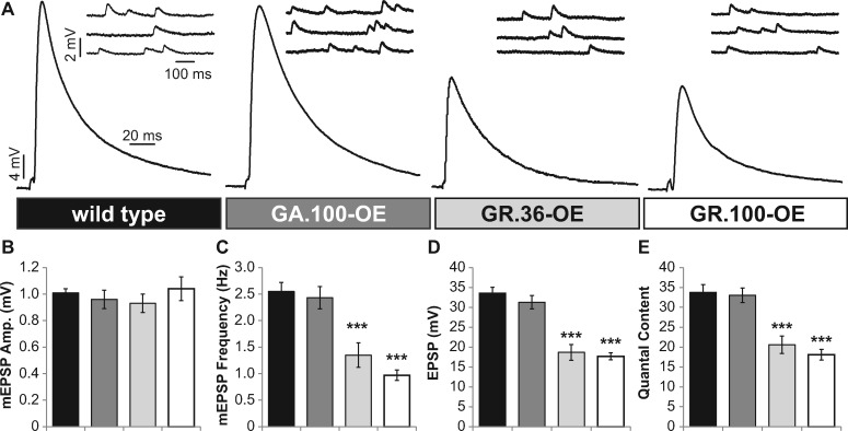
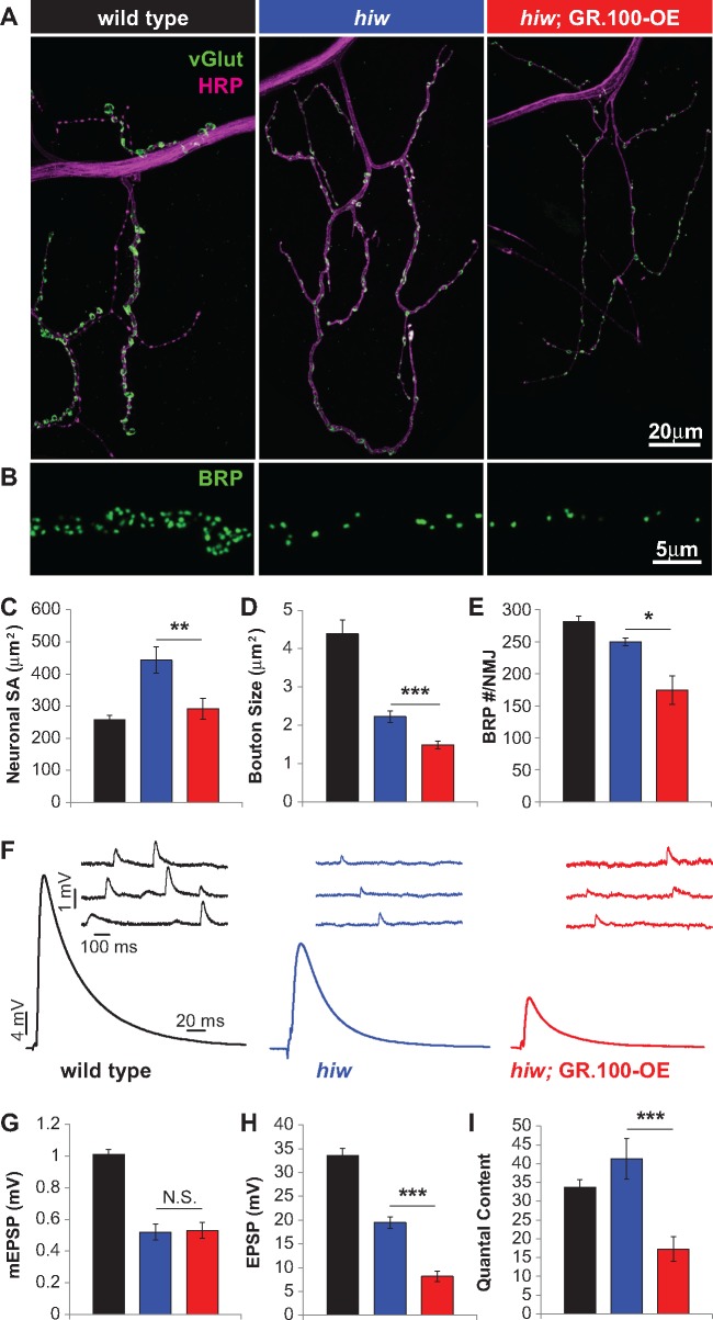
Amyotrophic lateral sclerosis (ALS) is debilitating neurodegenerative disease characterized by motor neuron dysfunction and progressive weakening of the neuromuscular junction (NMJ). Hereditary ALS is strongly associated with variants in the human C9orf72 gene. We have characterized C9orf72 pathology at the Drosophila NMJ and utilized several approaches to restore synaptic strength in this model. First, we demonstrate a dramatic reduction in synaptic arborization and active zone number at NMJs following C9orf72 transgenic expression in motor neurons. Further, neurotransmission is similarly reduced at these synapses, consistent with severe degradation. However, despite these defects, C9orf72 synapses still retain the ability to express presynaptic homeostatic plasticity, a fundamental and adaptive form of NMJ plasticity in which perturbation to postsynaptic neurotransmitter receptors leads to a retrograde enhancement in presynaptic release. Next, we show that these endogenous but dormant homeostatic mechanisms can be harnessed to restore synaptic strength despite C9orf72 pathogenesis. Finally, activation of regenerative signaling is not neuroprotective in motor neurons undergoing C9orf72 toxicity. Together, these experiments define synaptic dysfunction at NMJs experiencing ALS-related degradation and demonstrate the potential to activate latent plasticity as a novel therapeutic strategy to restore synaptic strength.


Figures

Figure 1
Figure 1
Overexpression of a C9orf72 repeat expansion transgene in motor neurons reduces the number of synaptic boutons. (A) Schematic illustrating various transgenes of human disease variants used to model ALS by transgenic expression in Drosophila. UAS-SOD1-A4V (top) is a mutant version of human SOD1 associated with ALS pathogenesis (6). UAS-(G4C2)3X, 30X(5), 8X, and 58X(9) (middle) are all versions of the human C9orf72 repeat expansion predicted to produce repetitive RNA complexes as well as dipeptide repeats (DPRs) through non-ATG initiated translation (RANT). UAS-Poly-GR.36 and GR.100(15) (bottom) are variations of the C9orf72 repeat expansion that produce only glycine-arginine DPRs through standard ATG translation. (B) Bouton counts at the muscle 6/7 larval NMJ in wild type (w1118) and following overexpression of the indicated ALS transgene in motor neurons. One copy of the motor neuron driver OK6-Gal4 was used to drive one copy of the UAS in most cases; two copies of the motor neuron driver OK371-Gal4 was used to drive two copies of UAS in 2(8X) and 2(58X), as previously reported(9). (C) Quantification of muscle surface area in indicated genotypes. (D) Quantification of bouton numbers normalized to muscle size. GR.36-OE and GR.100-OE show a decreased synapse area to muscle size ratio, consistent with NMJ degeneration. Error bars indicate ±SEM. Statistical comparisons to wild type were made using a 2-tailed Student‘s t-test. *P ≤ 0.05; **P 0.01; ***P = 0.001; detailed statistical information for represented data (mean values, SEM, n, p) is shown in Supplementary Material, Table S1.
Figure 2
Figure 2
Overexpression of GR.100 in motor neurons leads to reductions in presynaptic membrane surface area and active zone number at the NMJ. (A) Representative images of muscle 4 NMJs in wild type, GA.100-OE (w; OK6-Gal4/UAS-poly-GA.100), GR.36-OE (w; OK6-Gal4/UAS-poly-GR.36), and GR.100-OE (w; OK6-Gal4/UAS-poly-GR.100) DPR overexpression in motor neurons. NMJs were immunostained with anti-HRP to label the neuronal membrane and anti-vGlut to label synaptic vesicles (used to define synaptic boutons). (B) Representative images of active zones labeled with anti-BRP in the indicated genotypes. Quantification of neuronal membrane surface area (C), bouton size (D), total BRP puncta number per NMJ (E), BRP density (F), BRP size (G), and normalized mean intensity of BRP (H) in the indicated genotypes. (I) Representative images of M6/7 boutons immunostained with anti-DLG (postsynaptic scaffold) and anti-BRP in the indicated genotypes. White arrows indicate boutons where DLG and BRP are appropriately apposed (wild type) and where postsynaptic DLG puncta lack opposed BRP structures (GR.100-OE), indicating retraction. (J) Quantification of retracted NMJs in wild type and GR.100-OE larvae. Error bars indicate ±SEM. Statistical comparisons to wild type were made using a 2-tailed Student‘s t-test. *P ≤ 0.05; **P 0.01; ***P = 0.001; detailed statistical information for represented data (mean values, SEM, n, p) is shown in Supplementary Material, Table S1.
Figure 3
Figure 3
Postsynaptic glutamate receptor levels are increased following presynaptic expression of GR repeats. (A) Representative images of muscle 4 NMJs in the indicated genotypes immunostained with antibodies against three postsynaptic glutamate receptor subunits: GluRIIA, GluRIIB, and GluRIID. Quantification of total glutamate receptor puncta number (B), glutamate receptor size (C), and normalized mean intensity (D) in the indicated genotypes. Error bars indicate ±SEM. Statistical comparisons to wild type were made using a 2-tailed Student‘s t-test. *P ≤ 0.05; **P 0.01; ***P = 0.001; detailed statistical information for represented data (mean values, SEM, n, p) is shown in Supplementary Material, Table S1.
Figure 4
Figure 4
Presynaptic overexpression of GR repeats degrades synaptic strength at the NMJ. (A) Representative electrophysiological traces of evoked (EPSP) and spontaneous (mEPSP) responses from wild type, GA.100-OE, GR.36-OE, and GR.100-OE NMJs. Quantification of mEPSP amplitude (B), mEPSP frequency (C), EPSP amplitude (D), and quantal content (E) for the indicated genotypes. Error bars indicate ±SEM. Statistical comparisons to wild type were made using a 2-tailed Student‘s t-test. *P ≤ 0.05; **P 0.01; ***P = 0.001; detailed statistical information for represented data (mean values, SEM, n, p) is shown in Supplementary Material, Table S1.
Figure 5
Figure 5
Homeostatic control of presynaptic function persists despite GR induced NMJ degradation. Representative EPSP and mEPSP traces of wild type (A) and GR.100-OE (B) under baseline conditions and following acute application of the postsynaptic glutamate receptor antagonist philanthotoxin (PhTx). Following application, mEPSP amplitude is reduced, but EPSP amplitudes are maintained at baseline levels due to a homeostatic increase in presynaptic release (quantal content). Similarly, a chronic reduction in mEPSP amplitude can be induced through genetic reduction of glutamate receptor levels by postsynaptic knock down of the glutamate receptor subunit GluRIII (GluRIII-RNAi: w; OK6-Gal4/+; BG57-Gal4, GluRIII-RNAi). Note that both wild type and GR.100-OE NMJs exhibit reduced mEPSP amplitude following PhTx application or GluRIII-RNAi, but maintain baseline EPSP levels, demonstrating robust homeostatic compensation. (C) Quantification of mEPSP and quantal content values for the indicated genotypes following PhTx application, normalized to baseline values (absence of PhTx application). (D) Quantification of mEPSP and quantal content values for the indicated genotypes in combination with GluRIII-RNAi, normalized to control genotypes (absence of GluRIII-RNAi). Note that GR.100 (Muscle) indicates a control for muscle-only expression of GR.100 (UAS-Poly-GR.100/+; BG57-Gal4/+). Error bars indicate ±SEM. Detailed statistical information for additional controls, represented data, and absolute values (mean values, SEM, n, p) are shown in Supplementary Material, Table S1.
Figure 6
Figure 6
Postsynaptic overexpression of Tor triggers presynpatic homeostatic potentiation and restores synaptic strength in GR.100-OE NMJs. (A) Representative EPSP and mEPSP traces from wild type, postsynaptic Tor overexpression (Tor-OE: w; OK6-Gal4/+; MHC-Gal4/UAS-Tor), GR.100-OE control (w; OK6-Gal4/GR.100; MHC-Gal4/+), and GR.100-OE +TOR-OE (w; OK6-Gal4/GR.100; MHC-Gal4/UAS-Tor) recorded in 0.3 mM extracellular calcium. Note that Tor-OE triggers increased EPSP amplitude due to an enhancement in presynaptic release, restoring wild-type levels of synaptic strength in GR.100-OE, while mEPSP amplitudes are unchanged. Quantification of mEPSP amplitude (B), EPSP amplitude (C), and quantal content (D) in the indicated genotypes. Error bars indicate ±SEM. Statistical comparisons were made using a 2-tailed Student‘s t-test. *P ≤ 0.05; **P 0.01; ***P = 0.001; detailed statistical information for represented data (mean values, SEM, n, p) is shown in Supplementary Material, Table S1.
Figure 7
Figure 7
Postsynaptic overexpression of GluRIIA increases quantal size and restores synaptic strength and locomotor behavior in GR.100-OE. (A) Representative images of glutamate receptor immunostaining at NMJs of wild type, postsynaptic overexpression of GluRIIA (GluRIIA-OE: MHC-GluRIIA), and postsynaptic overexpression of GluRIIA with GR.100-OE (GluRIIA-OE + GR.100-OE: MHC-GluRIIA; OK6-Gal4/UAS-GR.100). Note that GluRIIA-OE increases GluRIIA expression and reduces GluRIIB expression. (B) Quantification of mean intensity levels of glutamate receptor staining from the indicated genotypes. (C) Representative EPSP and mEPSP traces from indicated genotypes. Note that GluRIIA-OE causes an increase in postsynaptic sensitivity to glutamate, resulting in enhanced quantal size, no change in quantal content, and a concomminant increase in EPSP amplitude. Quantification of mEPSP amplitude (D), EPSP amplitude (E), and quantal content (F) in the indicated genotypes. (G) Schematic showing larval mobility assay. Single larvae are allowed to crawl freely on an agarose plate placed on top of 5 mm grid paper. The number of gridlines crossed in 2 minutes is manually scored. (H) Locomotor behavior is improved following elevated postsynaptic receptor expression in GR.100-OE NMJs. Quantification for the mobility assay for wild type, GR.36-OE, GR.100-OE, and GluRIIA-OE third-instar larvae without and following GluRIIA overexpression. Error bars indicate ±SEM. Statistical comparisons were made using a 2-tailed Student‘s t-test. *P ≤ 0.05; **P 0.01; ***P = 0.001; ns = not significant, P > 0.05. Detailed statistical information for represented data (mean values, SEM, n, p) is shown in Supplementary Material, Table S1.
Figure 8
Figure 8
Constitutive regenerative signaling is not neuroprotective against GR.100-OE induced degeneration. (A) Representative images of wild type, highwire (hiw) mutant, and hiw; GR.100-OE (hiw; OK6-Gal4/UAS-GR.100) NMJs. Presynaptic neuronal membrane is immunostained with anti-HRP (magenta) and anti-vGlut (green). (B) Representative images of BRP puncta in these genotypes. Quantification of neuronal membrane surface area (C), bouton size (D), and BRP puncta number per NMJ (E) in the indicated genotypes. (F) Representative EPSP and mEPSP traces of the indicated genotypes. Quantification of mEPSP amplitude (G), EPSP amplitude (H), and quantal content (I) in the indicated genotypes. Error bars indicate ±SEM. Statistical comparisons were made using a 2-tailed Students‘s t-test. *P ≤ 0.05; **P 0.01; ***P = 0.001; ns = not significant, P > 0.05. Detailed statistical information for represented data (mean values, SEM, n, p) is shown in Supplementary Material, Table S1.

Similar articles

Cited by

References

    1. Renton A.E., Majounie E., Waite A., Simón-Sánchez J., Rollinson S., Gibbs J.R., Schymick J.C., Laaksovirta H., van Swieten J.C., Myllykangas L.. et al. (2011) A hexanucleotide repeat expansion in C9ORF72 is the cause of chromosome 9p21-linked ALS-FTD. Neuron, 72, 257–268. - PMC - PubMed
    1. DeJesus-Hernandez M., Mackenzie I.R., Boeve B.F., Boxer A.L., Baker M., Rutherford N.J., Nicholson A.M., Finch N.A., Flynn H., Adamson J.. et al. (2011) Expanded GGGGCC hexanucleotide repeat in noncoding region of C9ORF72 causes chromosome 9p-linked FTD and ALS. Neuron, 72, 245–256. - PMC - PubMed
    1. Rosen D.R., Siddique T., Patterson D., Figlewicz D.A., Sapp P., Hentati A., Donaldson D., Goto J., O'Regan J.P., Deng H.-X.. et al. (1993) Mutations in Cu/Zn superoxide dismutase gene are associated with familial amyotrophic lateral sclerosis. Nature, 362, 59–62. - PubMed
    1. Chew J., Gendron T.F., Prudencio M., Sasaguri H., Zhang Y.-J., Castanedes-Casey M., Lee C.W., Jansen-West K., Kurti A., Murray M.E.. et al. (2015) Neurodegeneration. C9ORF72 repeat expansions in mice cause TDP-43 pathology, neuronal loss, and behavioral deficits. Science, 348, 1151–1154. - PMC - PubMed
    1. Xu Z., Poidevin M., Li X., Li Y., Shu L., Nelson D.L., Li H., Hales C.M., Gearing M., Wingo T.S.. et al. (2013) Expanded GGGGCC repeat RNA associated with amyotrophic lateral sclerosis and frontotemporal dementia causes neurodegeneration. Proc. Natl. Acad. Sci. U. S. A., 110, 7778–7783. - PMC - PubMed

Publication types

MeSH terms

LinkOut - more resources