

Progressive Deformation of the Optic Nerve Head and Peripapillary Structures by Graded Horizontal Duction
- PMID: 28973373
- PMCID: PMC5627675
- DOI: 10.1167/iovs.17-22596
Progressive Deformation of the Optic Nerve Head and Peripapillary Structures by Graded Horizontal Duction
Abstract
Purpose: We investigated the effect of graded range of horizontal duction on the shape of the peripapillary Bruch's membrane (ppBM) and optic nerve head (ONH).
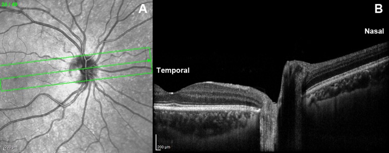
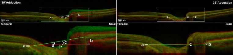
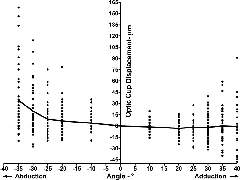
Methods: In 50 eyes of 25 normal subjects, the ONH and peripapillary retina were imaged by optical coherence tomography (OCT) in central gaze and incremental angles of add- and abduction. Displacements of the Bruch's membrane opening (BMO), optic cup (OC), and change in ONH angle in eccentric gazes were compared to those of central gaze, in add- and abduction.
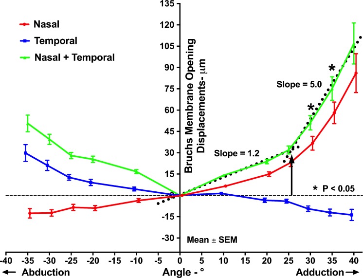
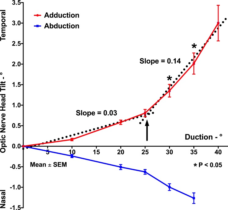
Results: With increasing duction, the nasal edge of the BMO (nBMO) shifted progressively anteriorly in adduction and posteriorly in abduction, while the temporal edge of the BMO (tBMO) shifted posteriorly in adduction and anteriorly in abduction. The summed absolute nBMO and tBMO displacements in 30° and 35° adduction significantly exceeded those in comparable abduction angles (P < 0.005 for both). The ONH progressively tilted temporally in adduction and nasally in abduction; absolute ONH tilt in adduction was significantly greater than that in abduction for 30° and 35° ductions (P < 0.005 for both). BMO displacement and ONH tilt in adduction exhibited bilinear behavior, with greater effects for both at angles exceeding 26°. The OC shifted significantly farther anteriorly in abduction than adduction at every angle from 10° to 35°.
Conclusions: Horizontal duction deforms the ONH and ppBM, but more in adduction than in abduction, and increasingly so for angles greater than 26°. This behavior is consistent with optic nerve sheath tethering for adduction exceeding 26°.
Figures
References
-
- Park KH, Tomita G, Liou SY, Kitazawa Y. . Correlation between peripapillary atrophy and optic nerve damage in normal-tension glaucoma. Ophthalmology. 1996; 103: 1899– 1906. - PubMed
-
- Uchida H, Ugurlu S, Caprioli J. . Increasing peripapillary atrophy is associated with progressive glaucoma. Ophthalmology. 1998; 105: 1541– 1545. - PubMed
-
- Jonas JB. . Clinical implications of peripapillary atrophy in glaucoma. Curr Opin Ophthalmol. 2005; 16: 84– 88. - PubMed
-
- Wang X, Rumpel H, Lim WE,et al. Finite element analysis predicts large optic nerve head strains during horizontal eye movements. Invest Ophthalmol Vis Sci. 2016; 57: 2452– 2462. - PubMed
Publication types
MeSH terms
Grants and funding
LinkOut - more resources
Full Text Sources
