. 2017 Nov 15;26(22):4506-4518.
doi: 10.1093/hmg/ddx338.

TUBB4A mutations result in specific neuronal and oligodendrocytic defects that closely match clinically distinct phenotypes

Affiliations

TUBB4A mutations result in specific neuronal and oligodendrocytic defects that closely match clinically distinct phenotypes

Julian Curiel et al. Hum Mol Genet. .

Abstract

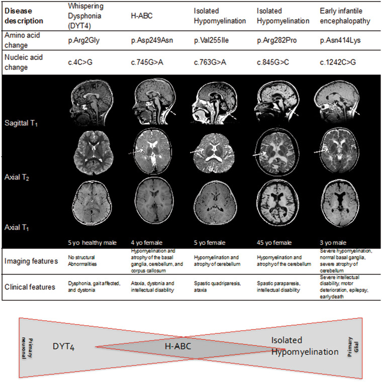
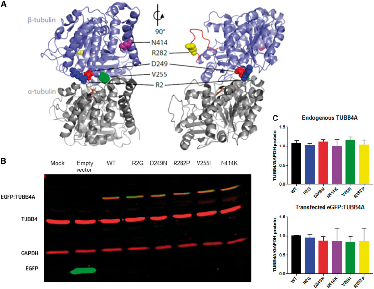
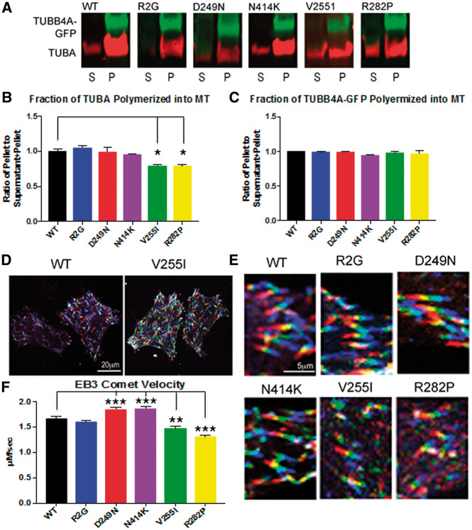
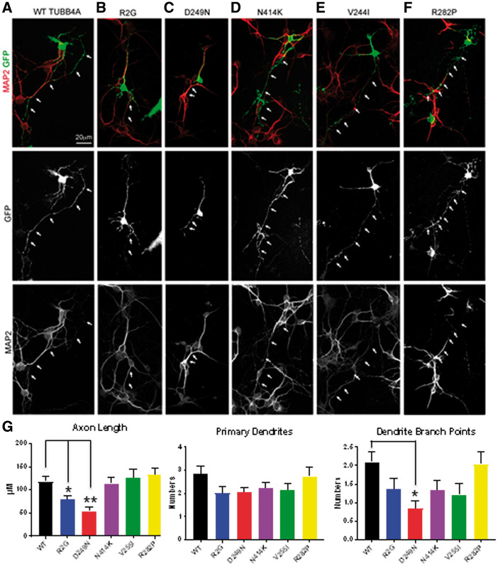
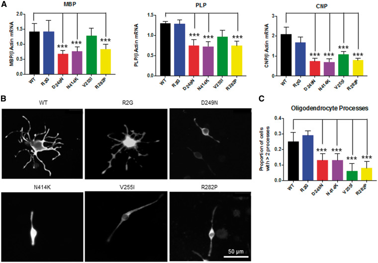
Hypomyelinating leukodystrophies are heritable disorders defined by lack of development of brain myelin, but the cellular mechanisms of hypomyelination are often poorly understood. Mutations in TUBB4A, encoding the tubulin isoform tubulin beta class IVA (Tubb4a), result in the symptom complex of hypomyelination with atrophy of basal ganglia and cerebellum (H-ABC). Additionally, TUBB4A mutations are known to result in a broad phenotypic spectrum, ranging from primary dystonia (DYT4), isolated hypomyelination with spastic quadriplegia, and an infantile onset encephalopathy, suggesting multiple cell types may be involved. We present a study of the cellular effects of TUBB4A mutations responsible for H-ABC (p.Asp249Asn), DYT4 (p.Arg2Gly), a severe combined phenotype with hypomyelination and encephalopathy (p.Asn414Lys), as well as milder phenotypes causing isolated hypomyelination (p.Val255Ile and p.Arg282Pro). We used a combination of histopathological, biochemical and cellular approaches to determine how these different mutations may have variable cellular effects in neurons and/or oligodendrocytes. Our results demonstrate that specific mutations lead to either purely neuronal, combined neuronal and oligodendrocytic or purely oligodendrocytic defects that closely match their respective clinical phenotypes. Thus, the DYT4 mutation that leads to phenotypes attributable to neuronal dysfunction results in altered neuronal morphology, but with unchanged tubulin quantity and polymerization, with normal oligodendrocyte morphology and myelin gene expression. Conversely, mutations associated with isolated hypomyelination (p.Val255Ile and p.Arg282Pro) and the severe combined phenotype (p.Asn414Lys) resulted in normal neuronal morphology but were associated with altered oligodendrocyte morphology, myelin gene expression, and microtubule dysfunction. The H-ABC mutation (p.Asp249Asn) that exhibits a combined neuronal and myelin phenotype had overlapping cellular defects involving both neuronal and oligodendrocyte cell types in vitro. Only mutations causing hypomyelination phenotypes showed altered microtubule dynamics and acted through a dominant toxic gain of function mechanism. The DYT4 mutation had no impact on microtubule dynamics suggesting a distinct mechanism of action. In summary, the different clinical phenotypes associated with TUBB4A reflect the selective and specific cellular effects of the causative mutations. Cellular specificity of disease pathogenesis is relevant to developing targeted treatments for this disabling condition.


Figures

Figure 1.
Figure 1.
MRI features in individuals with TUBB4A mutations. Sagittal T1 (top), Axial T2 (middle) and Axial T1 images (bottom) images are shown. Individuals are shown corresponding to the mutations tested in cellular models. The R2G mutation associated with DYT4 and whispering dysphonia/dystonia is not shown as there are no gross structural abnormalities associated with it. Individual 1 was a normal 12-year-old female to illustrate normal MRI findings. Individual 2 was a 13-year-old male with classic H-ABC and D249N mutation. Individual 3 (V255I) was a 5-year-old female with isolated hypomyelination on neuroimaging and clinical features of spastic quadriparesis and ataxia. Individual 4 (R282P) is a 45-year-old female with isolated hypomyelination on neuroimaging and clinical features of spastic quadreplegia. Individual 5 (N414K) is a 3-year-old male with severe early onset encephalopathy, severe intellectual disability, motor deterioration, epilepsy, and early death. Note the absence of a putamen in individual 2 with preserved putamen in individuals 3, 4 and 5 (thin arrow). Note cerebellar atrophy present in all affected cases (dotted arrow) as well as abnormal T2 signal in the white matter. The schematic below represents the relative contribution of oligodendrocyte versus neuronal mechanisms of disease to the phenotype. MRIs for D249N, V255I, and R282P modified from Pizzino et al. (2014).
Figure 2.
Figure 2.
Neuropathology of TUBB4A-related diseases. Images in the top left box are D249N mutation pathology figures and images in the top right box are N414K mutation pathology figures. (A) In classical H-ABC (D249N mutation), the cerebellar cortex is severely atrophic with massive loss of granular neurons; stain against neurofilaments (NF) (B) shows swelling of Purkinje cell dendrites and axons (asterix). (C) In early onset encephalopathy (N414K mutation), the cerebellum also shows severe cortical atrophy with enlargement of the sulci and thinning of the granular layer; stain against NF (D) shows swellings of Purkinje cell axons and dendrites (asterix). (E) On macroscopic examination of a classical H-ABC brain, the putamen and, to a lesser degree, the caudate nucleus is not recognizable (arrows) on this coronal brain slice cut at the level of the anterior hippocampus. (F) This corresponds microscopically with loss of striatal neurons. Note also the small perivascular calcification (arrow). (G) In the individual with the severe combined phenotype, the putamen and caudate nucleus are preserved (arrows), and (H) there is no loss of striatal neurons. (I–L) Microscopic examination of the cerebral white matter reveals lack of oligodendrocytes (dark round nuclei) (I) and presence of axonal swelling and spheroids (I, J; arrow in J) in classical H-ABC, whereas the number of oligodendrocytes is increased in the severe combined phenotype (K) and no spheroids are present (L). Note the white matter neuron normally expressing NF. (M–P) In both H-ABC (M,N) and the severe combined phenotype (R,S), the white matter also shows strong activation of rod-shaped microglia (O,P) with accruing of some plump macrophages in the perivascular spaces (bv) (P) and moderate isomorphic reactive astrogliosis with scattered hypertrophic cells in the parenchyma and around blood vessels (N,P). The bottom box has immunohistochemical stain against the major myelin protein proteolipid protein (PLP) from D249N (Q), control (R), and N414K (S) brain slices. (R) PLP staining in frontal lobe of normal brain. (Q,S) Images show severe lack of myelin in the frontal lobe in classical H-ABC (Q) and an even more profound lack in the severe combined phenotype (S).
Figure 3.
Figure 3.
3D Mapping of TUBB4A mutations and their impact on protein stability. (A) Schematic of αβ-tubulin heterodimer structure. TUBB4A (sky blue) is shown bound to alpha tubulin (gray) with the GTP (brown) binding site and the M-loop (red). [JC1] The amino acids R2 (dark blue), D249 (red), V255 (green), R282 (yellow), and N414 (purple) are shown as spheres. The structural model of human TUBB4A (UniProt accession P04350) was generated by ProMod3 v1.0.2 through the SWISS-MODEL web service (47) using the PDB structure 1JFF as a template (29). Pymol (Schrödinger, LLC.) was used to generate the images. (B) Representative immunoblot of protein lysates from HEK293 cells transfected with wild type and mutant tubulin for protein stability assays. Immunoblots were simultaneously probed with anti TUBB4 (red), anti GAPDH (red) as a loading control and anti-GFP (green) antibodies. The transfected TUBB4A migrates higher than endogenous TUBB4 due to the presence of the GFP tag and appears as a yellow band due to co-staining of both anti TUBB4 and anti GFP antibodies. (C) Quantitation of endogenous and GFP tagged TUBB4A levels relative to GAPDH show no significant alterations between different mutations. (n = 3 independent experiments for each construct).
Figure 4.
Figure 4.
TUBB4A mutations cause abnormal tubulin polymerization (A) The microtubule pelleting assay is used to show the amount of tubulin incorporated into microtubules under conditions that favor polymerization. Polymerized tubulin is contained in the pellet (P) and unpolymerized tubulin is contained in the supernatant (S) and is quantified using Western blot staining and imaged with Licor software. The red channel depicts alpha tubulin proteins. The green channel demonstrates TUBB4A-GFP. Results are normalized to WT TUBB4A-GFP. (B) Quantification of the fractionation of tubulin in the microtubule pellet vs. total tubulin (supernatant and pellet) shows TUBB4A-GFP containing mutations R2G, D249N, and N414K did not alter the fraction of WT tubulin in microtubules (red channel). However, TUBB4A-GFP with mutations in V255I and R282P, which cause primarily hypomyelinating phenotypes, resulted in statistically significant decrease in the fraction of tubulin in the microtubule pellet. (Error bars represent standard error of the mean, n = 3, *P < 0.05, calculated by one way ANOVA). (C) Quantification of the fraction of TUBB4A-GFP polymerized in microtubules shows no alterations resulting from mutations as compared with wild type TUBB4A. All of TUBB4A-GFP, for all six conditions, is incorporated into the tubulin polymer. (D) Live imaging of EB3-cherry is visualized by viewing successive frames as different channels, red, green, and blue, creating a ‘rainbow’ effect. More rapid polymerizations result in a longer rainbow with less overlap between the colors and slower polymerizations results in a shorter rainbow with greater overlap in colors manifested as white. Thus, when there is less overlap in channels as seen in the transfection with wild type TUBB4A-GFP (left) polymerization is faster and when comets are shorter and appear white microtubule elongation is slower as in the case of the V255I (right). Scale bar - 20 μm. (E) High power view of EB3 ‘rainbow’ comets show that TUBB4A mutations associated with an isolated hypomyelination phenotype (V255I and R282P) cause a decrease in the length of the EB3 ‘rainbow’ with greater amounts of overlap (white), indicating a slower rate of polymerization. Again, a scale bar 5 μm is shown. (F) Quantification of the EB3 comet velocity demonstrates increased velocity of the EB3-cherry comet in D249N (1.85 µm/s), and N414K (1.87 µm/s) when compared with WT TUBB4A-GFP (1.67 µm/s) velocities. Mutations V255I (1.47 µm/s) and R282P (1.31 µm/s), which cause primarily hypomyelinating phenotypes, both had slower EB3 comet velocities indicating a slower rate of microtubule elongation when compared with wild type TUBB4A. (Error bars represent standard error of the mean, n = 2 at least 80 comets were counted per condition, **P < 0.01, ***P < 0.001, calculated by one-way ANOVA).
Figure 5.
Figure 5.
Neuronal morphology is altered in TUBB4A mutations causing neuronal phenotypes. Cerebellar granule neurons were cultured and transfected wild type and mutant TUBB4A-GFP. Neuronal morphology was visualized by GFP (green channel), and MAP2 immunostaining (red channel) is used to differentiate dendrites vs. axons (white arrows). A scale bar (10 μm) is shown. (G) Quantification of morphology of cerebellar granule neurons is shown including axon length, and dendrite numbers and branch point. Error bars represent standard error of the mean, n = 4, at least 15 transfected neurons were counted for a total of >60 neurons per condition, *P < 0.05, **P < 0.01, calculated by one way ANOVA).
Figure 6.
Figure 6.
Tubulin mutations impact myelin gene expression and branching in differentiated Oli-neu cells. (A) Relative expression levels of mRNA for the myelin genes Mbp, Plp and Cnp obtained by qRT-PCR from differentiated Oli-neu cells transfected with the different tubulin plasmids. Values were normalized to β Actin taking mean value for the mock transfection as 1. Error bars represent standard error of the mean, n ≥ 3 independent experiments for each construct, ***P < 0.001, calculated by one-way ANOVA with Dunnet’s post hoc test. (B) Representative images of the Oli-neu cells transfected with the different GFP tagged-tubulin mutations. Immuno-staining using the GFP antibody is shown. Scale - 50 μM. (C) Proportion of the cells with more than two processes. Error bars are 95% confidence intervals. n = 3, more than 150 cells were counted for each replicate, ***P < 0.001, calculated by Z-test.

References

    1. Hamilton E.M., Polder E., Vanderver A., Naidu S., Schiffmann R., Fisher K., Raguž A.B., Blumkin L., van Berkel C.G.M., Waisfisz Q.. et al. (2014) Hypomyelination with atrophy of the basal ganglia and cerebellum: further delineation of the phenotype and genotype-phenotype correlation. Brain, 137, 1921–1930. - PMC - PubMed
    1. Schiffmann R., van der Knaap M.S. (2009) Invited article: an MRI-based approach to the diagnosis of white matter disorders. Neurology, 72, 750–759. - PMC - PubMed
    1. Lohmann K., Wilcox R.A., Winkler S., Ramirez A., Rakovic A., Park J.S., Arns B., Lohnau T., Groen J., Kasten M.. et al. (2013) Whispering dysphonia (DYT4 dystonia) is caused by a mutation in the TUBB4 gene. Ann. Neurol., 73, 537–545. - PMC - PubMed
    1. Hersheson J., Mencacci N.E., Davis M., MacDonald N., Trabzuni D., Ryten M., Pittman A., Paudel R., Kara E., Fawcett K.. et al. (2013) Mutations in the autoregulatory domain of beta-tubulin 4a cause hereditary dystonia. Ann. Neurol., 73, 546–553. - PMC - PubMed
    1. Kumar K.R., Vulinovic F., Lohmann K., Park J.S., Schaake S., Sue C.M., Klein C. (2015) Mutations in TUBB4A and spastic paraplegia. Mov. Disord., 30, 1857–1858. - PubMed

Publication types

MeSH terms

Supplementary concepts