

Trichothiodystrophy causative TFIIEβ mutation affects transcription in highly differentiated tissue
- PMID: 28973399
- PMCID: PMC5886110
- DOI: 10.1093/hmg/ddx351
Trichothiodystrophy causative TFIIEβ mutation affects transcription in highly differentiated tissue
Abstract
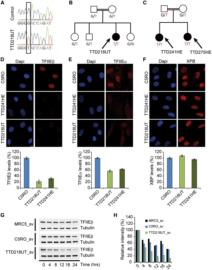
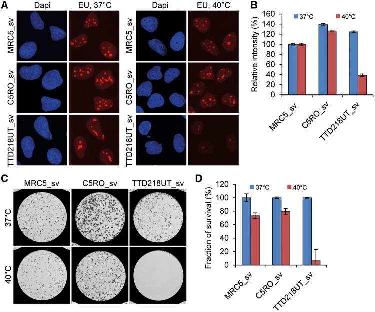
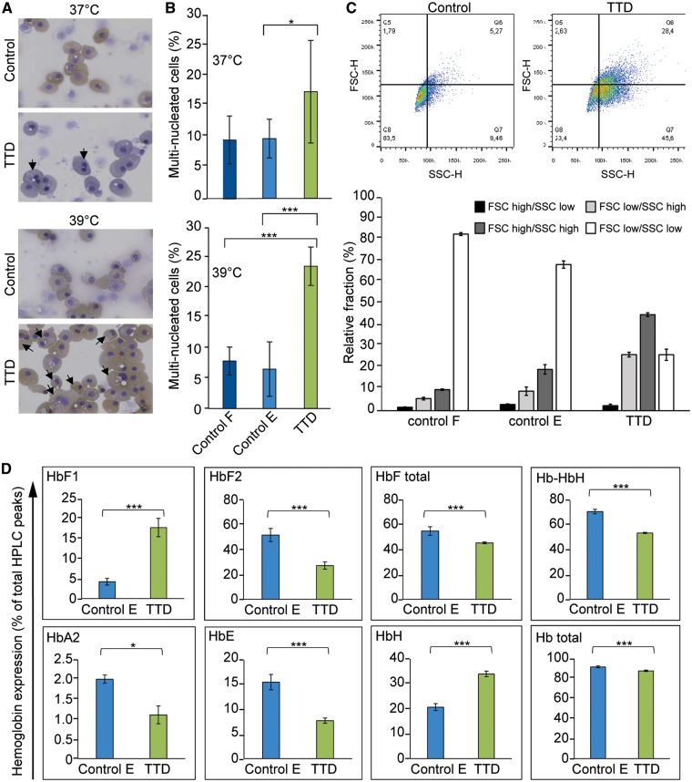
The rare recessive developmental disorder Trichothiodystrophy (TTD) is characterized by brittle hair and nails. Patients also present a variable set of poorly explained additional clinical features, including ichthyosis, impaired intelligence, developmental delay and anemia. About half of TTD patients are photosensitive due to inherited defects in the DNA repair and transcription factor II H (TFIIH). The pathophysiological contributions of unrepaired DNA lesions and impaired transcription have not been dissected yet. Here, we functionally characterize the consequence of a homozygous missense mutation in the general transcription factor II E, subunit 2 (GTF2E2/TFIIEβ) of two unrelated non-photosensitive TTD (NPS-TTD) families. We demonstrate that mutant TFIIEβ strongly reduces the total amount of the entire TFIIE complex, with a remarkable temperature-sensitive transcription defect, which strikingly correlates with the phenotypic aggravation of key clinical symptoms after episodes of high fever. We performed induced pluripotent stem (iPS) cell reprogramming of patient fibroblasts followed by in vitro erythroid differentiation to translate the intriguing molecular defect to phenotypic expression in relevant tissue, to disclose the molecular basis for some specific TTD features. We observed a clear hematopoietic defect during late-stage differentiation associated with hemoglobin subunit imbalance. These new findings of a DNA repair-independent transcription defect and tissue-specific malfunctioning provide novel mechanistic insight into the etiology of TTD.
© The Author 2017. Published by Oxford University Press.
Figures
Similar articles
-
GTF2E2 Mutations Destabilize the General Transcription Factor Complex TFIIE in Individuals with DNA Repair-Proficient Trichothiodystrophy.Am J Hum Genet. 2016 Apr 7;98(4):627-42. doi: 10.1016/j.ajhg.2016.02.008. Epub 2016 Mar 17. Am J Hum Genet. 2016. PMID: 26996949 Free PMC article.
-
A mutation in the XPB/ERCC3 DNA repair transcription gene, associated with trichothiodystrophy.Am J Hum Genet. 1997 Feb;60(2):320-9. Am J Hum Genet. 1997. PMID: 9012405 Free PMC article.
-
Bi-allelic TARS Mutations Are Associated with Brittle Hair Phenotype.Am J Hum Genet. 2019 Aug 1;105(2):434-440. doi: 10.1016/j.ajhg.2019.06.017. Am J Hum Genet. 2019. PMID: 31374204 Free PMC article.
-
Trichothiodystrophy view from the molecular basis of DNA repair/transcription factor TFIIH.Hum Mol Genet. 2009 Oct 15;18(R2):R224-30. doi: 10.1093/hmg/ddp390. Hum Mol Genet. 2009. PMID: 19808800 Review.
-
Trichothiodystrophy: Photosensitive, TTD-P, TTD, Tay syndrome.Adv Exp Med Biol. 2010;685:106-10. doi: 10.1007/978-1-4419-6448-9_10. Adv Exp Med Biol. 2010. PMID: 20687499 Review.
Cited by
-
The life and death of RNA across temperatures.Comput Struct Biotechnol J. 2022 Aug 8;20:4325-4336. doi: 10.1016/j.csbj.2022.08.008. eCollection 2022. Comput Struct Biotechnol J. 2022. PMID: 36051884 Free PMC article. Review.
-
RNA Polymerase II pausing temporally coordinates cell cycle progression and erythroid differentiation.medRxiv [Preprint]. 2023 Mar 7:2023.03.03.23286760. doi: 10.1101/2023.03.03.23286760. medRxiv. 2023. Update in: Dev Cell. 2023 Oct 23;58(20):2112-2127.e4. doi: 10.1016/j.devcel.2023.07.018. PMID: 36945604 Free PMC article. Updated. Preprint.
-
Molecular genetics of β-thalassemia: A narrative review.Medicine (Baltimore). 2021 Nov 12;100(45):e27522. doi: 10.1097/MD.0000000000027522. Medicine (Baltimore). 2021. PMID: 34766559 Free PMC article. Review.
-
Nucleolar TFIIE plays a role in ribosomal biogenesis and performance.Nucleic Acids Res. 2021 Nov 8;49(19):11197-11210. doi: 10.1093/nar/gkab866. Nucleic Acids Res. 2021. PMID: 34581812 Free PMC article.
-
Evaluation of AXIN1 and AXIN2 as targets of tankyrase inhibition in hepatocellular carcinoma cell lines.Sci Rep. 2021 Apr 2;11(1):7470. doi: 10.1038/s41598-021-87091-4. Sci Rep. 2021. PMID: 33811251 Free PMC article.
References
-
- Stefanini M., Botta E., Lanzafame M., Orioli D. (2010) Trichothiodystrophy: from basic mechanisms to clinical implications. DNA Repair (Amst), 9, 2–10. - PubMed
-
- Theil A.F., Hoeijmakers J.H., Vermeulen W. (2014) TTDA: big impact of a small protein. Exp. Cell Res., 329, 61–68. - PubMed
-
- Compe E., Egly J.M. (2012) TFIIH: when transcription met DNA repair. Nat Rev Mol. Cell Biol., 13, 343–354. - PubMed
-
- Vermeulen W., Bergmann E., Auriol J., Rademakers S., Frit P., Appeldoorn E., Hoeijmakers J.H., Egly J.M. (2000) Sublimiting concentration of TFIIH transcription/DNA repair factor causes TTD-A trichothiodystrophy disorder. Nat. Genet., 26, 307–313. - PubMed
Publication types
MeSH terms
Substances
Grants and funding
LinkOut - more resources
Full Text Sources
Other Literature Sources
Research Materials
