Comparative Study
. 2017 Sep 29;45(17):10284-10292.
doi: 10.1093/nar/gkx658.

Structural insights of lincosamides targeting the ribosome of Staphylococcus aureus

Affiliations
Comparative Study

Structural insights of lincosamides targeting the ribosome of Staphylococcus aureus

Donna Matzov et al. Nucleic Acids Res. .

Abstract

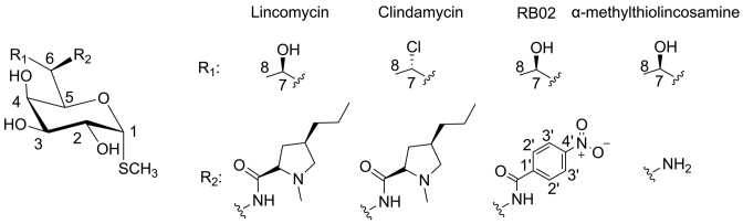
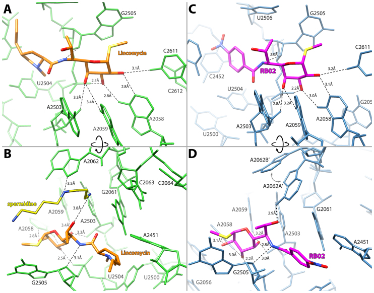
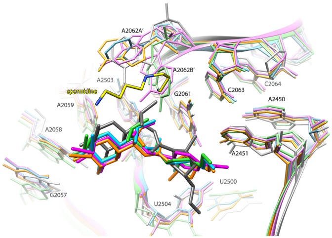
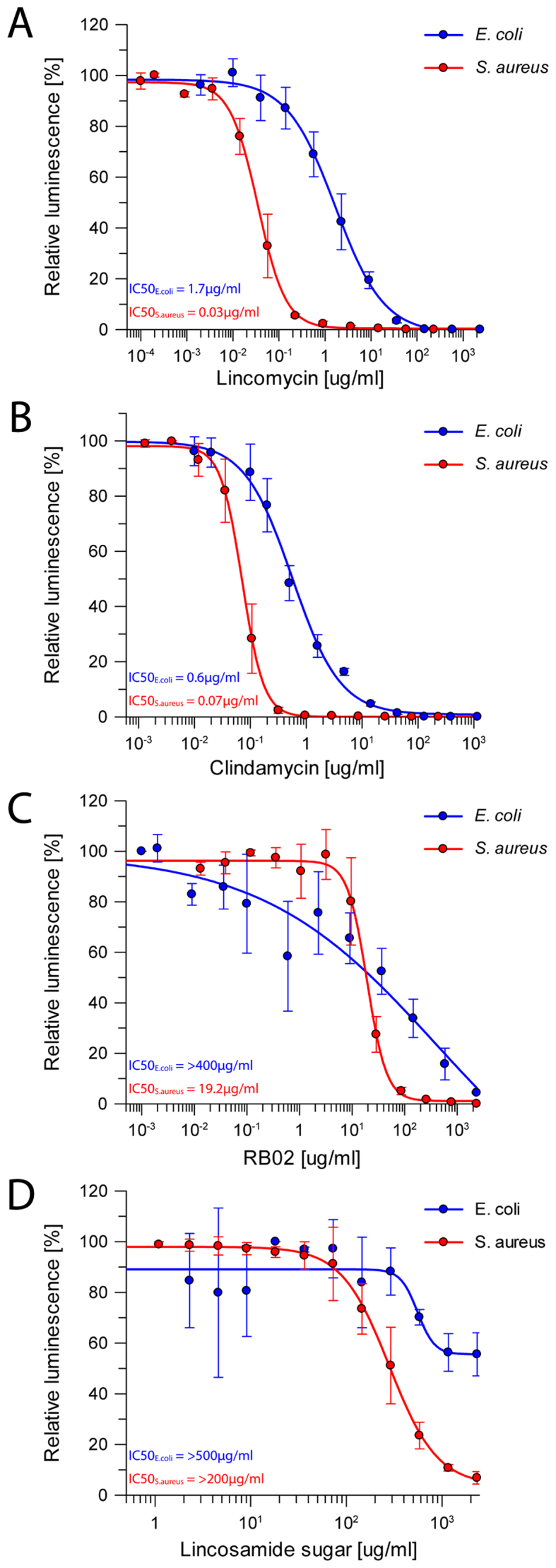
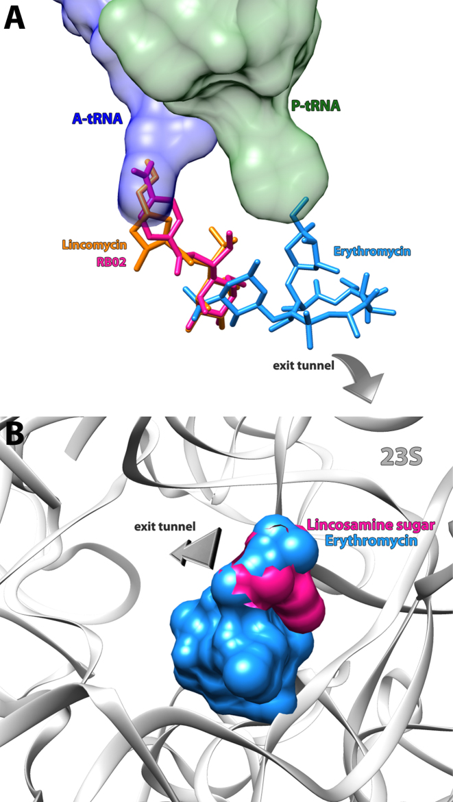
Antimicrobial resistance within a wide range of pathogenic bacteria is an increasingly serious threat to global public health. Among these pathogenic bacteria are the highly resistant, versatile and possibly aggressive bacteria, Staphylococcus aureus. Lincosamide antibiotics were proved to be effective against this pathogen. This small, albeit important group of antibiotics is mostly active against Gram-positive bacteria, but also used against selected Gram-negative anaerobes and protozoa. S. aureus resistance to lincosamides can be acquired by modifications and/or mutations in the rRNA and rProteins. Here, we present the crystal structures of the large ribosomal subunit of S. aureus in complex with the lincosamides lincomycin and RB02, a novel semisynthetic derivative and discuss the biochemical aspects of the in vitro potency of various lincosamides. These results allow better understanding of the drugs selectivity as well as the importance of the various chemical moieties of the drug for binding and inhibition.


Figures

Figure 1.
Figure 1.
Chemical structures of lincosamides. Lincomycin (natural antibiotic) and its semi-synthetic derivatives clindamycin and RB02. The chemical structure of Lincomycin’s sugar moiety, α-methylothiolincosamine is also shown.
Figure 2.
Figure 2.
Lincosamides interaction with their binding site within the SA50S complex. (A and B) Lincomycin (orange) and the network of hydrogen bonds (black dashes): it forms with the 23S rRNA (green) and spermidine (yellow). (C and D) RB02 (magenta) and the network of hydrogen bonds (black dashes) and electrostatic interactions (black dots): it forms with the 23S rRNA (blue). The two conformations of A2062 are designated as A’ and B’ with an arrow suggestion the movement between conformations.
Figure 3.
Figure 3.
Overlay of the structures of various ribosome–lincosamides complexes. SA50S–linc (orange; this study), SA50S–RB02 (magenta; this study), H50S-CLY (PDB ID: 1YJN) (green), D50S-CLY (gray) (PDB ID: 1JZX) and E70SCLY (sky blue) (PDB ID: 3OFZ). The color coding of the rRNA components of the various lincosamide-binding pockets is the same as that of the corresponding licosamides molecules.
Figure 4.
Figure 4.
Protein synthesis inhibition by lincosamides. Inhibition results of increasing concentrations of (A) lincomycin, (B) clindamycin, (C) RB02 and (D) α-methylothiolincosamine measured by the luminescence resulting from the translation of firefly luciferase on both S. aureus in vitro translation system (red) and E. coli in vitro system (blue). The error bars represent the standard deviations from the mean for triplicate experiments and the luminescence is normalized relative to that measured in the absence of any inhibitor, which was assigned as 100%.
Figure 5.
Figure 5.
Binding sites for antibiotics in the PTC and peptide exit tunnel. (A) Erythromycin (PDB ID: 1JZY) (blue), lincomycin (orange) and RB02 (pink) are shown as stick models. Ribbons denote the sugar phosphate backbone of 23S rRNA (gray), the acceptor ends of A-site tRNA (blue) and P-site tRNA (green). Lincomycin and RB02 are bound at the PTC and interferes with A-site tRNA positioning and erythromycin is bound at the entrance of the exit tunnel. Superimposing these structures shows that the sugar moiety of the lincosamides overlaps the desosamine sugar of erythromycin elucidating the MLSB cross-resistance. (B) Surface representation of erythromycin (PDB ID: 1JZY) (blue) superimposed on α-methylothiolincosamine (pink) in their binding pockets. The sugar phosphate backbone of 23S rRNA is denoted as ribbons (colored in gray).

Similar articles

Cited by

References

    1. World Health Organization Antimicrobial Resistance: Global Report on Surveillance. 2014; Geneva: World Health Organization.
    1. Kurosu M., Siricilla S., Mitachi K.. Advances in MRSA drug discovery: where are we and where do we need to be. Expert Opin. Drug Discov. 2013; 8:1095–1116. - PMC - PubMed
    1. Lowy F.D. Staphylococcus aureus infections. N. Engl. J. Med. 1998; 339:520–532. - PubMed
    1. Marcinak J.F., Frank A.L.. Treatment of community-acquired methicillin-resistant Staphylococcus aureus in children. Curr. Opin. Infect. Dis. 2003; 16:265–269. - PubMed
    1. Johnson M.D., Decker C.F.. Antimicrobial agents in treatment of MRSA infections. Dis. Mon. 2008; 54:793–800. - PubMed

Publication types

MeSH terms

LinkOut - more resources