Comparative Study
. 2017 Sep 29;45(17):10242-10258.
doi: 10.1093/nar/gkx663.

Direct modulation of T-box riboswitch-controlled transcription by protein synthesis inhibitors

Affiliations
Comparative Study

Direct modulation of T-box riboswitch-controlled transcription by protein synthesis inhibitors

Vassiliki Stamatopoulou et al. Nucleic Acids Res. .

Abstract

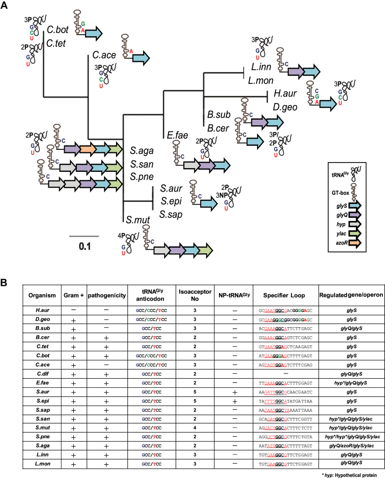
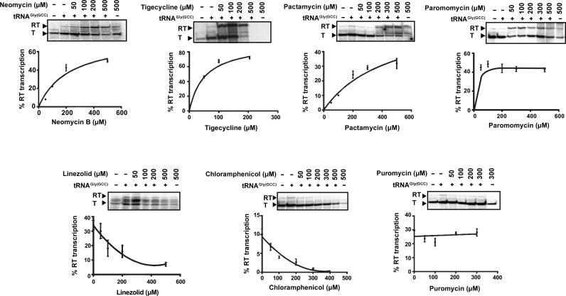
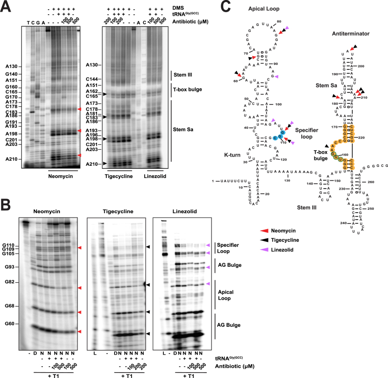
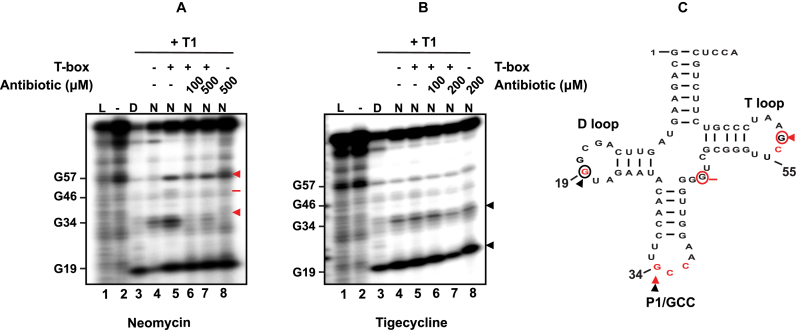
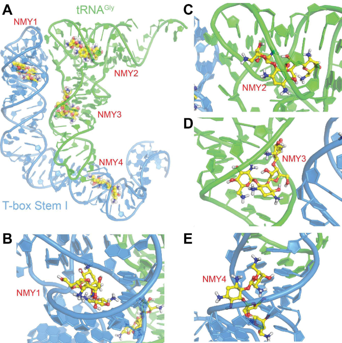
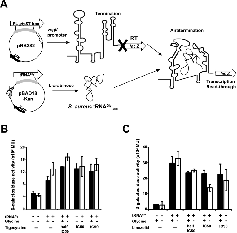
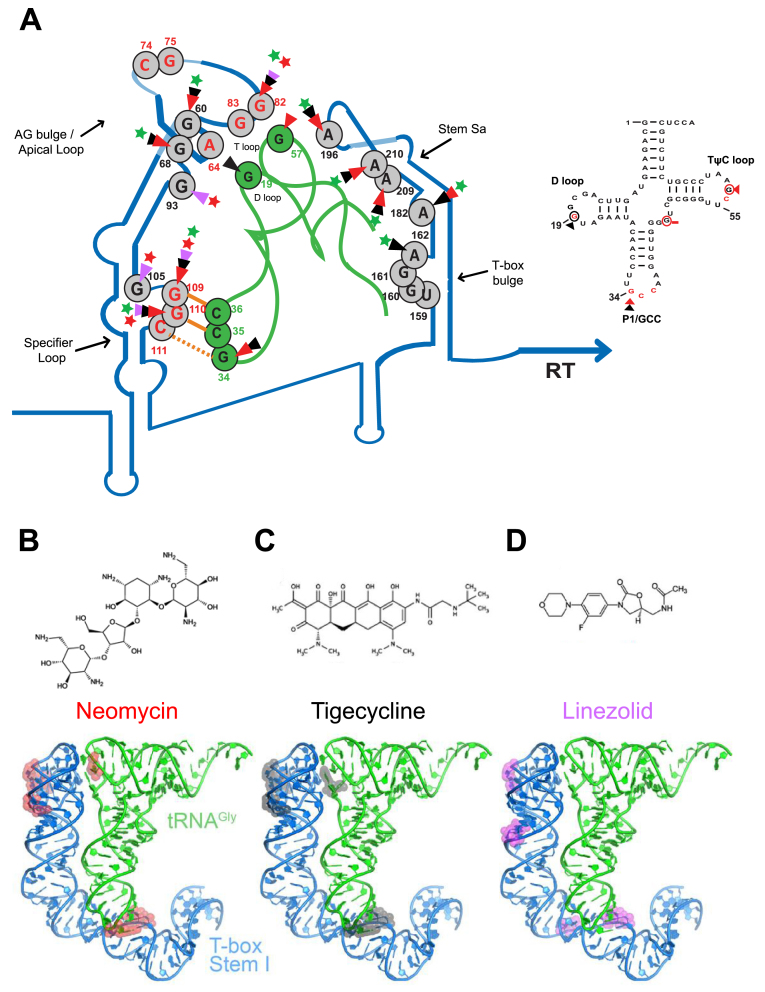
Recently, it was discovered that exposure to mainstream antibiotics activate numerous bacterial riboregulators that control antibiotic resistance genes including metabolite-binding riboswitches and other transcription attenuators. However, the effects of commonly used antibiotics, many of which exhibit RNA-binding properties, on the widespread T-box riboswitches, remain unknown. In Staphylococcus aureus, a species-specific glyS T-box controls the supply of glycine for both ribosomal translation and cell wall synthesis, making it a promising target for next-generation antimicrobials. Here, we report that specific protein synthesis inhibitors could either significantly increase T-box-mediated transcription antitermination, while other compounds could suppress it, both in vitro and in vivo. In-line probing of the full-length T-box combined with molecular modelling and docking analyses suggest that the antibiotics that promote transcription antitermination stabilize the T-box:tRNA complex through binding specific positions on stem I and the Staphylococcal-specific stem Sa. By contrast, the antibiotics that attenuate T-box transcription bind to other positions on stem I and do not interact with stem Sa. Taken together, our results reveal that the transcription of essential genes controlled by T-box riboswitches can be directly modulated by commonly used protein synthesis inhibitors. These findings accentuate the regulatory complexities of bacterial response to antimicrobials that involve multiple riboregulators.


Figures

Figure 1.
Figure 1.
(A) Phylogenetic tree constructed after analysis of the predicted GT-boxes based on the mRNA leader region from the SL until the T-box conserved sequence, along with the SL specificity (indicated with C for the GGC triplet, A for the GGA and G for the GGG) and the tRNAGly isoacceptor specificity. The number of proteinogenic (P) and non-proteinogenic (NP) isoacceptors and their corresponding anticodon triplet is shown (G for GCC, U for UCC, and C for CCC). (B) GT-boxes distribution among different pathogens, the potential SL regions and the tRNA anticodon triplets and the number of tRNAGly isoacceptors. In the specifier loop column, the red underlined sequences indicate the potential SL sequence and the bold letters correspond to nucleotides of the SL codon-like triplets for glycine. H.aur: Herpetosiphon aurantiacus, D.geo: Deinococcus geothermalis, B.sub: Bacillus subtilis, B.cer: Bacillus cereus, C.tet: Clostridium tetani, C.bot: Clostridium botulinum, C.ace: Clostridium acetobutylicum, C.dif: Clostridium difficile, E.fae: Enterococcus faecalis, S.aur: Staphylococcus aureus, S.epi: Staphylococcus epidermidis, S.sap: Staphylococcus saprophyticus, S.san: Streptococcus sanguinis, S.mut: Streptococcus mutans, S.pne: Streptococcus pneumoniae, S.aga: Streptococcus agalactiae, L.inn: Listeria innocua, L.mon: Listeria monocytogenes.
Figure 2.
Figure 2.
Dose-response curves showing the effect of increasing concentration of antibiotics on the S. aureus glyS T-box transcription antitermination, in vitro. The values for the curves were extracted after analysis of representative autoradiograms (insets) as described in Materials and Methods. All reactions were performed twice in duplicates and error bars represent ± SD from the corresponding experiments. T and RT correspond to transcription termination and transcription readthrough, respectively.
Figure 3.
Figure 3.
Probing analysis of the whole S. aureus glyS T-box in the presence of neomycin B, tigecycline, linezolid. (A) Chemical probing analysis of the full-length S. aureus glyS T-box (20 pmol) in the presence or the absence of tRNAGlyGCC (100 pmol) and/or increasing concentrations of neomycin B, tigecycline or linezolid. TbGl_6 primer was used for the terminator/antiterminator primer extension analysis. Red, black or magenta arrows indicate DMS base modification in the presence neomycin B, tigecycline and linezolid, respectively. (B) Enzymatic probing analysis; (D) indicates denaturing conditions, (N) native conditions and (L) alkaline hydrolysis ladder. Red, black or magenta arrows correspond to base protection from RNase T1 cleavage in the presence of neomycin B, tigecycline and linezolid, respectively. (C) Illustration of the S. aureus glyS T-box secondary structure. The binding sites of neomycin B, tigecycline and linezolid on the glyS T-box are shown; Red, black and magenta arrows indicate interacting points of neomycin B, tigecycline and linezolid, respectively. Yellow labelled nucleotides indicate the conserved T-box bulge region.
Figure 4.
Figure 4.
Probing analysis of tRNAGlyGCC in the presence of neomycin B (A) or tigecycline (B). Enzymatic probing analysis of the tRNAGlyGCC (20 pmol) in the presence of the full-length S. aureus glyS T-box (60 pmol) and/or increasing concentrations of neomycin B or tigecycline. Red and black arrows indicate base protection from RNase T1 cleavage in the presence of neomycin B and tigecycline, respectively. Red bar shows a weak base protection in the presence of neomycin B. (D) indicates denaturing conditions, (N) native conditions and (L) alkaline hydrolysis ladder. (Bottom panel) The secondary structure of the S. aureus tRNAGlyGCC is shown (C); based on the available crystal structure, red nucleotides are important for interaction with the Stem I (25). Red circled nucleotides and red arrows show the binding sites of neomycin B and black circled nucleotides and black arrows the binding sites of tigecycline on the tRNAGlyGCC.
Figure 5.
Figure 5.
Molecular modelling of antibiotics’ binding sites onto the glyS T-box:tRNA complex. (A) Schematic representation of the T-box stem I complex with tRNA, illustrating potential interaction sites of neomycin (NMY). The structure of the complex is based on the X-ray structure of T-box riboswitch of Oceanobacillus iheyensis glyQ (blue) in complex with its cognate tRNA (green) from PDB ID 4LCK (25) NMY is shown as CPK model with yellow C, blue N and red O atoms. Close-up views of the four binding sites NMY1 (B), NMY2 (C), NMY3 (D) and NMY4 (E).
Figure 6.
Figure 6.
In vivo glyS T-box-mediated transcription antitermination assay. (A) Schematic representation of the glyS T-box-dependent lacZ expression in E. coli. The full length (FL) glyS T-box and the tRNAGlyGCC were cloned into pRB382 and pBAD18 plasmid, respectively. Both constructs or pRB382-T-box alone were transformed into a ΔlacZ E. coli strain. T corresponds to T-box terminator conformation and anti-T to T-box antiterminator conformation. Diagram of the β-galactosidase activity in the presence of tigecycline (B) and linezolid (C). The bars represent β-gal activity relative to cell density, in Miller Units. Error bars represent ± SD values from three independent experiments. The β-galactosidase activity was measured after 4 h of culture in either rich (+Gly) or minimal media (–Gly) in the presence or in the absence of tigecycline or linezolid (concentrations tested half-IC50, IC50 and IC90) and after expression induction of S. aureus tRNAGlyGCC.
Figure 7.
Figure 7.
Illustration of the proposed model of neomycin B, tigecycline and linezolid binding sites on the S. aureus glyS T-box:tRNAGlyGCC complex (A) and the secondary structure of the S. aureus tRNAGlyGCC. The protected bases on the S. aureus glyS T-box:tRNAGlyGCC in the presence of neomycin B (B), tigecycline (C) and linezolid (D) are indicated with different colored arrows; red arrows correspond to neomycin B interaction, black arrows to tigecycline and magenta arrows to linezolid. Green or red stars show the enhancement or blocking of the glyS T-box read-through transcription by the antibiotics tested, respectively. Orange lines show the GCC codon-anticodon like interaction and the orange dashed line the wobble pairing. Numbers indicate the position of each nucleotide on the S. aureus glyS T-box or the tRNAGlyGCC (in green). Red nucleotides at the apical loop show the bases that interact with the tRNA elbow based on the crystal structure (25) (Right panel); based on the available crystal structure, red nucleotides are important for interaction with the Stem I. Red circled nucleotides and red arrows correspond to the binding sites of neomycin B, and black circled nucleotides and black arrows show and tigecycline on the tRNAGlyGCC.

Similar articles

Cited by

References

    1. Breaker R.R. Riboswitches and the RNA world. Cold Spring Harb. Perspect. Biol. 2012; 4:a003566. - PMC - PubMed
    1. Sherwood A.V., Henkin T.M.. Riboswitch-mediated gene regulation: novel RNA architectures dictate gene expression responses. Annu. Rev. Microbiol. 2016; 70:361–374. - PubMed
    1. Mellin J.R., Cossart P.. Unexpected versatility in bacterial riboswitches. Trends Genet. 2015; 31:150–156. - PubMed
    1. Breaker R.R. Prospects for riboswitch discovery and analysis. Mol. Cell. 2011; 43:867–879. - PMC - PubMed
    1. Henkin T.M. Riboswitch RNAs: using RNA to sense cellular metabolism. Genes Dev. 2008; 22:3383–3390. - PMC - PubMed

Publication types

MeSH terms