Review
. 2017 Oct 1;38(5):379-431.
doi: 10.1210/er.2017-00073.

Insulin Receptor Isoforms in Physiology and Disease: An Updated View

Affiliations
Review

Insulin Receptor Isoforms in Physiology and Disease: An Updated View

Antonino Belfiore et al. Endocr Rev. .

Abstract

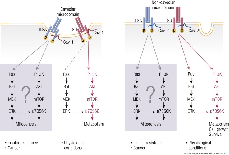
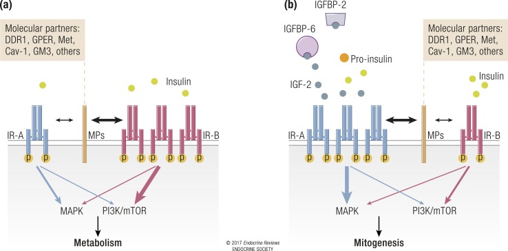
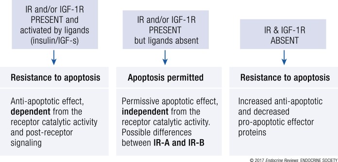
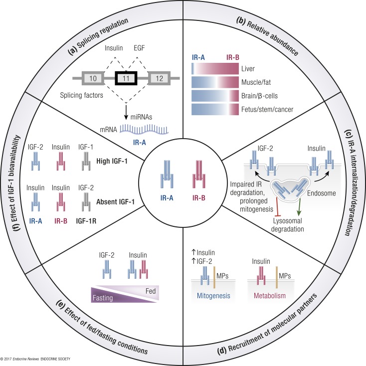
The insulin receptor (IR) gene undergoes differential splicing that generates two IR isoforms, IR-A and IR-B. The physiological roles of IR isoforms are incompletely understood and appear to be determined by their different binding affinities for insulin-like growth factors (IGFs), particularly for IGF-2. Predominant roles of IR-A in prenatal growth and development and of IR-B in metabolic regulation are well established. However, emerging evidence indicates that the differential expression of IR isoforms may also help explain the diversification of insulin and IGF signaling and actions in various organs and tissues by involving not only different ligand-binding affinities but also different membrane partitioning and trafficking and possibly different abilities to interact with a variety of molecular partners. Of note, dysregulation of the IR-A/IR-B ratio is associated with insulin resistance, aging, and increased proliferative activity of normal and neoplastic tissues and appears to sustain detrimental effects. This review discusses novel information that has generated remarkable progress in our understanding of the physiology of IR isoforms and their role in disease. We also focus on novel IR ligands and modulators that should now be considered as an important strategy for better and safer treatment of diabetes and cancer and possibly other IR-related diseases.


Figures

Figure 1.
Figure 1.
The structure of the IR (αβ)2 homodimer. (a) Location of domains within the IR αβ polypeptide. Interchain disulfide bonds are indicated by solid green lines, N-linked glycosylation sites by white dots, and O-linked glycosylation sites are indicated by brown dots. The N termini of the chains are labeled in red (α or β), and the interchain disulfide bonds are shown as green lines. C-tail, C-terminal tail of the IR β chain; JM, juxtamembrane segment; L2, second leucine-rich repeat domain; TM, transmembrane segment. (b) Λ-shaped assembly of the IR ectodomain. One monomer is depicted as a ribbon, with the domains colored and labeled as in (a); the second is depicted as a white molecular surface. The depiction is based on PDB entry 4ZXB (11).
Figure 2.
Figure 2.
Structural biology of the interaction of insulin with its primary binding site on the receptor. (a) Overview of the insulin plus μIR complex, demonstrating the displacement of the insulin B-chain C-terminal segment (purple ribbon) away from the hormone core. (b) Rearrangement of αCT on the L1-β2 surface upon insulin binding. The C-terminal IR residues His710 and Phe714 are highlighted to show the change in the length and position of αCT upon hormone binding. (c) Engagement of the insulin core by IR residues Phe714 and His 710, highlighting insulin residues that are within 4 Å of these two receptor residues. (d) Steric overlap between bound insulin and the FnIII-1 and FnIII-2 domains upon superposition of the insulin plus μIR complex onto the structure of the apo-IR ectodomain. (e) Detail of the location of the insulin aromatic triplet PheB24-PheB25-TyrB26 within the insulin plus μIR complex. (f) Structure of IGF in complex with the IR L1-CR domain and the IGF-1R αCT segment (colored orange, light brown, and black, respectively), that is, a “hybrid” microreceptor complex, overlaid onto that of the insulin plus μIR complex (white). Unless otherwise indicated, the IR domains are colored as in Fig. 1, the insulin A chain is colored light blue, and the insulin B chain is colored purple.
Figure 3.
Figure 3.
Schematic representation of regulators of IR isoform expression. The figure summarizes the principal IR regulators acting at the promoter (transcription factors) and mRNA level (splicing factors and miRNAs). Transcription factors act by promoting (in red) or blocking (in blue) IR gene transcription. Several splicing factors and miRNAs are involved in the posttranscriptional regulation of IR expression. Once the IR mRNA is formed, splicing factors remove introns and let exons bind together. They regulate the differential splicing of exon 11, thereby generating IR-A (ex11) or IR-B (ex11+). Several miRNAs can bind to the 3′-UTR of the IR mRNA, favoring its degradation.
Figure 4.
Figure 4.
Schematic model for alternative IR splicing regulation by splicing factors. IR sequences encoding exons 10, 11, and 12 and introns 10 and 11 are shown. (a) Some splicing factors regulate exon 11 inclusion, thereby modulating a preferential expression of isoform B of the IR. hnRNP F binds to both ends (5′ and 3′) of intron 10. Mbnl1 recognizes an intronic splicing enhancer (ISE) element within intron 11 and binds to two other regions localized in intron 10 and in exon 11. SRp20 and SF2/ASF bind the exonic splicing enhancer (ESE) element within exon 11. RBM4 binds GC-rich sequences in intron 10 and acts synergistically with other IR splicing regulators. (b) Factors regulating IR-A formation. CUGBP1 binds to two silencer sequences, one located at the 3′ end of intron 10 (ISS) and the other one in exon 11 (ESS). hnRNP A1 binds similarly to the 5′ splice site of both intron 10 and intron 11. hnRNP H favors exon 11 skipping by binding a region within intron 10.
Figure 5.
Figure 5.
IR signal diversification and partitioning by caveolins. (a) IR signaling in caveolae: under physiological conditions, insulin promotes phasic IR-B interaction with cav-1 at caveolar necks and consequent activation of metabolic effects, such as glucose transport and glycogen synthesis. In cells overexpressing IR-A, such as cancer cells, IR-B association with cav-1 may be compromised and switched in favor of the IR-A/cav-1 interaction, which may be biased toward mitogenic stimuli. (b) IR signaling in noncaveolar microdomains: in cav-2– and IR-A–enriched cells, such as certain cancer cells, the cav-2/IR-A interaction may elicit prolonged IR-A phosphorylation with enhanced mTOR/Stat-3 activation and preferential activation of mitogenic and prosurvival stimuli.
Figure 6.
Figure 6.
Schematic representation of the proposed IR signal diversification by ligand-induced crosstalk with molecular partners. (a) In cells and tissues with predominant IR-B expression, IR-B activation by insulin is normally phasic (postprandial) and favors “metabolic” downstream signaling, which may be affected by the IR-B crosstalk with tissue-specific molecular partners (MPs) such as Met, GPER, Cav1/2, DDR1, and GM3. These molecules, in turn, may autonomously activate additional signaling. (b) In cells predominantly expressing IR-A and producing IGF-2, such as fetal or cancer cells, IR-A activation by IGF-2 elicits a steady IR-A interaction with molecular partners (MPs), favoring “nonmetabolic” effects, including mitogenesis and cell migration. In insulin-resistant patients, hyperinsulinemia, and perhaps proinsulin, may elicit similar effects. The line thickness indicates the strength of signaling pathway activation.
Figure 7.
Figure 7.
Three-state model for the IR and IGF-1R control of cell apoptosis. The schematic representation of the complex regulation of cell apoptosis by the IR and IGF-1R in the presence or absence of ligands is shown. Unliganded IR-B might have a more rapid effect on apoptosis.
Figure 8.
Figure 8.
IR isoforms affect insulin/IGF signaling diversification. (a) Splicing regulation: IR isoform relative abundance is regulated by numerous splicing factors and miRNAs. Growth factors such as insulin and EGF favor IR-A generation. (b) Relative abundance: The relative abundance of IR isoforms is also regulated by tissue-specific factors. IR-A is predominant in fetal, stem, and cancer cells. (c) IR-A Internalization/degradation: Unlike insulin, IGF-2 binding to IR-A does not target the receptor to lysosomal degradation, allowing for prolonged mitogenic effects. (d) Recruitment of molecular partners: Ligand binding recruits various molecular partners (MPs) to the activated receptor. It can be hypothesized that IR-A tonic activation by IGF-2 or hyperinsulinemia may induce prolonged MP interaction and biased signaling. (e) Effect of fed/fasting conditions: According to ligand binding affinities, it can be predicted that during fasting (low circulating insulin) IGF-2 is the predominant IR-A ligand, whereas in fed conditions (high circulating insulin) both IR isoforms are engaged by insulin. (f) Effect of IGF-1 bioavailability: IR-A is predicted to be the major IGF-2 receptor when IGF-1 is present and saturates the IGF-1R. In contrast, IR-A would preferably bind insulin when IGF-1 bioavailability is low or absent.

References

    1. Belfiore A, Frasca F, Pandini G, Sciacca L, Vigneri R. Insulin receptor isoforms and insulin receptor/insulin-like growth factor receptor hybrids in physiology and disease. Endocr Rev. 2009;30(6):586–623. - PubMed
    1. Pandini G, Frasca F, Mineo R, Sciacca L, Vigneri R, Belfiore A. Insulin/insulin-like growth factor I hybrid receptors have different biological characteristics depending on the insulin receptor isoform involved. J Biol Chem. 2002;277(42):39684–39695. - PubMed
    1. Boucher J, Tseng YH, Kahn CR. Insulin and Growth Insulin-like. Factor-1 Receptors Act as Ligand-specific Amplitude Modulators of a Common Pathway Regulating Gene Transcription. J Biol Chem. 2010;285(22):17235–17245. - PMC - PubMed
    1. Menting JG, Whittaker J, Margetts MB, Whittaker LJ, Kong GK-W, Smith BJ, Watson CJ, Žáková L, Kletvíková E, Jiráček J, Chan SJ, Steiner DF, Dodson GG, Brzozowski AM, Weiss MA, Ward CW, Lawrence MC. How insulin engages its primary binding site on the insulin receptor. Nature. 2013;493(7431):241–245. - PMC - PubMed
    1. Menting JG, Lawrence CF, Kong GK-W, Margetts MB, Ward CW, Lawrence MC. Structural Congruency of Ligand Binding to the Insulin and Insulin/Type 1 Insulin-like Growth Factor Hybrid Receptors. Structure. 2015;23(7):1271–1282. - PubMed

Publication types