. 2017 Dec 1;26(23):4606-4616.
doi: 10.1093/hmg/ddx342.

Loss of the imprinted, non-coding Snord116 gene cluster in the interval deleted in the Prader Willi syndrome results in murine neuronal and endocrine pancreatic developmental phenotypes

Affiliations

Loss of the imprinted, non-coding Snord116 gene cluster in the interval deleted in the Prader Willi syndrome results in murine neuronal and endocrine pancreatic developmental phenotypes

Lisa Cole Burnett et al. Hum Mol Genet. .

Abstract

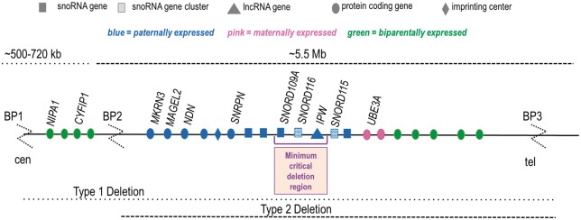
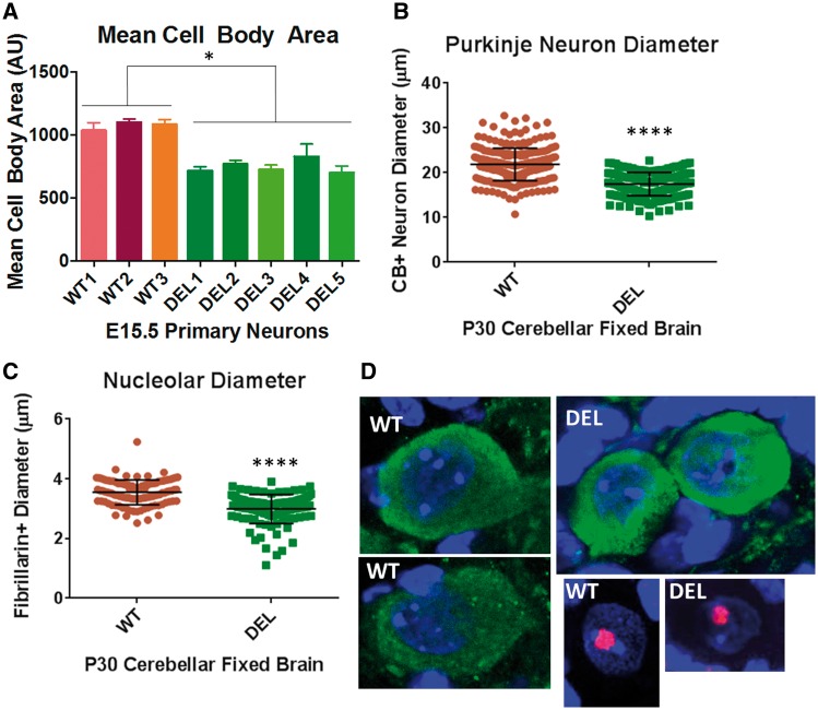
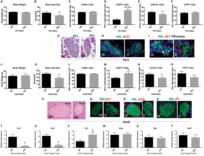
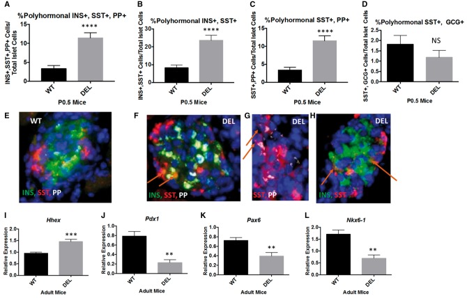
Global neurodevelopmental delay is a prominent characteristic of individuals with Prader-Willi syndrome (PWS). The neuromolecular bases for these delays are unknown. We identified neuroanatomical changes in the brains of mice deficient for a gene in the minimal critical deletion region for PWS (Snord116p-/m+). In Snord116p-/m+ mice, reduced primary forebrain neuron cell body size is apparent in embryonic day 15.5 fetuses, and persists until postnatal day 30 in cerebellar Purkinje neurons. Snord116 is a snoRNA gene cluster of unknown function that can localize to the nucleolus. In cerebellar Purkinje neurons from postnatal day 30 Snord116p-/m+ mice the reduction in neuronal cell body size was associated with decreased neuronal nucleolar size. We also identified developmental changes in the endocrine pancreas of Snord116p-/m+ animals that persist into adulthood. Mice lacking Snord116 have smaller pancreatic islets; within the islet the percentage of δ-cells is increased, while the percentage of α-cells is reduced. The α-cell markers, Sst and Hhex, are upregulated in Snord116p-/m+ isolated islets while Ins1, Ins2, Pdx1, Nkx6-1, and Pax6 are downregulated. There is a 3-fold increase in the percentage of polyhormonal cells in the neonatal pancreata of Snord116p-/m+ mice, due primarily to an increase in cells co-positive with somatostatin. Snord116 may play a role in islet cell lineage specification. The Snord116 gene cluster is important for developmental processes in the brain as well as the endocrine pancreas.


Figures

Figure 1.
Figure 1.
The imprinted Prader-Willi region on 15q11-13.
Figure 2.
Figure 2.
Loss of paternal Snord116 in Snord116p−/m+ mice results in smaller neuronal nucleolus and cell body size. (A) Tuj1+ primary neurons isolated from E15.5 embryos of Snord116p−/m+ mice also display an ∼31% reduction in mean cell body area as compared with wild type (WT). (B) There is a 21% reduction in the mean Purkinje neuron diameter in Snord116p−/m+ fixed brain sections from P30 animals. (C) Purkinje neuron nucleolar diameter is 16% less in P30 Snord116p−/m+ mice. (D) Representative images of calbindin D28K (green) positive neurons and fibrillarin (red) positive nucleoli from cerebellar sections of P30 WT and Snord116p−/m+ (DEL) fixed brains; nuclei are marked in blue by Hoechst stain. All images were taken using a 63X objective with 1.1X zoom.
Figure 3.
Figure 3.
Snord116p−/m+ mice display developmental changes in the endocrine pancreas that persist into adulthood. (A) There is no difference in the body weight of P0.5 Snord116p−/m+ neonates compared with WT littermates. (B) There is a 23% decrease in the mean islet size (number of nuclei per islet) of P0.5 Snord116p−/m+ mice compared with WT littermates. (C) The proportion of insulin-positive cells in the islet is unchanged; while the absolute number trends towards a 14% reduction, this does not reach statistical significance (Supplementary Material, Fig. S6). (D) There is a 3-fold increase in the proportion of somatostatin (SST) positive cells in the islet in Snord116p−/m+ mice. (E) The percentage of glucagon (GCG) positive cells is decreased in islets of Snord116p−/m+animals. (F) There is no difference in the percentage of pancreatic polypeptide (PP) positive cells. (G) Representative images of the hematoxylin and eosin (H&E) stain sections from which the number of nuclei per islet was quantified in P0.5 mice. (H,I) Representative images of insulin, glucagon, somatostin, and pancreatic polypeptide immunofluorescent staining. (J) Adult Snord116p−/m+ mice are generally smaller than WT littermates. However, to minimize somatic size-related artifacts in comparisons of islet endocrine cell type populations, body weight-matched adult wild type and Snord116p−/m+ mice were selected. (K) The mean islet size is decreased by 32%. In adult Snord116p−/m+ mice compared with WT. (L) In adult Snord116p−/m+the proportion of insulin positive cells in the islet is unchanged in adult mice. (M) The percentage of somatostatin positive cells are increased 1.5 fold. (N) The percentage of glucagon positive cells is decreased by 30% in Snord116p−/m+ mice. (O) The proportion of pancreatic polypeptide positive cells is not altered in adult Snord116p−/m+ mice. (P) Representative H&E images used for quantification of mean islet size. (Q–S) Representative images of the immunofluoresent staining for insulin, somatostatin, glucagon, and pancreatic polypeptide. (T,U) Ins1 and Ins2 gene expression levels are decreased in isolated islets from Snord116p−/m+ adult mice compared with WT. (V) Sst gene expression levels are 2-fold increased in Snord116p−/m+ isolated islets. (W–Y) There is no difference in the gene expression levels of Gcg, Ppy, or Ghrl in adult isolated islets.
Figure 4.
Figure 4.
The increase in δ-cells in Snord116p−/m+ mice is associated with an increased percentage of polyhormonal endocrine cells in the neonatal pancreas and altered transcription factor expression levels. (A) There is a 3.4-fold increase in the percentage of polyhormonal cells positive for insulin, somatostatin, pancreatic polypeptide in P0.5 Snord116p−/m+ pancreata compared with WT littermates. (B) There is a 2.7-fold increase in polyhormonal cells co-staining for insulin and somatostatin in Snord116p−/m+ neonatal pancreata. (C) Similarly, there is a 3.4-fold increase in somatostatin and pancreatic polypeptide double positive cells in P0.5 Snord116p−/m+ pancreata. (D) There is no difference in the percentage of glucagon and somatostatin double-positive cells. E-H) Representative images of polyhormonal cells in WT (E) and Snord116p−/m+ pancreata (F–H). (I) Hhex expression is 1.5-fold higher in isolated islets from adult Snord116p−/m+ mice compared with WT littermates. (J) Pdx1 gene expression levels are 72% lower in Snord116p−/m+ isolated islets. (K) Pax6 is 46% lower in isolated islets from adult Snord116p−/m+ mice compared with WT littermates. (L) Nkx6-1 gene expression levels are 60% lower in Snord116p−/m+ isolated islets compared with WT.

References

    1. Angulo M.A., Butler M.G., Cataletto M.E. (2015) Prader-Willi syndrome: a review of clinical, genetic, and endocrine findings. J. Endocrinol. Invest., 38, 1249–1263. - PMC - PubMed
    1. Butler M.G., Lee P.D.K., Whitman B.Y. (2006) Management of Prader-Willi Syndrome. Springer Science Business Media, Inc, New York.
    1. Miller J.L., Couch J.A., Schmalfuss I., He G., Liu Y., Driscoll D.J. (2007) Intracranial abnormalities detected by three-dimensional magnetic resonance imaging in Prader-Willi syndrome. Am. J. Med. Genet. A, 143A, 476–483. - PubMed
    1. Miller J.L., Couch J., Schwenk K., Long M., Towler S., Theriaque D.W., He G., Liu Y., Driscoll D.J., Leonard C.M. (2009) Early childhood obesity is associated with compromised cerebellar development. Dev. Neuropsychol., 34, 272–283. - PMC - PubMed
    1. Leung K.N., Vallero R.O., DuBose A.J., Resnick J.L., LaSalle J.M. (2009) Imprinting regulates mammalian snoRNA-encoding chromatin decondensation and neuronal nucleolar size. Hum. Mol. Genet., 18, 4227–4238. - PMC - PubMed

Publication types

MeSH terms