. 2017 Dec 1;26(23):4657-4667.
doi: 10.1093/hmg/ddx347.

A human patient-derived cellular model of Joubert syndrome reveals ciliary defects which can be rescued with targeted therapies

Affiliations

A human patient-derived cellular model of Joubert syndrome reveals ciliary defects which can be rescued with targeted therapies

Shalabh Srivastava et al. Hum Mol Genet. .

Abstract

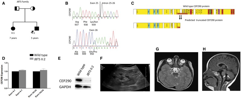
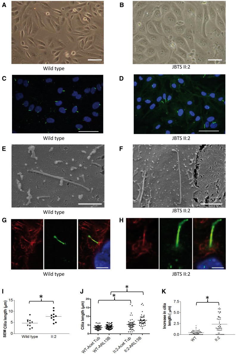
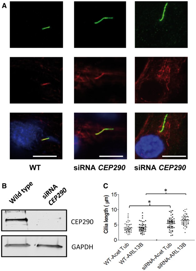
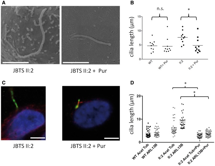
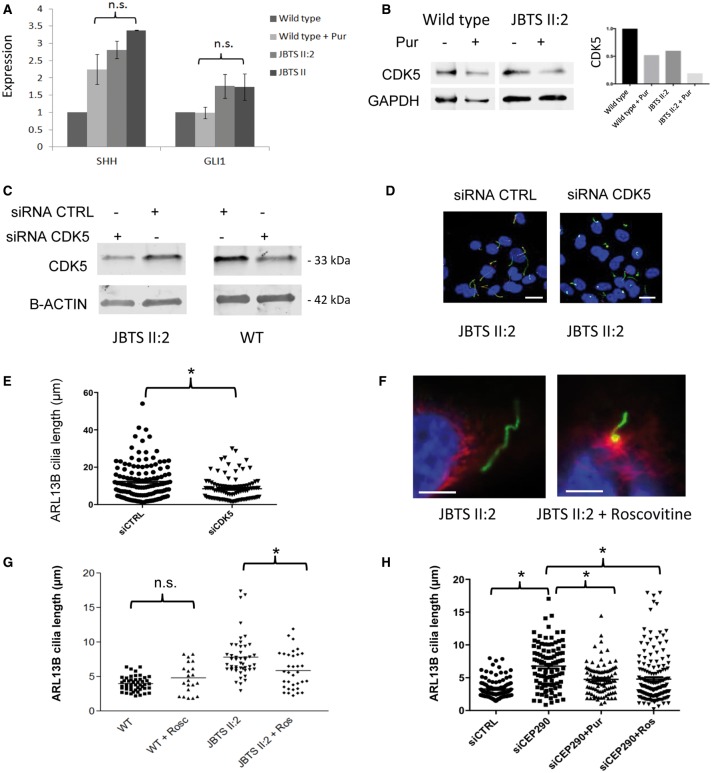
Joubert syndrome (JBTS) is the archetypal ciliopathy caused by mutation of genes encoding ciliary proteins leading to multi-system phenotypes, including a cerebello-retinal-renal syndrome. JBTS is genetically heterogeneous, however mutations in CEP290 are a common underlying cause. The renal manifestation of JBTS is a juvenile-onset cystic kidney disease, known as nephronophthisis, typically progressing to end-stage renal failure within the first two decades of life, thus providing a potential window for therapeutic intervention. In order to increase understanding of JBTS and its associated kidney disease and to explore potential treatments, we conducted a comprehensive analysis of primary renal epithelial cells directly isolated from patient urine (human urine-derived renal epithelial cells, hURECs). We demonstrate that hURECs from a JBTS patient with renal disease have elongated and disorganized primary cilia and that this ciliary phenotype is specifically associated with an absence of CEP290 protein. Treatment with the Sonic hedgehog (Shh) pathway agonist purmorphamine or cyclin-dependent kinase inhibition (using roscovitine and siRNA directed towards cyclin-dependent kinase 5) ameliorated the cilia phenotype. In addition, purmorphamine treatment was shown to reduce cyclin-dependent kinase 5 in patient cells, suggesting a convergence of these signalling pathways. To our knowledge, this is the most extensive analysis of primary renal epithelial cells from JBTS patients to date. It demonstrates the feasibility and power of this approach to directly assess the consequences of patient-specific mutations in a physiologically relevant context and a previously unrecognized convergence of Shh agonism and cyclin-dependent kinase inhibition as potential therapeutic targets.


Figures

Figure 1.
Figure 1.
Pedigree, genetic and clinical imaging data of JBTS family H67. (A) Pedigree diagram showing two affected siblings (squares, males; circles females). (B) Sequence chromatograms showing compound heterozygous changes in CEP290 c.2817G>T; p.K939N (predicted to affect splice donor site) and c.2848insC; p.Q950Pfs*6. (C) Domain structure of CEP290 protein (2479 amino acids) with predicted coiled coil domains (CC) numbered and shown in yellow; tropomyosin homology domain (TM), RepA/Rep+ protein KID (KID); bipartite nuclear localization signal (NLS_BP); ATP/GTP-binding site motif A (P-loop). The extent of homology with SMC proteins is indicated by an orange bar. The predicted truncation of CEP290 at amino acid positions 939 and 950 are shown. (D) qPCR showing relative level of CEP290 transcript measured at the 5’ (Exon 6-7) and the 3’ (Exon 48-49) ends. RNA samples from patient II: 2 hURECs show a similar level of transcript to wild type (WT, normalized to 1) indicating a lack of nonsense mediated decay (Standard error of means indicated by bars). Experiments performed in triplicate. (E) Western blot (cropped image) showing reduced expression of full-length wild type CEP290 protein from hURECs derived from II: 2 using a C-terminal directed CEP290 antibody. Experiments performed in triplicate. (F) Renal ultrasound scan of II: 2 showing cystic change within the kidney. (G) Axial MRI image of brain showing molar tooth sign in II: 1. (H) Sagittal MRI image of brain shows hypoplasia of the cerebellar vermis and enlarged mid-fourth ventricle (white arrows) in II: 1.
Figure 2.
Figure 2.
Renal epithelial cilia derived from a patient with CEP290 mutations demonstrate length and morphological defects. (A and B) Wild type and patient JBTS II: 2 hURECs form a typical cobblestone epithelial cell layer when grown in 2D culture (Scale bar 100 µm). (C and D) Wild type and patient JBTS II: 2 hURECs following immunofluorescence imaging using anti-ARL13B (green) to identify ciliary membrane and DAPI to identify nuclei. (Scale bar 50 µm). (E and F) Scanning electron microscopy (SEM) images reveal abnormally long cilia in patient JBTS II: 2 cells with distal tip anomalies (Scale bar 2 µm). (G and H) Immunofluorescence staining using anti-alpha-acetylated tubulin (red) to identify axoneme and anti-ARL13B (green) to identify ciliary membrane in wild type and JBTS II: 2 cells (Scale bar 5um). (I) Dot plot with means (4.7 versus 7.7 µm) to show ciliary length measured by SEM. (n = 10 for each group, *P < 0.05, Unpaired Student’s t-test). (J) Dot plots with means indicated to show ciliary length measured by immunofluorescence imaging using antibodies against alpha-acetylated tubulin (Acet Tub) and ARL13B. (n = 49 for WT cilia and n = 49 for II: 2 cilia, *P < 0.0001, Unpaired Student’s t-test). (K) Dot plots with means indicated to show the increase in cilia length of each cilia (data from panel j) as determined by measuring axonemal length under immunofluorescence imaging using antibodies against alpha-acetylated tubulin (Acet Tub) and ARL13B. (*P < 0.0001, Unpaired Student’s t-test).
Figure 3.
Figure 3.
CEP290 knockdown by siRNA in hURECs reveals a ciliary length phenotype. (A) Immunofluorescence imaging of cilia identified by anti-alpha-acetylated tubulin (Acet Tub red) and anti-ARL13B (ARL13B, green). Scale bar 10 μm. (B) Western blot (cropped image) showing loss of full length CEP290 protein in wild type hURECs following knockdown of CEP290 using directed siRNA. (C). Dot plots to show ciliary length in wild type (WT) untreated and siRNA CEP290 treated hURECs measured by immunofluorescence imaging using antibodies against alpha-acetylated tubulin (Acet Tub) and ARL13B. (n = 47 for WT cilia and n = 52 for siRNA treated cilia, *P < 0.0001, Unpaired Students t-test).
Figure 4.
Figure 4.
Rescue of CEP290 mutant cilia length defects in hURECs using purmorphamine treatment. (A) Scanning electron microscopy (SEM) images showing cilia in patient JBTS II: 2 hURECs before and after purmorphamine (Pur) treatment. Scale bar 2 µm. (B) Dot plots with means showing ciliary lengths measured by SEM in wild type (WT) and patient JBTS II: 2 hURECs before and after treatment with purmorphamine (Pur). (n = 10 for each group, *P < 0.0001, Unpaired Student t-test; n.s. not significant). Untreated WT and II: 2 cilia lengths are replicated from Figure 2I. (C) Immunofluorescence images showing cilia in patient JBTS II: 2 hURECs before and after purmorphamine treatment. Scale bar 5 µm. (D) Dot plots with means to show ciliary length determined by immunofluorescence imaging in wild type (WT) patient JBTS II: 2 hURECs untreated and treated with purmorphamine using anti-alpha-acetylated tubulin (Acet Tub) and anti-ARL3B (ARL13B). (Cilia counts n = 36 for WT, n = 35 for II: 2 untreated and n = 40 for II: 2 treated; *P < 0.0001, ANOVA).
Figure 5.
Figure 5.
CDK5 inhibition via purmorphamine, siRNA and roscovitine rescues ciliary phenotype in CEP290 deficient cells. (A) Expression of SHH and GLI1 RNA transcripts in hURECs from wild type controls and JBTS II: 2 before and after treatment with purmorphamine (Pur). Expression was normalized to control genes GAPDH and HPRT. Error bars indicate standard deviation; n.s., not significant, ANOVA. Experiments performed in triplicate. (B) Western blot (cropped image) of CDK5 levels following treatment with purmorphamine in wild type and JBTS II: 2 hURECs. Quantification (right) normalized to GAPDH expression. Experiments were performed in triplicate. (C) Western blot (cropped image) of siRNA CDK5 and control (CTRL) siRNA treatment of JBTS II: 2 and wild type (WT) hURECs shows a reduction in CDK5 protein expression. Beta-actin (B-ACTIN) loading control. Experiments performed in triplicate. (D) Low power immunofluorescence images (alpha-acetylated tubulin, red; ARL13B green, DAPI, blue) of hURECs from JBTS II: 2 after treatment with control siRNA (siCTRL) or siRNA CDK5 (siCDK5) (scale bar 10 µm). (E) Dot plot with means to show quantification of cilia length of hURECs from JBTS II: 2 (shown in d) after treatment with control siRNA (siCTRL) or siRNA CDK5 (siCDK5) (*P < 0.001, unpaired Student's t-test, n = 129 siCTRL, n = 95 siCDK5, median values siCTRL 10.43 µm, siCDK5 7.356 µm). (F) High power immunofluorescence images (alpha-acetylated tubulin, red; ARL13B green, DAPI, blue) of hURECs from JBTS II: 2 before and after treatment with roscovitine. Scale bar 5 µm. (G) Dot plot with means to show quantification of cilia length using ARL13B in wild type (WT, n = 48), wild type treated with roscovitine (WT + Rosc, n = 21), JBTS II: 2 hURECs (n = 48) and JBTS II: 2 treated with roscovitine (Ros, n = 34) (n.s. not significant, *P = 0.004, unpaired Student's t-test). (H) Dot plot with means to show quantification of cilia length using ARL13B in control siRNA (siCTRL, n = 109), siRNA CEP290 (siCEP290, n = 100), siRNA CEP290 + purmorphamine (siCEP290+Pur, n = 95) and siRNA Cep290 + roscovitine (siCEP290 + Ros, n = 162) treated hURECs (*P < 0.0001, ANOVA).

References

    1. Simms R.J., Hynes A.M., Eley L., Sayer J.A. (2011) Nephronophthisis: a genetically diverse ciliopathy. Int. J. Nephrol., 2011, 527137.. - PMC - PubMed
    1. Sayer J.A., Otto E.A., O'Toole J.F., Nurnberg G., Kennedy M.A., Becker C., Hennies H.C., Helou J., Attanasio M., Fausett B.V.. et al. (2006) The centrosomal protein nephrocystin-6 is mutated in Joubert syndrome and activates transcription factor ATF4. Nat. Genet., 38, 674–681. - PubMed
    1. Valente E.M., Silhavy J.L., Brancati F., Barrano G., Krishnaswami S.R., Castori M., Lancaster M.A., Boltshauser E., Boccone L., Al-Gazali L.. et al. (2006) Mutations in CEP290, which encodes a centrosomal protein, cause pleiotropic forms of Joubert syndrome. Nat. Genet., 38, 623–625. - PubMed
    1. Valente E.M., Brancati F., Boltshauser E., Dallapiccola B. (2013) Clinical utility gene card for: Joubert syndrome–update 2013. Eur. J. Hum. Genet. EJHG, 21. - PMC - PubMed
    1. Perrault I., Delphin N., Hanein S., Gerber S., Dufier J.L., Roche O., Defoort-Dhellemmes S., Dollfus H., Fazzi E., Munnich A.. et al. (2007) Spectrum of NPHP6/CEP290 mutations in Leber congenital amaurosis and delineation of the associated phenotype. Hum. Mutat., 28, 416. - PubMed

MeSH terms

Supplementary concepts