. 2017 Nov 15;26(22):4441-4450.
doi: 10.1093/hmg/ddx331.

Alpha-synuclein induces the unfolded protein response in Parkinson's disease SNCA triplication iPSC-derived neurons

Affiliations

Alpha-synuclein induces the unfolded protein response in Parkinson's disease SNCA triplication iPSC-derived neurons

Sabrina M Heman-Ackah et al. Hum Mol Genet. .

Abstract

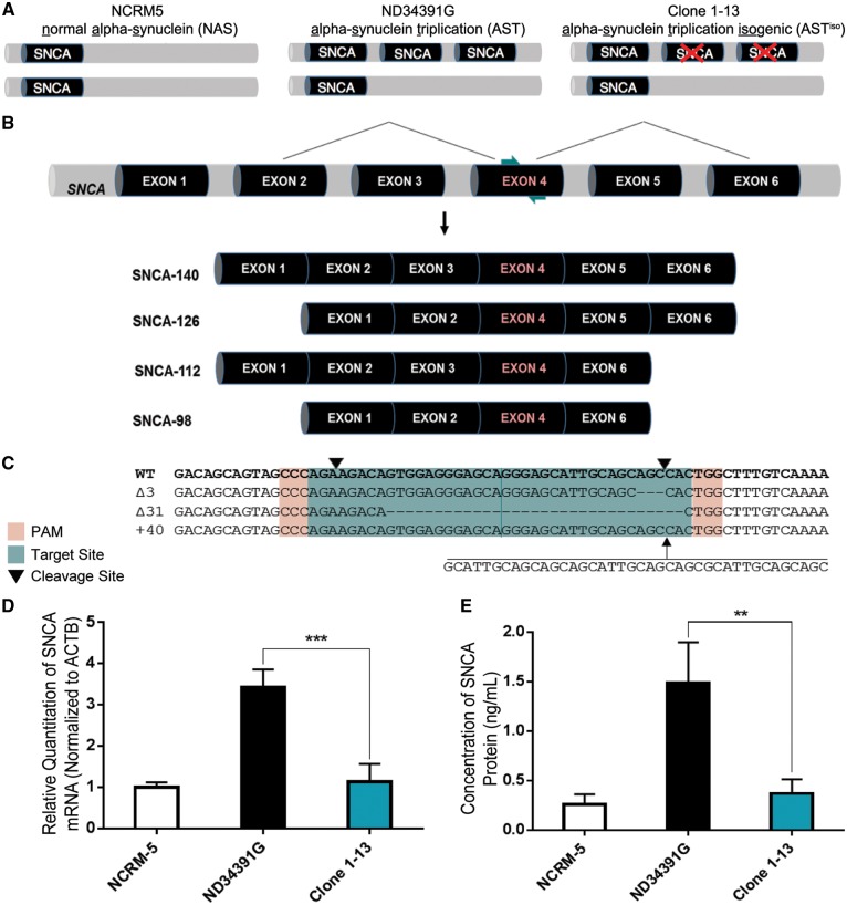
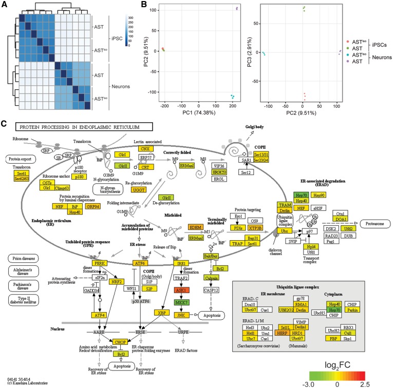
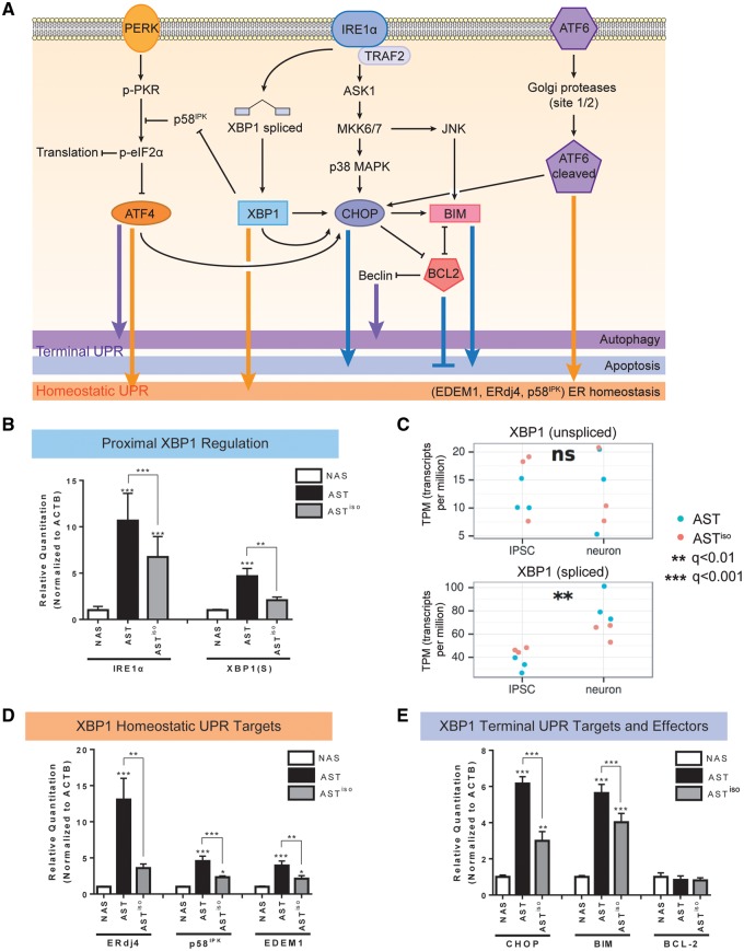
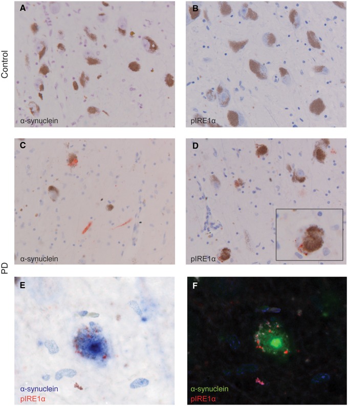
The recent generation of induced pluripotent stem cells (iPSCs) from a patient with Parkinson's disease (PD) resulting from triplication of the α-synuclein (SNCA) gene locus allows unprecedented opportunities to explore its contribution to the molecular pathogenesis of PD. We used the double-nicking CRISPR/Cas9 system to conduct site-specific mutagenesis of SNCA in these cells, generating an isogenic iPSC line with normalized SNCA gene dosage. Comparative gene expression analysis of neuronal derivatives from these iPSCs revealed an ER stress phenotype, marked by induction of the IRE1α/XBP1 axis of the unfolded protein response (UPR) and culminating in terminal UPR activation. Neuropathological analysis of post-mortem brain tissue demonstrated that pIRE1α is expressed in PD brains within neurons containing elevated levels of α-synuclein or Lewy bodies. Having used this pair of isogenic iPSCs to define this phenotype, these cells can be further applied in UPR-targeted drug discovery towards the development of disease-modifying therapeutics.


Figures

Figure 1.
Figure 1.
Double Knockout of SNCA in iPSCs Derived from a PD Patient with SNCA Triplication via Double-Nicking CRISPR/Cas9. (A) Schematic representation of normal alpha synuclein (NAS), alpha-synuclein triplication (AST) and alpha-synuclein triplication isogenic (ASTiso) iPS lines. The triplication involves a large Mb region including the SNCA gene. (B) Position of sgRNAs (teal arrows) guiding Cas9 nickases to SNCA exon 4, common to all four α-synuclein transcript isoforms. (C) Wild-type, 3 bp deletion, 31 bp deletion, and 40 bp insertion alleles identified by sequencing of Clone 1-13. PAMs are highlighted in red, sgRNAs in teal and downward pointing black arrows represent cleavage sites. (D) qRT-PCR for total SNCA mRNA shows a normalization of α-synuclein transcript levels in Clone 1-13 iPSCs. Data are represented as mean ± SEM of biological triplicates. (E) ELISA shows a normalization of α-synuclein protein levels in Clone 1-13 iPSCs. Data are represented as mean ± SEM of biological triplicates. **P ≤  0.01, ***P ≤  0.001. See also Supplementary Material, Figs S1 and S2 and Table S4.
Figure 2.
Figure 2.
Comparative transcriptomic analysis reveals an ER stress phenotype specific to SNCA triplication iPSC-derived neurons. (A) Cross-correlation and clustering analysis between datasets. Color denotes correlation coefficient. The primary source of variation is between iPSCs and iPSC-derived neurons, followed by AST and ASTiso genotypes in neurons. (B) Principal component analysis of differentially expressed genes. The first principal component (PC1) accounts for 74.38% of the variance, largely due to differentiation stage. PC2 accounts for the differences in AST and ASTiso genotypes in neurons (9.51% variance) and PC3 similarly accounts for genotype differences in iPSCs (2.91% variance). Cell types are indicated as follows: red = ASTiso iPSC, green = AST iPSC, blue = ASTiso neurons and purple = AST neurons. (C) Protein processing in the endoplasmic reticulum KEGG pathway diagram highlighting fold changes in gene expression between AST and ASTiso iPSC-derived neurons. Color indicates log2 gene expression change (red is higher in AST neurons). Data show biological triplicates, **Q < 0.01, ***Q < 0.01, ns = not significant. See also Supplementary Material, Figs S3–S5, Tables S1 and S2.
Figure 3.
Figure 3.
Transcript and protein expression validation of IRE1α/XBP1-induced UPR activation in SNCA triplication iPSC-derived neurons. (A) Schematic of signaling pathways downstream of the UPR. PERK, IRE1α and ATF6 branches are indicated, and the effects of downstream effectors on homeostatic, autophagic and apoptotic outcomes are indicated. (B, D, E) Validation of mRNA expression level differences in IRE1α/XBP1 pathway components by qRT-PCR. Levels of IRE1α and spliced XBP1 [XBP1(S)] (B), the homeostatic UPR targets ERdj4, p58IPK and EDEM1 (D), and the terminal UPR targets CHOP, BIM and BCL-2 (E) at the mRNA level were evaluated by qRT-PCR in NAS, AST and ASTiso iPSC-derived neurons. Data are represented as mean ± SEM of biological triplicates. (C) Gene expression from RNAseq data (in transcripts per million, TPM) of unspliced and spliced XBP1 transcripts across differentiation stage (iPSC or neuron) and genotype (blue = AST, red = ASTiso). *P ≤ 0.5, ** P ≤ 0.01, *** P ≤ 0.001, ns = not significant. See also Supplementary Material, Figs S5–S7 and Table S4.
Figure 4.
Figure 4.
Immunohistochemical detection of α-synuclein and pIRE1α in substantia nigra pars compacta of representative PD and control cases. (A,B) Immunoreactivity for α-synuclein and pIRE-1α is absent in the control case. (C) α-synuclein positive structures, Lewy neurites and bodies, (red) are observed in the PD case. (D) pIRE1α immunoreactive granules (red) are observed in neurons in the substantia nigra of the PD case. Inset shows a higher magnification of a single neuron. (E) Double-immunohistochemistry for pIRE1α (red) and α-synuclein (blue) shows double-labelling of a melanin-containing dopaminergic neuron. (F) Double-staining deconvoluted with spectral imaging showing α-synuclein (green), pIRE1α (red), haematoxylin (blue) and neuromelanin (white). Scale bar: 50 µm in A-D; 20 µm in the inset (D) and E, F., n = 5 PD cases, n = 6 controls. See also Supplementary Material, Table S3.

References

    1. Singleton A.B., Farrer M., Johnson J., Singleton A., Hague S., Kachergus J., Hulihan M., Peuralinna T., Dutra A., Nussbaum R. (2003) Alpha-synuclein locus triplication causes Parkinson's disease. Science, 302, 841.. - PubMed
    1. Chartier-Harlin M.C., Kachergus J., Roumier C., Mouroux V., Douay X., Lincoln S., Levecque C., Larvor L., Andrieux J., Hulihan M.. et al. (2004) Alpha-synuclein locus duplication as a cause of familial Parkinson's disease. Lancet, 364, 1167–1169. - PubMed
    1. Ibanez P., Bonnet A.M., Debarges B., Lohmann E., Tison F., Pollak P., Agid Y., Durr A., Brice A. (2004) Causal relation between alpha-synuclein gene duplication and familial Parkinson's disease. Lancet, 364, 1169–1171. - PubMed
    1. Farrer M., Kachergus J., Forno L., Lincoln S., Wang D.S., Hulihan M., Maraganore D., Gwinn-Hardy K., Wszolek Z., Dickson D.. et al. (2004) Comparison of kindreds with parkinsonism and alpha-synuclein genomic multiplications. Ann. Neurol., 55, 174–179. - PubMed
    1. Singleton A.C. J. (2007) Dawson T. (ed.), In Parkinson's Disease: Genetics and Pathogenesis. Informa Healthcare USA, Inc, New York, NY, in press., pp. 19–33.

Publication types

MeSH terms