. 2017 Oct 10;114(41):E8721-E8730.
doi: 10.1073/pnas.1707572114. Epub 2017 Sep 25.

Catheterization alters bladder ecology to potentiate Staphylococcus aureus infection of the urinary tract

Affiliations

Catheterization alters bladder ecology to potentiate Staphylococcus aureus infection of the urinary tract

Jennifer N Walker et al. Proc Natl Acad Sci U S A. .

Abstract

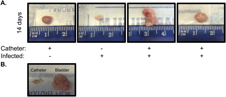
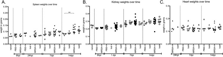
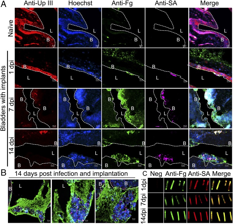
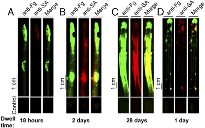
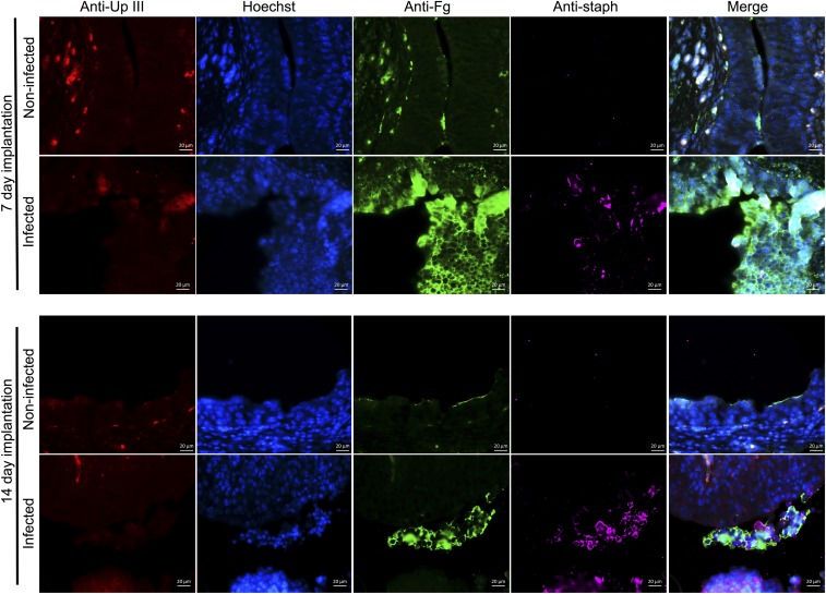
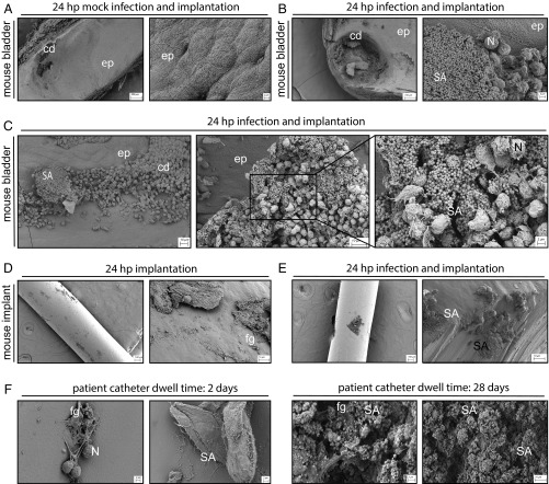
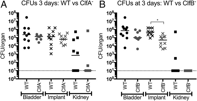
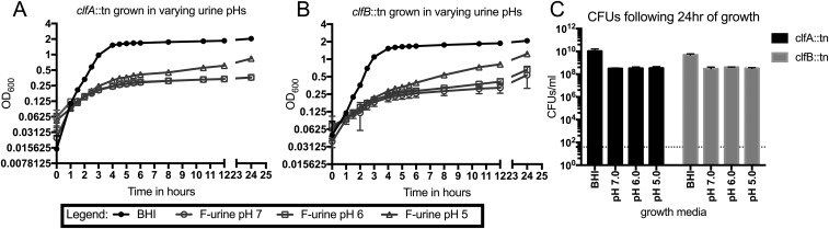
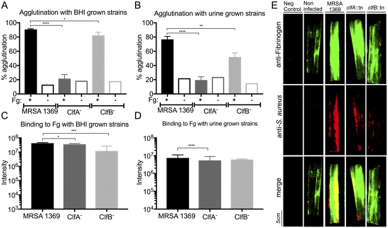
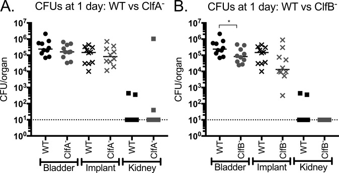
Methicillin-resistant Staphylococcus aureus (MRSA) is an emerging cause of catheter-associated urinary tract infection (CAUTI), which frequently progresses to more serious invasive infections. We adapted a mouse model of CAUTI to investigate how catheterization increases an individual's susceptibility to MRSA UTI. This analysis revealed that catheterization was required for MRSA to achieve high-level, persistent infection in the bladder. As shown previously, catheter placement induced an inflammatory response resulting in the release of the host protein fibrinogen (Fg), which coated the bladder and implant. Following infection, we showed that MRSA attached to the urothelium and implant in patterns that colocalized with deposited Fg. Furthermore, MRSA exacerbated the host inflammatory response to stimulate the additional release and accumulation of Fg in the urinary tract, which facilitated MRSA colonization. Consistent with this model, analysis of catheters from patients with S. aureus-positive cultures revealed colocalization of Fg, which was deposited on the catheter, with S. aureus Clumping Factors A and B (ClfA and ClfB) have been shown to contribute to MRSA-Fg interactions in other models of disease. We found that mutants in clfA had significantly greater Fg-binding defects than mutants in clfB in several in vitro assays. Paradoxically, only the ClfB- strain was significantly attenuated in the CAUTI model. Together, these data suggest that catheterization alters the urinary tract environment to promote MRSA CAUTI pathogenesis by inducing the release of Fg, which the pathogen enhances to persist in the urinary tract despite the host's robust immune response.

Keywords: ClfB–fibrinogen interactions; MRSA CAUTI; host–pathogen interactions.


Conflict of interest statement

The authors declare no conflict of interest.

Figures

Fig. S1.
Fig. S1.
Growth of S. aureus strains in rich media or human urine of varying pH. (AF) Growth curves of JE2-MRSA (A), SSTI-MRSA (B), Co-MRSA (C), MRSA 1369 (D), PUTS-1 (E), and TOP6-555 (F) in BHI or urine pH 7.0, 6.0, and 5.0 as measured by OD600. (G) CFUs of JE2-MRSA, SSTI-MRSA, Co-MRSA, MRSA 1369, PUTS-1, and TOP6-555 following 24 h of growth in BHI or urine pH 7.0, 6.0, and 5.0. The number of independent data points represented in each graph (AF) is six and (G) four. Error bars represented in each graph (AF) are the mean and error with standard deviation and (G) mean with standard deviation.
Fig. 1.
Fig. 1.
Mouse model of S. aureus CAUTI. MRSA 1369 (A), PUTS-1 (B), and TOP6-555 (C) all require an implant to colonize bladders (circles) and kidneys (squares), and all implants (Xs) are colonized at 24 h post infection (hpi). Open and closed symbols denote nonimplanted mice and mice that retained the implant, respectively. Statistical significance was assessed using the Mann–Whitney U test, *P < 0.01, **P < 0.001.
Fig. 2.
Fig. 2.
Catheterization facilitates persistent, invasive MRSA CAUTI. (A) Mice with an implant maintain persistent bladder colonization. Mice that lose the implant display significantly lower bacterial burdens. (B) MRSA persistently colonizes the implant. (C) MRSA requires an implant to persistently colonize kidneys. (D) Bacteria were also detected in the blood at 6 h post implantation and infection (hpii), with most mice controlling the bacteremia by 1 dpii. (E and F) Mice also displayed dissemination to the spleen (E) and heart (F) at early time points but were able to control the disseminated infection by 14 dpii. Open and closed symbols denote mice that lost the implant and mice that retained the implant over the course of the experiment, respectively. Statistical significance was assessed using the Mann–Whitney U test, *P < 0.05, **P < 0.005.
Fig. 3.
Fig. 3.
MRSA CAUTI induces a localized inflammatory response. (A) Implants induce inflammation, which can be measured as an increase in bladder weight over time. Infection with MRSA 1369 following implantation further increases bladder inflammation. Light-gray circles represent naïve mice, dark-gray circles denote mock-infected implanted mice, and black circles are implanted-infected mice. Open circles denote mice that did not receive or that lost an implant, and closed circles represent mice that retained the implant over the course of the experiment. Statistical significance was assessed using the Mann–Whitney U test, *P < 0.05, **P < 0.005. (B and C) Cytokines increased >twofold (B) or >10-fold (C) in implanted MRSA-infected mice compared with implanted mock-infected control mice over a 2-wk time course. There are at least five independent data points represented in B and C. Error bars represent the mean with the standard deviation. ND, not determined.
Fig. S2.
Fig. S2.
Mouse bladders and catheters after a 2-wk experiment. (A) Mice infected and implanted for 14 d exhibit enlarged, abnormal bladders compared with implanted control mice or infected mice that lost the implant over the course of the experiment. (B) Mouse bladder following 14 d of implantation and infection with MRSA 1369 and the retrieved implant.
Fig. S3.
Fig. S3.
Organ weights of naïve, implanted, and implanted-infected mice over a 2-wk experiment. Spleens become enlarged by day 14 (A). Kidney (B) and heart (C) weights are unaffected by MRSA 1369 CAUTI. Closed symbols denote mice that maintained an implant for the duration of the experiment; open symbols represent mice that did not have an implant or lost the implant over the course of the infection. Light gray represents naïve animals; dark gray represents implanted, mock-infected mice; black denotes implanted, infected mice. Statistical analysis was performed using the Mann–Whitney U test, **P < 0.001.
Fig. 4.
Fig. 4.
Implants create a permissive environment for MRSA. (A) Representative images of a naïve mouse bladder or mouse bladders with implants at 1, 7, and 14 dpi. Sections were stained for uroplakin III (red), a protein that lines the umbrella cells of the bladder, cell nuclei (blue), fibrinogen (green), and MRSA (pink). Dotted white lines denote the separation of bladder tissue (B) from lumen (L). Infected mice with implants at day 1 have similar bladder morphology to naïve mice. However, by day 7, uroplakin III staining diminishes, while cell nuclei become smaller and staining becomes diffuse, which persists through day 14. This change in uroplakin III and cell nuclei staining is indicative of urothelial cell damage that results in exfoliation of the umbrella cells and an expansion of the underlying transitional layer. This, along with Fg coating the umbrella cells, is important for healing the damaged epithelium. However, MRSA predominately colocalizes with Fg accumulating on the epithelium, suggesting the protein plays a role in MRSA pathogenesis. (B) Representative images of mouse bladders at 14 d post infection and implantation. Large Fg aggregates form, encasing MRSA. (C) MRSA colocalizes with Fg deposited on implants at 1, 7, and 14 dpi. (Scale bars, 20 μm.)
Fig. 5.
Fig. 5.
S. aureus colocalizes with Fg deposited on patients’ catheters. Immunofluorescence staining of catheters 6 (A), 12 (B), 57 (C), and 64 (D) (Table S2) from patients with S. aureus urine or catheter positive cultures indicates Fg was deposited and, despite receiving appropriate antibiotic treatment, S. aureus was found on all catheters colocalizing with deposited Fg.
Fig. S4.
Fig. S4.
Representative images comparing implanted, mock-infected and implanted, MRSA-infected bladders at 7 and 14 d. Bladders are stained for uroplakin III (red), a protein expressed on the surface of umbrella cells, cell nuclei (blue), Fg (green), and MRSA (pink). Implantation induces inflammation resulting in the exfoliation of umbrella cells (decrease in red staining), expansion of the underlying transitional cells (smaller cell nuclei, with more diffuse staining), and accumulation of Fg along the bladder cell surface (green staining). Infection with MRSA 1369 exacerbates inflammation and Fg accumulation compared with implantation alone. (Scale bars, 20 μm.)
Fig. 6.
Fig. 6.
Representative SEM images of mouse bladders, implants, and human catheters. Tissues, implants, and catheters were processed following implantation, implantation and infection (mouse), or dwell time (human). (AC) Mouse bladders, with each pair showing low-, medium-, and (C) high-magnification images as indicated. (Scale bars: 200, 100, and 2 μm, respectively.) (D and E) Implants, with each pair showing medium- and high-magnification images, as indicated. (Scale bars: 100 and 10 μm, respectively.) Three mice were used per group. (F) Human catheters, with each pair showing two representative images from catheters recovered from the patients analyzed in Fig. 5. [Scale bars: 2 and 10 μm (Far Right).] Labeled are the epithelium (ep); epithelial cell damage from the implant (cd); MRSA (SA); immune cell infiltrates, likely neutrophils (N); and proteins, likely fibrinogen (fg).
Fig. 7.
Fig. 7.
Fg-binding protein ClfB, but not ClfA, contributes to CAUTI. (A) The clfA mutant displayed similar bladder, implant, and kidney burdens to the wild type. (B) ClfB exhibited significantly reduced bacterial titers on the implant at day 3 compared with WT. Statistical analysis was performed using the Mann–Whitney U test, *P < 0.005.
Fig. S5.
Fig. S5.
Growth of the ClfA and ClfB strains in rich media or urine. (A and B) CflA (A) and ClfB (B) display similar growth phenotypes to WT MRSA 1369 when grown in either rich media or human urine. (C) CFUs of both mutant strains following overnight growth in either rich media or human urine. At least four independent data points are represented. Error bars represented in each graph (A and B) are mean and error with standard deviation and (C) mean with standard deviation.
Fig. S6.
Fig. S6.
ClfA and ClfB are required for MRSA–Fg interactions. (A and B) The clfA and clfB mutants display agglutination defects compared with the wild type following a 3-h incubation with Fg when grown in BHI (A) or urine (B). (C and D) The clfA mutant also displayed a reduction in attachment to Fg-coated wells compared with the wild type when grown in either BHI (C) or urine (D). The clfB mutant is only defective for attachment to Fg-coated wells when grown in BHI (C) but not urine (D). (E) MRSA 1369 binds ex vivo to human urinary catheters and colocalizes with deposited Fg. The ClfA and ClfB strains display lower adherence to human urinary catheters compared with MRSA 1369, despite the catheter containing a similar amount of Fg. The number of independent data points was three for A and B and six for C and D. Statistics were calculated using the Student’s t test, *P < 0.05, **P < 0.005, ***P < 0.0005, and ****P < 0.0001. Error bars in each graph represent the mean with standard deviation.
Fig. S7.
Fig. S7.
MRSA 1369, ClfA, and ClfB UT colonization at day 1. (A) ClfA does not contribute to MRSA CAUTI, as the mutant displays similar bladder, implant, and kidney titers to WT. (B) The ClfB strain displays a moderate but significant bladder colonization defect at day 1 compared with WT, indicating the protein is important for MRSA CAUTI. Statistical analysis was performed using the Mann–Whitney U test, *P < 0.05.

References

    1. Barrett SP, et al. Antibiotic sensitivity of bacteria associated with community-acquired urinary tract infection in Britain. J Antimicrob Chemother. 1999;44:359–365. - PubMed
    1. Flores-Mireles AL, Walker JN, Caparon M, Hultgren SJ. Urinary tract infections: Epidemiology, mechanisms of infection and treatment options. Nat Rev Microbiol. 2015;13:269–284. - PMC - PubMed
    1. Zhanel GG, et al. NAUTICA Group Antibiotic resistance in outpatient urinary isolates: Final results from the North American Urinary Tract Infection Collaborative Alliance (NAUTICA) Int J Antimicrob Agents. 2005;26:380–388. - PubMed
    1. Al Mohajer M, Musher DM, Minard CG, Darouiche RO. Clinical significance of Staphylococcus aureus bacteriuria at a tertiary care hospital. Scand J Infect Dis. 2013;45:688–695. - PubMed
    1. Gilbert NM, et al. Urinary tract infection as a preventable cause of pregnancy complications: Opportunities, challenges, and a global call to action. Glob Adv Health Med. 2013;2:59–69. - PMC - PubMed

Publication types

MeSH terms