. 2017 Oct 17;114(42):11163-11168.
doi: 10.1073/pnas.1708319114. Epub 2017 Oct 2.

Discovery of an O-mannosylation pathway selectively serving cadherins and protocadherins

Affiliations

Discovery of an O-mannosylation pathway selectively serving cadherins and protocadherins

Ida Signe Bohse Larsen et al. Proc Natl Acad Sci U S A. .

Abstract

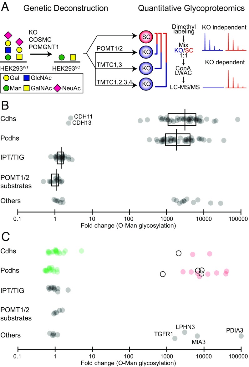
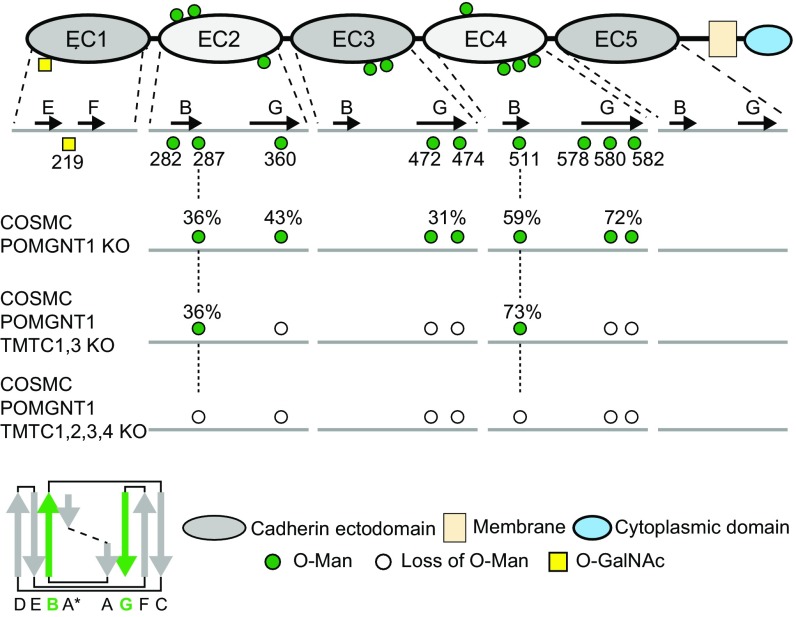
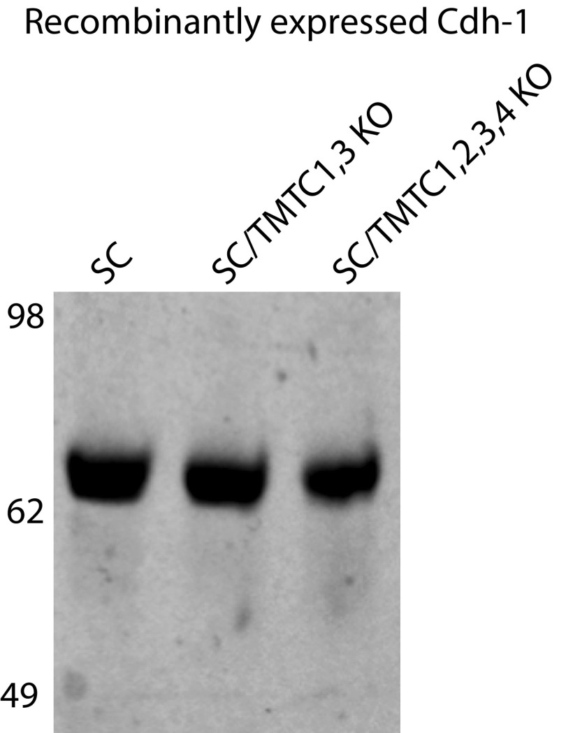
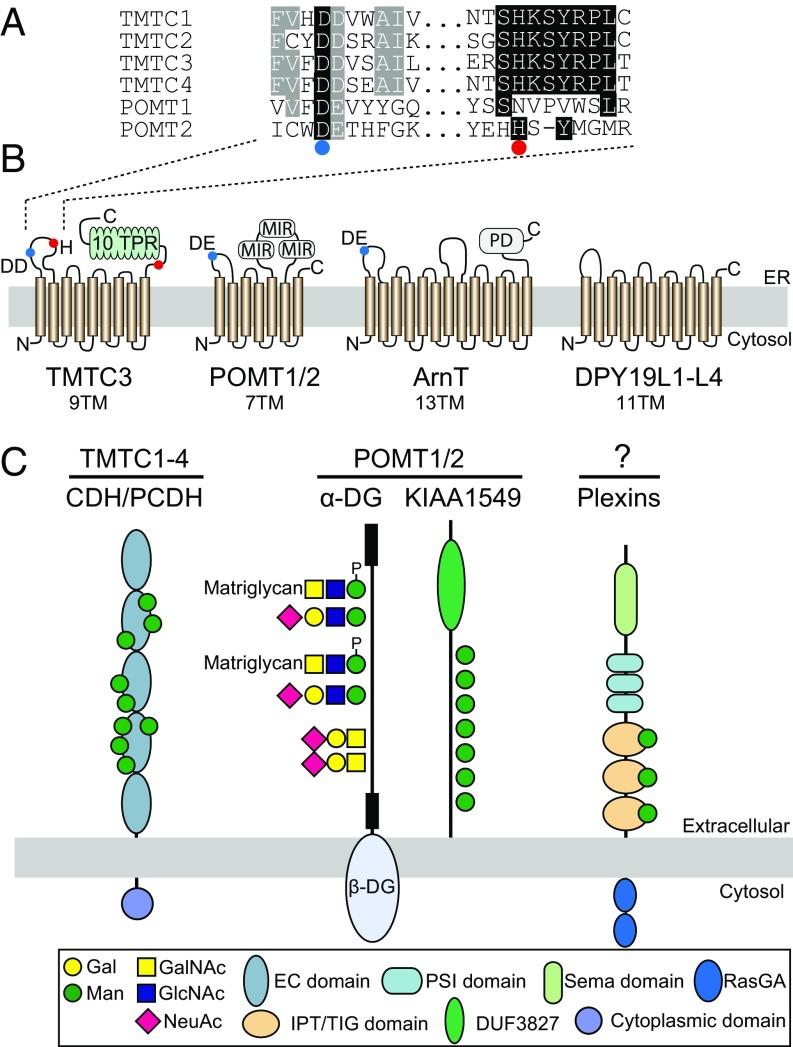
The cadherin (cdh) superfamily of adhesion molecules carry O-linked mannose (O-Man) glycans at highly conserved sites localized to specific β-strands of their extracellular cdh (EC) domains. These O-Man glycans do not appear to be elongated like O-Man glycans found on α-dystroglycan (α-DG), and we recently demonstrated that initiation of cdh/protocadherin (pcdh) O-Man glycosylation is not dependent on the evolutionary conserved POMT1/POMT2 enzymes that initiate O-Man glycosylation on α-DG. Here, we used a CRISPR/Cas9 genetic dissection strategy combined with sensitive and quantitative O-Man glycoproteomics to identify a homologous family of four putative protein O-mannosyltransferases encoded by the TMTC1-4 genes, which were found to be imperative for cdh and pcdh O-Man glycosylation. KO of all four TMTC genes in HEK293 cells resulted in specific loss of cdh and pcdh O-Man glycosylation, whereas combined KO of TMTC1 and TMTC3 resulted in selective loss of O-Man glycans on specific β-strands of EC domains, suggesting that each isoenzyme serves a different function. In addition, O-Man glycosylation of IPT/TIG domains of plexins and hepatocyte growth factor receptor was not affected in TMTC KO cells, suggesting the existence of yet another O-Man glycosylation machinery. Our study demonstrates that regulation of O-mannosylation in higher eukaryotes is more complex than envisioned, and the discovery of the functions of TMTCs provide insight into cobblestone lissencephaly caused by deficiency in TMTC3.

Keywords: O-glycosylation; gene editing; glycoproteomics; glycosyltransferase; mass spectrometry.


Conflict of interest statement

The authors declare no conflict of interest.

Figures

Fig. 1.
Fig. 1.
Genetic dissection and differential quantitative glycoproteome analysis identify TMTC1–4 as directing O-Man glycosylation of cdhs and pcdhs. (A) Graphical depiction of gene editing in HEK293 double SimpleCells (SC) with truncated O-Man and O-GalNAc glycans targeting TMTCs. For differential O-Man glycoproteome analyses, paired dimethyl labeled digests from HEK293SC and HEK293SC/TMTC1,2,3,4 or HEK293SC/TMTC1,3 cells were performed. We previously reported the differential O-Man glycoproteome analyses with HEK293SC/POMT1,2 (13). (B) Scatter plot of dimethyl labeled O-Man glycopeptide ratios (HEK293SC/HEK293SC/TMTC1,2,3,4) expressed on a log10 scale showing loss of O-Man glycopeptides (>100-fold change) derived from cdhs/pcdhs and other proteins, but not glycopeptides derived from IPT/TIG domains or previously identified POMT1/2 substrates (α-DG, KIAA1549, SUCO; <10-fold change) (13) in HEK293 cells with KO of TMTC1–4. Median ratios are plotted for unique O-Man glycopeptides. The box represents the interquartile range, and the vertical line represents the median value within each protein group. (C) Scatter plot of dimethyl labeled O-Man glycopeptide ratios (HEK293SC/HEK293SC/TMTC1,3) showing selective loss of O-Man glycopeptides derived from EC G-strands (red dots; >100-fold change) of cdhs and pcdhs, but not from B-strands (green dots; <10-fold change) in HEK293 cells with KO of TMTC1/3. White dots represent overlapping O-Man glycosylations on B-strand/loop regions. Additional proteins with TMTC1/3-dependent O-Man glycosylations are indicated at lower right: TGF-β receptor type-1 (TGFR1), Latrophilin-3 (LPHN3), Melanoma inhibitory activity protein 3 (MIA3) and protein disulfide-isomerase A3 (PDIA3).
Fig. 2.
Fig. 2.
Summary of O-Man glycosites identified on recombinant E-cdh expressed in HEK293 mutant cell lines. Schematic representation of E-cdh domain organization (Top) with known O-Man glycosylation sites (green circles). Selected β-strands are indicated by black arrows for each EC domain, and all O-Man and O-GalNAc glycosites identified previously are shown with positions and numbering relative to EC β-strands and amino acids of E-cdh (UniProt Knowledgebase ID code P12830). Below the schematic representation are the O-Man glycosites identified on the recombinantly expressed E-cdh in HEK293SC, HEK293SC/TMTC1,3, and HEK293SC/TMTC1,2,3,4 cells, respectively. The stoichiometry (i.e., site occupancy) of O-Man glycans was calculated from the LC peak areas for each peptide (with and without O-Man glycans) and is indicated above each glycosylation site (as percentages). White circles represent loss of O-Man glycosylation sites (not detectable). (Bottom Left) Canonical EC β-strand arrangement, with B- and G-strands indicated in green.
Fig. S1.
Fig. S1.
SDS/PAGE analysis of recombinant expressed E-cdh. E-cdh (5 µg) purified from HEK293SC, HEK293SC/TMTC1,3, and HEK293SC/TMTC1,2,3,4 cells was separated on a NuPAGE 4–12% Bis-Tris protein gel and visualized by InstantBlue.
Fig. 3.
Fig. 3.
A proposed model for differential genetic regulation of protein O-mannosylation in higher eukaryotes. (A) Sequence alignment of a segment from the first luminal loop of human TMTCs and POMTs. A conserved acidic amino acid motif DD/DE in the TMTCs, POMTs (32), and ArnT is indicated by a blue circle. Single amino acid mutations in TMTC3 causing cobblestone lissencephaly are indicated by a red circle. (B) The transmembrane (TM) organization of TMTCs is based on the predicted structure of TMTC3 (16). POMT1/2 is adapted from ref. . The ArnT structure is based on the crystal structure from C. metallidurans (23), and the structure of DPY19Ls is adapted from the Caenorhabditis elegans DPY-19 (29). (C) O-mannosylation in higher eukaryotes is predicted to be controlled by at least three distinct enzyme families. The TMTCs control the cdh superfamily, whereas the classical POMT1/2 controls α-DG and KIAA1549. O-Man glycans on α-DG are elongated by different core structures (45), but it is currently unknown if the O-Man glycans on KIAA1549 are further elongated to form core M1–M3 and/or other glycan structures. The POMT1/2-independent O-mannosylation of cdhs, plexins, and other proteins does not appear to be further elongated (13). A novel O-Man glycosylation capacity dedicated to plexins and IPT/TIG domains is predicted to exist.
Fig. S2.
Fig. S2.
MS-based relative quantification of the total proteolytic digest from paired isogenic KO cells using stable dimethyl isotopes. The technical/biological variability of the assay was estimated by comparing the proteome-level fold change of peptide M/L ratios. (AC) Total tryptic digests of HEK293SC and HEK293SC/TMTC1,2,3,4 KO cells; (D) total chymotryptic digest of HEK293SC and HEK293SC/TMTC1,2,3,4 KO cells, and (E) total tryptic digest of HEK293SC and HEK293SC/TMTC1,3 KO cells. The bar-chart plots demonstrate that >97% of the quantified peptides in all datasets show <10-fold change. The data are centered around log10(M/L) = 0, which further shows that the proteolytic digest from paired isogenic KO cells have been accurately mixed in 1:1 ratios.

References

    1. Lommel M, Strahl S. Protein O-mannosylation: Conserved from bacteria to humans. Glycobiology. 2009;19:816–828. - PubMed
    1. Loibl M, Strahl S. Protein O-mannosylation: What we have learned from baker’s yeast. Biochim Biophys Acta. 2013;1833:2438–2446. - PubMed
    1. Halim A, Anonsen JH. Microbial glycoproteomics. Curr Opin Struct Biol. 2017;44:143–150. - PubMed
    1. Neubert P, et al. Mapping the O-mannose glycoproteome in Saccharomyces cerevisiae. Mol Cell Proteomics. 2016;15:1323–1337. - PMC - PubMed
    1. Barresi R, Campbell KP. Dystroglycan: From biosynthesis to pathogenesis of human disease. J Cell Sci. 2006;119:199–207. - PubMed

Publication types