. 2017 Oct 17;114(42):11199-11204.
doi: 10.1073/pnas.1706193114. Epub 2017 Oct 2.

CRISPR-Cas9-based treatment of myocilin-associated glaucoma

Affiliations

CRISPR-Cas9-based treatment of myocilin-associated glaucoma

Ankur Jain et al. Proc Natl Acad Sci U S A. .

Abstract

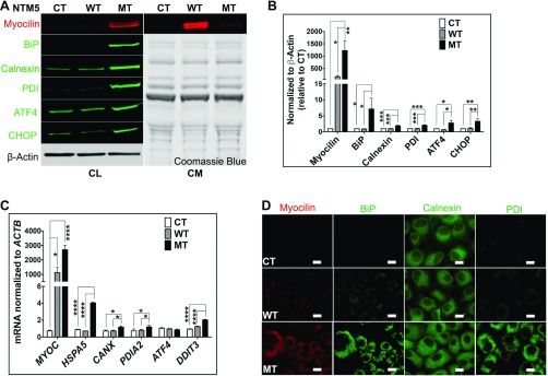
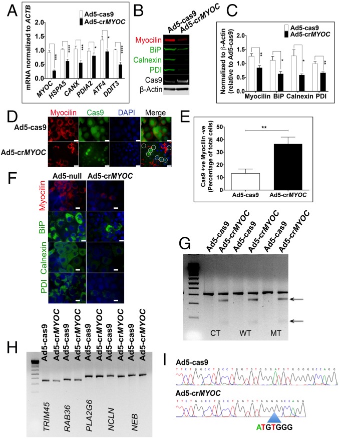
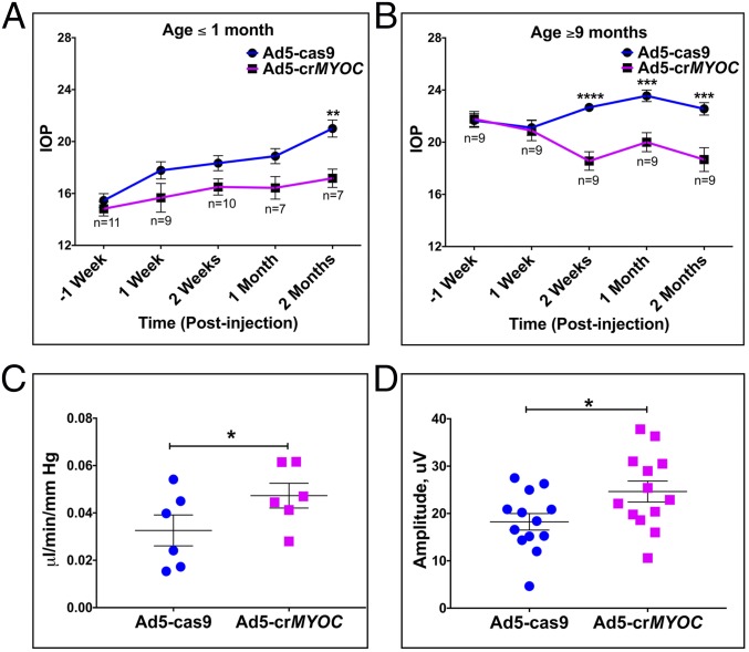
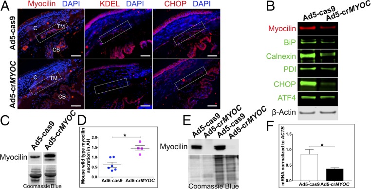
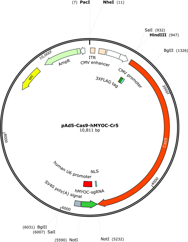
Primary open-angle glaucoma (POAG) is a leading cause of irreversible vision loss worldwide, with elevated intraocular pressure (IOP) a major risk factor. Myocilin (MYOC) dominant gain-of-function mutations have been reported in ∼4% of POAG cases. MYOC mutations result in protein misfolding, leading to endoplasmic reticulum (ER) stress in the trabecular meshwork (TM), the tissue that regulates IOP. We use CRISPR-Cas9-mediated genome editing in cultured human TM cells and in a MYOC mouse model of POAG to knock down expression of mutant MYOC, resulting in relief of ER stress. In vivo genome editing results in lower IOP and prevents further glaucomatous damage. Importantly, using an ex vivo human organ culture system, we demonstrate the feasibility of human genome editing in the eye for this important disease.

Keywords: CRISPR; genome editing; glaucoma; myocilin; trabecular meshwork.


Conflict of interest statement

The authors declare no conflict of interest.

Figures

Fig. S1.
Fig. S1.
Mutant myocilin accumulates inside TM cells. (A) Stably transfected NTM5 cell lines overexpress WT and mutant (MT) forms of human MYOC compared with naïve untransfected control (CT) cells (n = 4). While mutated myocilin accumulates in cells, as demonstrated by analysis of the cell lysate (Left; CL), the WT form is efficiently secreted into the conditioned medium (Right; CM). The accumulation of mutant myocilin causes ER stress, as shown by increased expression of BiP, Calnexin, PDI, ATF4, and CHOP proteins in MT cells. β-actin served as a loading control for cell lysate, and Coomassie blue gel stain was used to test equal loading of secreted proteins in conditioned medium. (B) Densitometry showing significantly higher levels of myocilin, BiP, Calnexin, PDI, ATF4, and CHOP in NTM5 cells overexpressing mutant MYOC (MT) (n = 4) compared with CT or WT cells. (C) qRT-PCR showing overexpression of MYOC mRNA in WT and MT cells and significantly increased expression of ER stress markers mRNA in MT cells (n = 4). (D) An NTM5 cell line overexpressing MT myocilin showing ER accumulation of MYOC (DsRed), BiP (green), Calnexin (green), and PDI (green) compared with CT or NTM5 cells overexpressing WT myocilin. (Scale bar: 100 µm.) Error bars represent SEM. *P < 0.05; **P < 0.01; ***P < 0.001; ****P < 0.0001, one-way ANOVA.
Fig. 1.
Fig. 1.
MYOC disruption by CRISPR-Cas9 in vitro. (A) Ad5-crMYOC treatment of stably transfected NTM5 cell lines overexpressing a mutant (MT) form of human MYOC decreases myocilin (MYOC), BiP (HSP5), Calnexin (CANX), PDI (PDIA2), ATF4 (ATF4), and CHOP (DDIT3) mRNA expression (n = 5). (B) Representative Western blot showing that Ad5-crMYOC decreases mutant myocilin and associated BiP, Calnexin, and PDI (n = 5). Cas9 and β-actin served as loading controls. (C) Densitometry showing significant decreases in myocilin, BiP, Calnexin, and PDI. (D) ICC showing that Ad5-crMYOC treatment decreases myocilin (n = 5). Most of the Cas9 (green)-positive cells are MYOC (red)-negative (denoted by circles in the last panel). (Scale bar: 20 µm.) (E) Quantification of Cas9-positive but myocilin-negative cells showing a significant increase in numbers after Ad5-crMYOC treatment. (F) Ad5-crMYOC–mediated decrease in myocilin (red) and associated BiP, Calnexin, and PDI levels (green; ICC). (Scale bar: 100 µm.) (G) GeneArt Genomic Cleavage assay showing cleavage bands from the MYOC PCR product (arrows) in Ad5-crMYOC–treated DNA in all three cell lines: control (CT), WT, and mutant (MT). (H) GeneArt genomic cleavage assay showing no cleavage bands from predicated off-target sites in PLA2G6, NCLN, and NEB, confirming gRNA specificity for the MYOC gene. (I) Clonal sequencing of the PCR product from Ad5-crMYOC treated samples revealing the most common 7-bp (ATGTGGG) deletion. The error bars represent SEM. *P < 0.05; **P < 0.01; ***P < 0.001; ****P < 0.0001, one-way ANOVA or paired Student’s t test.
Fig. 2.
Fig. 2.
CRISPR-Cas9 lowers IOP and prevents further glaucomatous damage. (A) Ad5-crMYOC (intravitreal injections; 2 × 10⁷ pfu/eye) prevents IOP elevation in ≤1-mo-old Tg-MYOCY437H mice (n = 7–11). (B) Ad5-crMYOC (intravitreal injections; 2 × 10⁷ pfu/eye) lowers IOP in older (age ≥9 mo) Tg-MYOCY437H mice (n = 9). (C) Improved or preserved outflow facility in eyes of 4- to 5-mo-old Tg-MYOCY437H mice at 1 mo after Ad5-crMYOC treatment. The mean facility in these eyes is 0.0468, compared with 0.0309 μL/min/mmHg in Ad5-cas9–treated eyes (n = 7). (D) Improved retinal ganglion cell function measured by pERG in eyes of 4- to 5-mo-old Tg-MYOCY437H mice at 1 mo after Ad5-crMYOC treatment (mean amplitude, 24.644 μV vs. 18.239 μV in Ad5-cas9–treated eyes; n = 13). Error bars represent SEM. *P < 0.05; **P < 0.01; ***P < 0.001; ****P < 0.0001, ANOVA with Bonferroni correction or paired Student’s t test.
Fig. 3.
Fig. 3.
CRISPR efficiency in vivo in mice and ex vivo in human eyes. (A) Representative image showing that Ad5-crMYOC treatment reduces myocilin (red), KDEL (red), and CHOP (red) levels in the TM (rectangular box) of Tg-MYOCY437H mice (n = 3). C, cornea; CB, ciliary body. (Scale bar: 50 µm.) (B) Representative image of WB samples from a TM ring showing that Ad5-crMYOC treatment decreases myocilin levels and associated BiP, Calnexin, PDI, ATF4, and CHOP protein levels (n = 9). (C) Representative image showing increased secretion of mouse WT myocilin into the aqueous humor of Ad5-crMYOC–treated eyes compared with Ad5-cas9–treated eyes. (D) Densitometry showing a significant increase in the levels of secreted mouse WT myocilin in Ad5-crMYOC–treated eyes of Tg-MYOCY437H mice (n = 4). (E) inhibition of myocilin secretion by Ad5-crMYOC–mediated myocilin knockdown in a human ex vivo anterior segment perfusion culture (n = 2). Coomassie blue staining was performed to ensure relatively equal loading. (F) Decreased myocilin (MYOC) mRNA expression in human ex vivo anterior segment perfusion culture by Ad5-crMYOC treatment. Error bars represent SEM. *P < 0.05.
Fig. S2.
Fig. S2.
H&E staining of anterior segment tissues of Ad5-crMYOC–injected mice. H&E staining was used to examine inflammation and gross abnormalities in the anterior segment due to adenovirus 5 (Ad) expression of Cas9. The left eyes of Tg-MYOCY437H mice were injected intravitreally with Ad5-cas9, and the contralateral eyes were injected with Ad5-crMYOC (n = 2 mice). Eyes were enucleated at 1 mo postinjection, and paraffin-embedded anterior segment sections were stained with H&E. The H&E staining reveals an open iridocorneal angle, with no ocular abnormalities associated with Ad5-cas9 or Ad5-crMYOC. We have previously shown that Ad5 injections cause mild inflammation in the anterior segment (36). (Left) Mild inflammation was associated with control Ad5-cas9 injection, as evidenced by a few inflammatory cells. (Right) Interestingly, Ad5-crMYOC produced less inflammation in the anterior segment tissues (n = 2). The rectangular box indicates the TM. (Scale bar: 100 µm.)
Fig. S3.
Fig. S3.
Ad5-crMYOC specifically targets human MYOC. The GeneArt genomic cleavage assay shows a cleavage band (from MYOC qPCR product; arrow) in Ad5-crMYOC–treated DNA in all NTM5 cell lines, but not in mouse kidney cell line IMCD3, confirming the gRNA specificity for the human MYOC gene. Cas9 expression appears similar in both Ad5-crMYOC–treated NTM5 and IMCD3 cells.
Fig. S4.
Fig. S4.
BrdU incorporation in skin fibroblasts. Skin fibroblasts from a control group (n = 3 cell strains) show inclusion of BrdU (red) in nuclei. There are relatively fewer BrdU-positive cells in fibroblasts from myocilin (Y437H) glaucoma patients (n = 3 cell strains). Quantification of BrdU- positive cells (per mm2 in ∼12 images per coverslip) reveals that Ad5-crMYOC treatment significantly increases the number of BrdU-positive cells in the Y437H group, suggesting better proliferation after mutant MYOC gene disruption. (Scale bar: 100 µm.) *P < 0.05.
Fig. S5.
Fig. S5.
Map of the shuttle vector used to generate the Ad5-cas9 virus.
Fig. S6.
Fig. S6.
Map of the shuttle vector used to generate the Ad5-crMYOC virus.

References

    1. Hollands H, et al. Do findings on routine examination identify patients at risk for primary open-angle glaucoma? The rational clinical examination systematic review. JAMA. 2013;309:2035–2042. - PubMed
    1. Weinreb RN, Aung T, Medeiros FA. The pathophysiology and treatment of glaucoma: A review. JAMA. 2014;311:1901–1911. - PMC - PubMed
    1. Alward WL, et al. Clinical features associated with mutations in the chromosome 1 open-angle glaucoma gene (GLC1A) N Engl J Med. 1998;338:1022–1027. - PubMed
    1. Meyer A, et al. [Linkage between juvenile glaucoma and chromosome 1q in 2 French families] C R Acad Sci III. 1994;317:565–570. French. - PubMed
    1. Richards JE, et al. Mapping of a gene for autosomal dominant juvenile-onset open-angle glaucoma to chromosome Iq. Am J Hum Genet. 1994;54:62–70. - PMC - PubMed

Publication types

Substances