. 2017 Oct 17;114(42):11235-11240.
doi: 10.1073/pnas.1706053114. Epub 2017 Oct 2.

Granulocyte-colony-stimulating factor (G-CSF) signaling in spinal microglia drives visceral sensitization following colitis

Affiliations

Granulocyte-colony-stimulating factor (G-CSF) signaling in spinal microglia drives visceral sensitization following colitis

Lilian Basso et al. Proc Natl Acad Sci U S A. .

Abstract

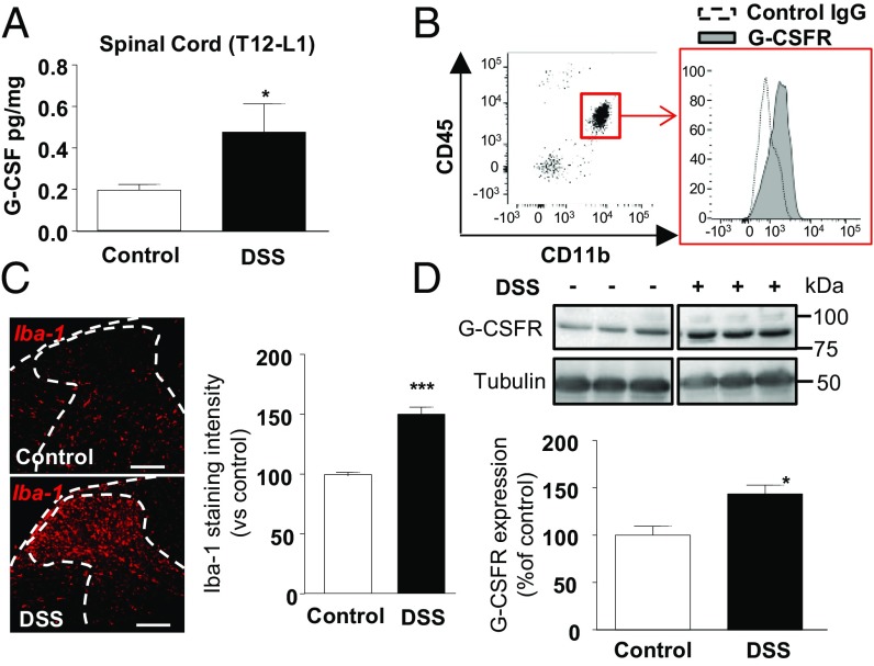
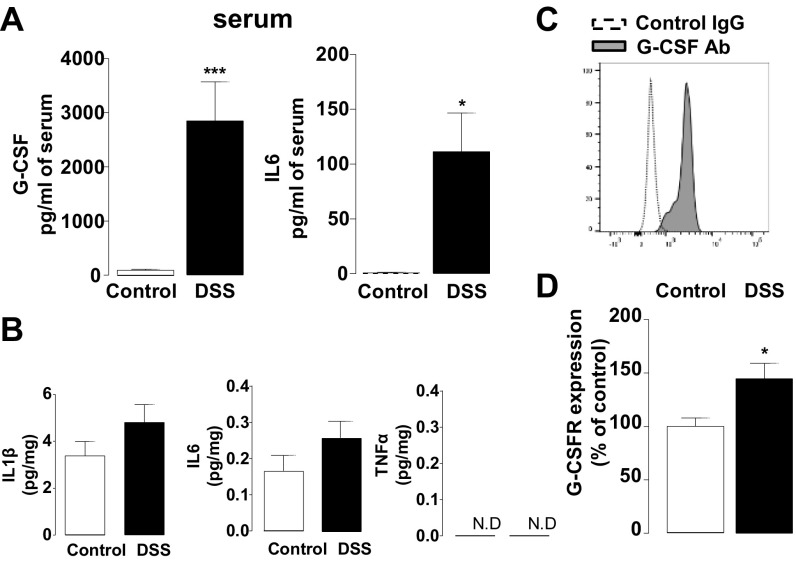
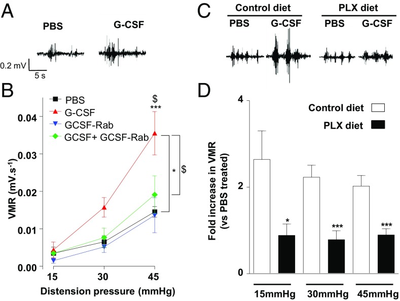
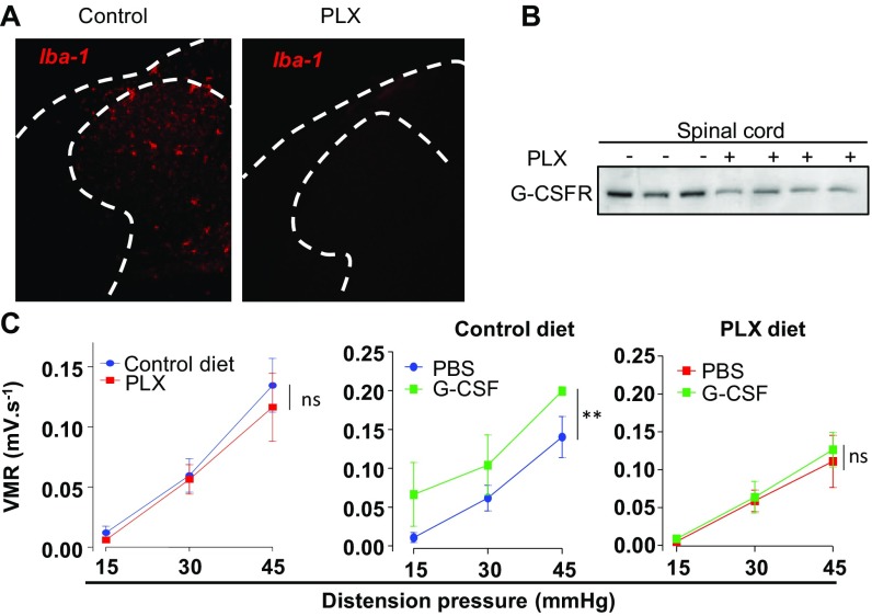
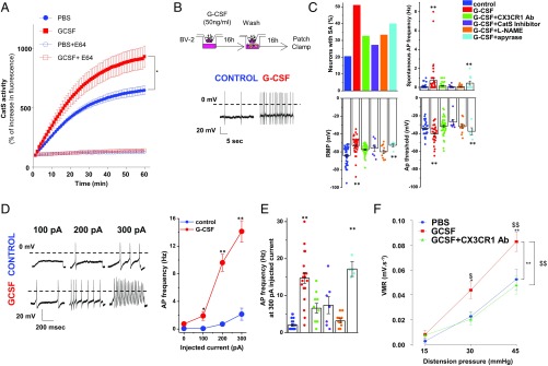
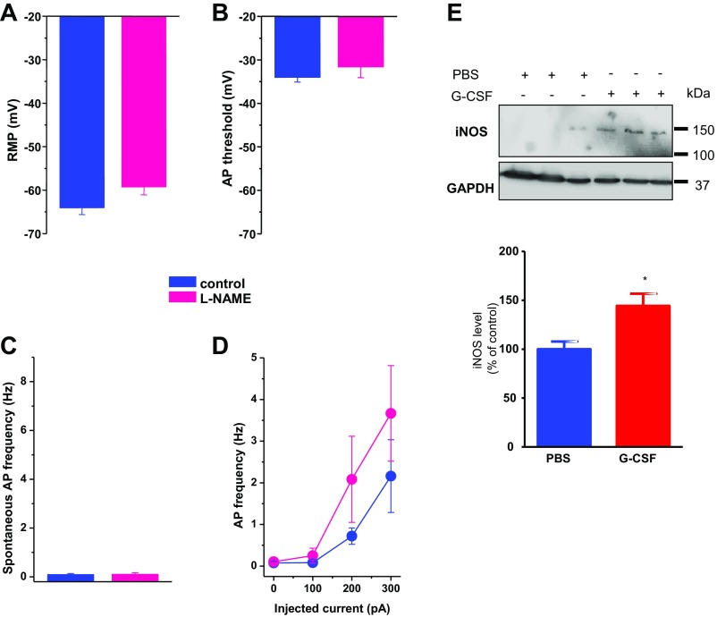
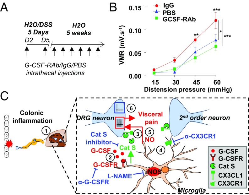
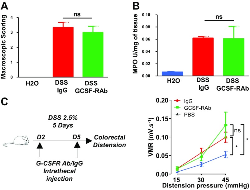
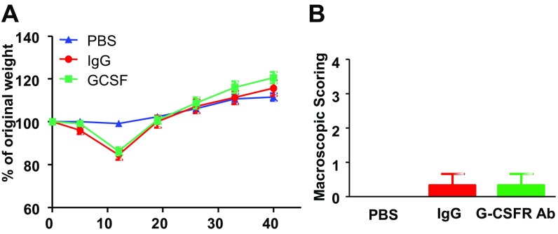
Pain is a main symptom of inflammatory diseases and often persists beyond clinical remission. Although we have a good understanding of the mechanisms of sensitization at the periphery during inflammation, little is known about the mediators that drive central sensitization. Recent reports have identified hematopoietic colony-stimulating factors as important regulators of tumor- and nerve injury-associated pain. Using a mouse model of colitis, we identify the proinflammatory cytokine granulocyte-colony-stimulating factor (G-CSF or Csf-3) as a key mediator of visceral sensitization. We report that G-CSF is specifically up-regulated in the thoracolumbar spinal cord of colitis-affected mice. Our results show that resident spinal microglia express the G-CSF receptor and that G-CSF signaling mediates microglial activation following colitis. Furthermore, healthy mice subjected to intrathecal injection of G-CSF exhibit pronounced visceral hypersensitivity, an effect that is abolished by microglial depletion. Mechanistically, we demonstrate that G-CSF injection increases Cathepsin S activity in spinal cord tissues. When cocultured with microglia BV-2 cells exposed to G-CSF, dorsal root ganglion (DRG) nociceptors become hyperexcitable. Blocking CX3CR1 or nitric oxide production during G-CSF treatment reduces excitability and G-CSF-induced visceral pain in vivo. Finally, administration of G-CSF-neutralizing antibody can prevent the establishment of persistent visceral pain postcolitis. Overall, our work uncovers a DRG neuron-microglia interaction that responds to G-CSF by engaging Cathepsin S-CX3CR1-inducible NOS signaling. This interaction represents a central step in visceral sensitization following colonic inflammation, thereby identifying spinal G-CSF as a target for treating chronic abdominal pain.

Keywords: G-CSF; colitis; microglia; nociceptors; visceral pain.


Conflict of interest statement

The authors declare no conflict of interest.

Figures

Fig. 1.
Fig. 1.
G-CSF and its receptor, expressed on microglia, are increased in the spinal cord during acute DSS-induced colitis. Acute colitis was induced in mice with 2.5% DSS for 7 d. After blood removal, G-CSF levels were determined in the thoracolumbar (T12–L1) spinal cord by luminex technology in control (white bar, n = 10) or colitis (black bar, n = 7) mice (A). Mononuclear cells of the spinal cord were isolated, and CD45low CD11b+ cells (microglia) were analyzed using flow cytometry for their expression of G-CSFR represented by increased binding of specific G-CSFR antibody (gray histogram, representative of three independent experiments) compared with control IgG (dotted histogram) (B). (C, Left) Iba-1 expression was quantified by immunostaining in spinal cord sections of control and DSS colitis mice. (C, Right) Intensity of the immunostaining was measured using Image J on a total of 35 sections from three independent experiments. (D) G-CSFR expression level was determined by Western blot in spinal cord of control (white bar, n = 6) or colitis (black bar, n = 6) mice. Statistical analyses were performed using Mann–Whitney U test; *P < 0.05; ***P < 0.001. (Scale bar: 100 μm.)
Fig. S1.
Fig. S1.
Inflammatory state of spinal cord during acute colitis. (A) Measure of G-CSF and IL6 in the serum of control (white bar, n = 10) or DSS colitis (black bar, n = 7) mice. (B) Measure of IL1β, IL6, and TNFα levels from the thoracolumbar (T12–L1) spinal cord of control (white bar, n = 10) or DSS colitis (black bar, n = 7) mice. (C) Spinal cord mononuclear cells were isolated, and CD45low CD11b+ cells (microglia) were analyzed using flow cytometry. Expression of G-CSF was determined after in vitro stimulation with phorbol myristate acetate (PMA) and ionomycin for 3 h and is represented by increased binding of specific G-CSF antibody (gray histogram) compared with control IgG (dotted histogram). This is a representative result from three independent experiments. (D) Expression of G-CSFR mRNA was assessed by qPCR in the thoracolumbar spinal cord of control (white bar, n = 4) or colitis (black bar, n = 5) mice.
Fig. 2.
Fig. 2.
G-CSF signaling in microglia induces visceral hypersensitivity. (A) Representative electromyogram recording elicited by 45 mmHg pressure at 12 h of intrathecal PBS or G-CSF (20 ng) injection. (B) VMR to colorectal distension after treatment with PBS (n = 4), G-CSF (20 ng; n = 8), anti–G-CSFR antibody (G-CSF-Rab 1 µg; n = 5; ***P < 0.001), or a combination of both (n = 5). (C) Representative electromyogram recording elicited by 45 mmHg pressure at 12 h of intrathecal PBS or G-CSF (20 ng) injection in mice that received control or the PLX 5622 diet for 2 wk. (D) VMR to colorectal distension after treatment with PBS (control diet: n = 15, PLX diet: n = 10) or G-CSF (control diet: n = 16, PLX diet: n = 11). Results are expressed as fold increase in VMR (±SEM) for control diet (white bar) or PLX diet (black bar) animals. Statistical analysis was performed using either repeated-measures two-way ANOVA and Bonferroni post-hoc test (B; *P < 0.05 G-CSF; ***P < 0.001 vs. PBS; $P < 0.05 G-CSF vs. G-CSF+G-CSF Receptor antibody) or the Mann–Whitney U test (D; *P < 0.05; ***P < 0.001).
Fig. S2.
Fig. S2.
Ablation of microglia by PLX 5622 has no effect on basal visceral sensitivity. Iba-1 expression was assessed by immunostaining (A), and G-CSFR expression was assessed by Western blot (B) in spinal cord of control-diet or PLX-fed mice. (C) Visceral sensitivity was measured in control (blue circle), G-CSF–treated (green square) or PLX mice (red square), and PLX mice treated with G-CSF (green square).
Fig. 3.
Fig. 3.
G-CSF–treated microglia increases excitability of DRG neurons through Cathepsin S-CX3CR1-iNOS signaling. (A) Mice were treated intrathecally with either PBS or G-CSF (20 ng) 12 h before evaluating Cathepsin S activity in thoracolumbar spinal cord lysate (n = 3 experiments). (B) Cartoon illustrating the coculture system experiment. BV2 microglia cells were plated into the upper chamber of a transwell and treated with G-CSF for 16 h. G-CSF was removed, and DRG neurons were plated in the lower chamber of the transwell for 16 h of coculture and then used for electrophysiological recordings. (Lower) A representative trace of spontaneous activity (SA) in a small-size TRPV1-sensitive DRG neuron cocultured with vehicle- or G-CSF–primed BV2 cells. (C) Exposure of BV2 microglia to G-CSF depolarizes the resting membrane potential (RMP) of DRG neurons and increases the percentage of neurons with SA (5/28 in control, 17/30 for G-CSF treated, 10/31 for G-CSF combined with CX3CR1 Ab-treated, 3/11 for G-CSF combined with Cat S inhibitor-treated, 5/15 for G-CSF combined with L-NAME, and 4/10 for G-CSF combined with Apyrase). (Right) G-CSF induces a decrease in the AP threshold and an increase in spontaneous AP discharge. These parameters are reversed by cotreatment with CX3CR1 Ab, Cathepsin S inhibitor, or L-NAME, but not apyrase. (D) Representative AP discharge evoked by 100-, 200-, and 300-pA current injections (1 s) in control (blue circle) and G-CSF (red circle) and for different conditions of treatment (E). (F) Measure of VMR to colorectal distension in mice injected with PBS (n = 9), G-CSF (20 ng; n = 12), or G-CSF + CX3CR1 Ab (5 µg; n = 9) 12 h before recording. Results indicate mean ± SEM (*P < 0.05; **P < 0.01). Statistical analysis was performed using one-way (C and E) or two-way ANOVA (A, D, and F) followed by Bonferroni post hoc test (*P < 0.05; **P < 0.01; $P < 0.05; $$P < 0.01).
Fig. S3.
Fig. S3.
Role of iNOS in G-CSF–induced hyperexcitability. Exposure to L-NAME (100 µM) alone has no effect on the resting membrane potential (A), the AP threshold (B), or spontaneous (C) and evoked (D) AP discharge. Results indicate mean ± SEM. Statistical analysis was performed using one-way ANOVA followed by Bonferroni post hoc test. (E) Western blot analysis of iNOS level from spinal cord samples (T12–L1) collected from G-CSF–treated mice. (Lower) Quantification of relative integrated density values from Western blots corrected from GAPDH and normalized to control PBS.
Fig. 4.
Fig. 4.
Blocking G-CSF receptor signaling in the spinal cord alleviates DSS-induced postinflammatory visceral hypersensitivity. (A) Colitis was induced with 2.5% DSS for 5 d, after which mice received only water for the next 5 wk. During the recovery period, mice were treated twice a week with either G-CSFR–blocking antibodies or control IgG. A healthy control group that did not receive DSS was intrathecally injected with PBS. (B) Visceromotor responses to colorectal distension post-DSS–induced colitis in G-CSF-Rab (green squares; n = 11) and control IgG (red circles; n = 6) treated mice compared with healthy mice injected with PBS (blue triangle; n = 4). Data are expressed as mean of VMR ± SEM. Statistical analysis was performed using repeated-measures two-way ANOVA and the Bonferroni post hoc test (*P < 0.05; **P < 0.01; ***P < 0.001). (C) Schematic representation of G-CSF–mediated visceral sensitization in the spinal cord. (1) Acute DSS colitis increases G-CSF in the spinal cord. (2) G-CSF binds to its receptor on microglia and (3) induces the secretion of Cathepsin S. (4) Cathepsin S triggers the release of soluble fractalkine that activates its receptor CX3CR1 at the microglial cell surface. (5) This drives a hyperexcitability of DRG neurons through a NO-dependent process and the establishment of visceral sensitization. (6) Inhibition of either G-CSFR or CX3CR1 prevents both DRG hyperexcitability and visceral hypersensitivity.
Fig. S4.
Fig. S4.
G-CSFR antibody does not alter disease recovery. Mice were treated for 7 d with 2.5% DSS and injected with either control IgG (n = 3) or G-CSF-RAb (n = 3) on days 2, 5, and 7 of DSS regimen. Macroscopic damages (A) and myeloperoxidase activity (B) were evaluated at day 7 of DSS. (C) Schematic representation of the experimental design to assess acute DSS-induced visceral hypersensitivity; colitis was induced by 2.5% DSS regimen and mice received intrathecal injection of either IgG (red circle n = 13) or G-CSF-RAb (green square n = 10). A control group receiving only water was injected with PBS IT (blue triangle n = 11) at similar time points. On day 5, visceral hypersensitivity was assessed by measuring visceromotor response to colorectal distension.
Fig. S5.
Fig. S5.
Chronic intrathecal treatment with G-CSF-RAb does not alter disease recovery post colitis. Colitis was induced by adding 2.5% DSS in the drinking water of mice. On day 5, DSS mice were on water regimen for 5 wk. During the recovery period, mice were treated twice a week with either G-CSFR–blocking antibody (blue square; n = 11) or control IgG (red circle; n = 6). A control group that was not exposed to DSS received intrathecal PBS (n = 4). Recovery of colitis was monitored by weighing mice weekly (A) and macroscopic scoring of colonic tissue damages (B) as previously described (46).

References

    1. Basso L, Bourreille A, Dietrich G. Intestinal inflammation and pain management. Curr Opin Pharmacol. 2015;25:50–55. - PubMed
    1. Farrokhyar F, Marshall JK, Easterbrook B, Irvine EJ. Functional gastrointestinal disorders and mood disorders in patients with inactive inflammatory bowel disease: Prevalence and impact on health. Inflamm Bowel Dis. 2006;12:38–46. - PubMed
    1. Teruel C, Garrido E, Mesonero F. Diagnosis and management of functional symptoms in inflammatory bowel disease in remission. World J Gastrointest Pharmacol Ther. 2016;7:78–90. - PMC - PubMed
    1. Young Casey C, Greenberg MA, Nicassio PM, Harpin RE, Hubbard D. Transition from acute to chronic pain and disability: A model including cognitive, affective, and trauma factors. Pain. 2008;134:69–79. - PubMed
    1. Woolf CJ. What is this thing called pain? J Clin Invest. 2010;120:3742–3744. - PMC - PubMed

Publication types

MeSH terms

Grants and funding

LinkOut - more resources