. 2017 Oct 3;10(1):159.
doi: 10.1186/s13045-017-0531-y.

The stem cell factor SALL4 is an essential transcriptional regulator in mixed lineage leukemia-rearranged leukemogenesis

Affiliations

The stem cell factor SALL4 is an essential transcriptional regulator in mixed lineage leukemia-rearranged leukemogenesis

Lina Yang et al. J Hematol Oncol. .

Abstract

Background: The stem cell factor spalt-like transcription factor 4 (SALL4) plays important roles in normal hematopoiesis and also in leukemogenesis. We previously reported that SALL4 exerts its effect by recruiting important epigenetic factors such as DNA methyltransferases DNMT1 and lysine-specific demethylase 1 (LSD1/KDM1A). Both of these proteins are critically involved in mixed lineage leukemia (MLL)-rearranged (MLL-r) leukemia, which has a very poor clinical prognosis. Recently, SALL4 has been further linked to the functions of MLL and its target gene homeobox A9 (HOXA9). However, it remains unclear whether SALL4 is indeed a key player in MLL-r leukemia pathogenesis.

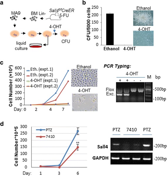
Methods: Using a mouse bone marrow retroviral transduction/ transplantation approach combined with tamoxifen-inducible, CreERT2-mediated Sall4 gene deletion, we studied SALL4 functions in leukemic transformation that was induced by MLL-AF9-one of the most common MLL-r oncoproteins found in patients. In addition, the underlying transcriptional and epigenetic mechanisms were explored using chromatin immunoprecipitation (ChIP) sequencing (ChIP-Seq), mRNA microarray, qRT-PCR, histone modification, co-immunoprecipitation (co-IP), cell cycle, and apoptosis assays. The effects of SALL4 loss on normal hematopoiesis in mice were also investigated.

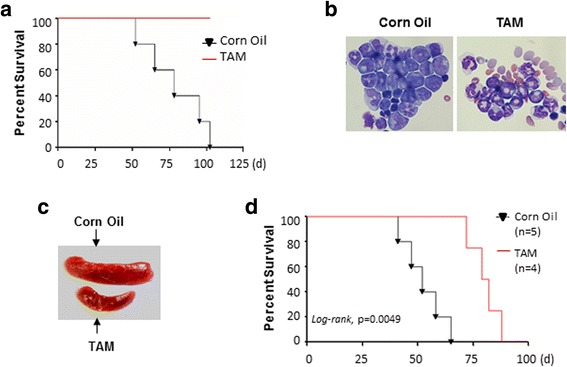
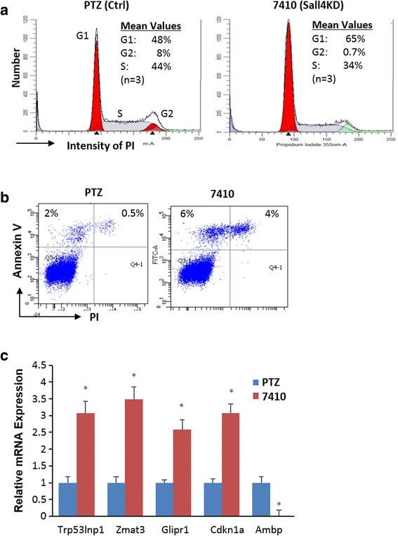
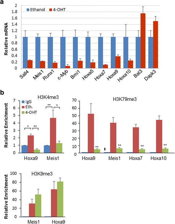
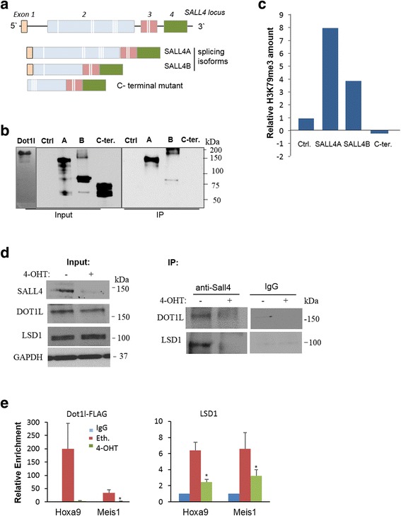
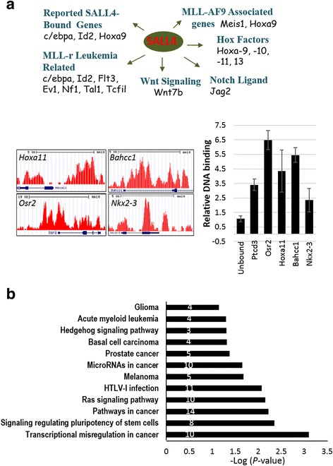
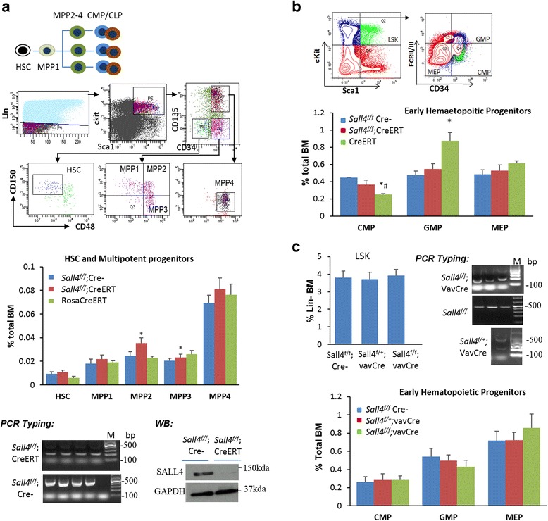
Results: In vitro and in vivo studies revealed that SALL4 expression is critically required for MLL-AF9-induced leukemic transformation and disease progression in mice. Loss of SALL4 in MLL-AF9-transformed cells induced apoptosis and cell cycle arrest at G1. ChIP-Seq assay identified that Sall4 binds to key MLL-AF9 target genes and important MLL-r or non-MLL-r leukemia-related genes. ChIP-PCR assays indicated that SALL4 affects the levels of the histone modification markers H3K79me2/3 and H3K4me3 at MLL-AF9 target gene promoters by physically interacting with DOT1-like histone H3K79 methyltransferase (DOT1l) and LSD1/KDM1A, and thereby regulates transcript expression. Surprisingly, normal Sall4 f/f /CreERT2 mice treated with tamoxifen or vav-Cre-mediated (hematopoietic-specific) Sall4 -/- mice were healthy and displayed no significant hematopoietic defects.

Conclusions: Our findings indicate that SALL4 critically contributes to MLL-AF9-induced leukemia, unraveling the underlying transcriptional and epigenetic mechanisms in this disease and suggesting that selectively targeting the SALL4 pathway may be a promising approach for managing human MLL-r leukemia.

Keywords: DOT1l; Epigenetic; Hematopoietic stem cells; Histone methylation; LSD1; Transcription factor.


Conflict of interest statement

Ethics approval and consent to participate

Not applicable.

Consent for publication

Not applicable.

Competing interests

The authors declare that they have no competing interests.

Publisher’s Note

Springer Nature remains neutral with regard to jurisdictional claims in published maps and institutional affiliations.

Figures

Fig. 1
Fig. 1
SALL4 is required for MLL-AF9-induced transformation in vitro. a Diagram of the in vitro procedures. b Relative CFU of MA9-transduced cells after ethanol or 4-OHT treatment during the fourth round plating are shown. An equal number of MA9-transformed cells were plated at each round. Representative colony images (×10) were captured using a Ti-S inverted phase/fluorescent microscope with an SPOT cooled 2.0-megapixel digital microscope camera system (Nikon, Tokyo, Japan). c Cells plucked from the third plating colonies were cultured in liquid and counted daily after ethanol or 4-OHT treatment. Proliferating cells were counted from two independent experiments. Representative culture images were shown (×20). The efficiency of 4-OHT-induced Sall4 excision was confirmed by PCR with primers as described previously [28]. M: DNA size marker. d PTRIPZ#7410 or control PTRIPZ (PTZ) vector infected MA9 cells were treated with doxycycline and viable cell numbers were counted at different days. Representative RT-PCR analysis of shRNA-induced Sall4 knockdown was shown using specific primers for mouse Sall4 or GAPDH (internal control) as described [12]. Error bars represent SD of three independent experiments. **p < 0.01
Fig. 2
Fig. 2
SALL4 is required for MLL-AF9-induced transformation in vivo. a Kaplan–Meier survival curve of mice transplanted with MA9-transduced Sall4 f/f CreERT2 cells, with tamoxifen (TAM) or oil treatment (n = 5 per group). BM infiltration of AML blast cells (b) and enlarged spleen (c) are seen in oil- but not TAM-treated mice. d Kaplan–Meier survival curve of mice transplanted with MA9-transduced ckit+ Sall4 f/f Cre ERT2 cells. TAM or oil was treated 4 weeks after cell transplantation
Fig. 3
Fig. 3
Loss of SALL4 caused apoptosis and cell cycle arrest in G1 phase in MLL-AF9-transformed cells. a PTRIPZ#7410 or PTZ vector infected MA9 cells were treated with doxycycline, stained with propidium iodide (PI), and their DNA content analyzed using flow cytometry. b Representative flow cytometry data showing Annexin V and PI stained apoptotic cells for indicated cells. c qRT-PCR analysis shows mRNA expression levels for indicated genes. Error bars represent SD of three independent experiments. *p < 0.05
Fig. 4
Fig. 4
Sall4 knockout affects expression levels and histone methylation status of important MLL-AF9 downstream genes. a qRT-PCR analysis shows downregulation of Meis1, HOX cluster, and multiple important MLL-AF9 target genes in cells after 5-day treatment with 4-OHT (n = 3). Values were normalized to GAPDH mRNA expression. b ChIP-qPCR assay has been conducted in MA9-transformed cells using anti-H3K79me2/3, anti-H3K4me3, and anti-H3K9me3 antibodies. Data showing that H3K79me2/3 and H3K4me3 enrichment levels at Hoxa9, Meis1, and other Hox gene promoter regions significantly decreased after 4-OHT treatment, which contrasts with the repressive marker H3K9me3. A 1.2-kb region upstream of the gene transcription start sites, as previously reported [18, 19, 26], were examined. Error bars represent SD of three independent experiments
Fig. 5
Fig. 5
SALL4 recruits DOT1L and LSD1 to target genes in MLL-AF9-transformed cells. a SALL4 isoforms and C-terminal mutant are shown schematically. White lines indicate zinc-finger motifs. b HA-tagged SALL4 isoforms, the C-terminal mutant, and an empty vector control were transfected into 293 T cells. Their expressions and DOT1l levels are shown by Western blotting (input). An anti-DOT1L antibody (Bethyl Laboratories) was used for Dynabeads Protein G immunoprecipitation (Life Technologies). Anti-HA immunoprecipitates were analyzed by Western blotting (IP). c Above different SALL4-DOT1L immune-complexes were pulled down by an anti-HA antibody. The amount of tri-methylated H3K79 in the complex extracts was measured using the fluorometric EpiQuik Global Pan-methyl Histone H3K79 Quantification Kit (Epigentek) using a SpectraMax M3 microplate reader at 530–590 nm. d Co-IP assays were conducted with MLL-AF9-transformed Lin-BM cells, with or without 4-OHT treatment. An anti-Sall4 antibody was used for IP, and the immunoprecipitated complexes were analyzed by Western blotting using indicated antibodies (n ≥ 2). e ChIP analysis demonstrates that in MA9-transformed cells, 4-OHT treatment caused reduced binding of Dot1l and Lsd1 at Hoxa9 and Meis1 promoter regions. Error bars represent SD of three independent experiments. *p < 0.05; **p < 0.01
Fig. 6
Fig. 6
Analysis of SALL4 binding genes. a ChIP-Seq-identified SALL4 binding genes are classified. Representative screenshots of ChIP-Seq results displayed from the UCSC genome browser are shown. ChIP-qPCR on indicated genes is indicated as fold change over the unbound control untr17. Ptcd3 serves as a positive control locus. b DAVID gene ontology and KEGG_Pathway analysis of SALL4-bound genes. Involved gene numbers are shown. The detected pathways are ordered by p value significance (transformed by -10log10 of p value; highest number is the most significant). Error bars represent SD of three independent experiments
Fig. 7
Fig. 7
The BM stem/progenitor populations corresponding to HSC and MPP1–MPP4 (a) and each HPC fractions (b) were FACS analyzed. Representative flow cytometry scheme and quantification of the BM HSPC compartments were shown from Sall4 f/f, Sall4 f/f CreERT2, and RosaCreERT2mice 14 days after TAM treatment (n = 3 per group). PCR typing (4 mouse samples per group) and representative Western blotting assays for SALL4 deletion in TAM-treated Sall4 f/f; CreERT2 mice are shown. c Representative flow cytometry scheme and quantification of the BM HSPC compartments from Sall4 f/fCre-, Sall4 f/+ /vavCre, and Sall4 f/f/vavCre mice (n = 4 per group, 2–3-weeks old). PCR typing showing Sall4 excision in indicated mouse BM cells. Error bars represent SD of at least three independent experiments. *p < 0.05 when compared to Sall4 f/f/CreERT; #p < 0.05 compared to Sall4 f/fCre-. LSK Lin-Sca1 + cKit+; GMP granulocyte-macrophage progenitor; CMP common myeloid progenitor; MEP megakaryocyte-erythroid progenitor

Similar articles

Cited by

References

    1. Kohlhase J, Schuh R, Dowe G, Kuhnlein RP, Jackle H, Schroeder B, Schulz-Schaeffer W, Kretzschmar HA, Kohler A, Muller U, et al. Isolation, characterization, and organ-specific expression of two novel human zinc finger genes related to the Drosophila gene spalt. Genomics. 1996;38(3):291–298. doi: 10.1006/geno.1996.0631. - DOI - PubMed
    1. Al-Baradie R, Yamada K, St Hilaire C, Chan WM, Andrews C, McIntosh N, Nakano M, Martonyi EJ, Raymond WR, Okumura S, et al. Duane radial ray syndrome (Okihiro syndrome) maps to 20q13 and results from mutations in SALL4, a new member of the SAL family. Am J Hum Genet. 2002;71(5):1195–1199. doi: 10.1086/343821. - DOI - PMC - PubMed
    1. Zhang J, Tam WL, Tong GQ, Wu Q, Chan HY, Soh BS, Lou Y, Yang J, Ma Y, Chai L, et al. Sall4 modulates embryonic stem cell pluripotency and early embryonic development by the transcriptional regulation of Pou5f1. Nat Cell Biol. 2006;8(10):1114–1123. doi: 10.1038/ncb1481. - DOI - PubMed
    1. Yang J, Chai L, Fowles TC, Alipio Z, Xu D, Fink LM, Ward DC, Ma Y. Genome-wide analysis reveals Sall4 to be a major regulator of pluripotency in murine-embryonic stem cells. Proc Natl Acad Sci U S A. 2008;105(50):19756–19761. doi: 10.1073/pnas.0809321105. - DOI - PMC - PubMed
    1. Wu Q, Chen X, Zhang J, Loh YH, Low TY, Zhang W, Sze SK, Lim B, Ng HH. Sall4 interacts with Nanog and co-occupies Nanog genomic sites in embryonic stem cells. J Biol Chem. 2006;281(34):24090–24094. doi: 10.1074/jbc.C600122200. - DOI - PubMed

Publication types