. 2017 Nov 1;25(11):2440-2451.
doi: 10.1016/j.ymthe.2017.09.010. Epub 2017 Sep 14.

Adenovirotherapy Delivering Cytokine and Checkpoint Inhibitor Augments CAR T Cells against Metastatic Head and Neck Cancer

Affiliations

Adenovirotherapy Delivering Cytokine and Checkpoint Inhibitor Augments CAR T Cells against Metastatic Head and Neck Cancer

Amanda Rosewell Shaw et al. Mol Ther. .

Abstract

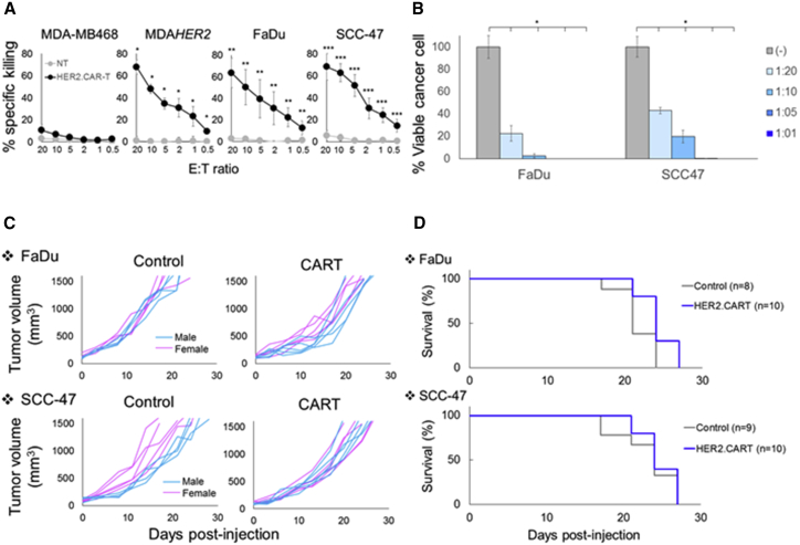
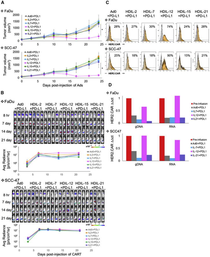
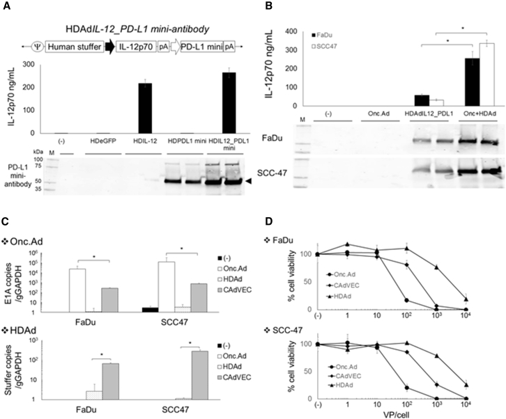
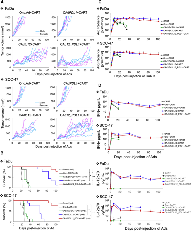
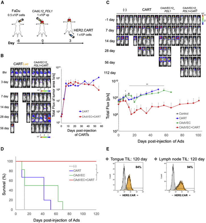
In solid tumors, chimeric antigen receptor (CAR)-modified T cells must overcome the challenges of the immunosuppressive tumor microenvironment. We hypothesized that pre-treating tumors with our binary oncolytic adenovirus (CAd), which produces local oncolysis and expresses immunostimulatory molecules, would enhance the antitumor activity of HER2-specific CAR T cells, which alone are insufficient to cure solid tumors. We tested multiple cytokines in conjunction with PD-L1-blocking antibody and found that Ad-derived IL-12p70 prevents the loss of HER2.CAR-expressing T cells at the tumor site. Accordingly, we created a construct encoding the PD-L1-blocking antibody and IL-12p70 (CAd12_PDL1). In head and neck squamous cell carcinoma (HNSCC) xenograft models, combining local treatment with CAd12_PDL1 and systemic HER2.CAR T cell infusion improved survival to >100 days compared with approximately 25 days with either approach alone. This combination also controlled both primary and metastasized tumors in an orthotopic model of HNSCC. Overall, our data show that CAd12_PDL1 augments the anti-tumor effects of HER2.CAR T cells, thus controlling the growth of both primary and metastasized tumors.

Keywords: CAR T cell therapy; adenovirotherapy; head and neck cancer; oncolytic virus; orthotopic animal model.


Figures

Figure 1
Figure 1
HNSCC Lines Resist HER2.CAR T Cells In Vivo (A) The ability of HER2.CAR T cells to lyse HNSCC lines was assessed using a 4-hr 51Cr release assay. Data are presented as means ± SD (n = 3 donors). *p < 0.002, **p < 0.005, ***p < 0.001. (B) FaDu and SCC-47 expressing ffLuc were cultured with increasing doses of HER2.CAR T cells. Viable cancer cells were analyzed at 120 hr by luciferase assay, and percent viability was calculated. Data are presented as means ± SD (n = 4). *p < 0.001. (C) FaDu and SCC-47 cells were transplanted into the right flanks of NSG mice (pink, female; blue, male). A total of 1 × 106 HER2.CAR T cells were systemically administered after the tumor volume reached 100 mm3. Tumor volumes were measured at different time points. (D) Kaplan-Meier survival curve after administration of HER2.CAR T cells. The end point was established at a tumor volume of >1,500 mm3. Data are presented as means ± SD (n = 8–10).
Figure 2
Figure 2
HDAd-Derived Cytokines Minimally Enhance the Anti-tumor Effect of HER2.CAR T Cells In Vitro and In Vivo (A) Schematic structure of HDAd encoding a cytokine expression cassette (HDAdCyto). A549 cells were infected with 100 vps/cell of HDAdCyto. Media were collected 48 hr post-infection, and cytokine levels in the media were measured by ELISA. Data are presented as means ± SD (n = 4). *p < 0.05, **p < 0.001. (B) HER2.CAR T cells expanded with IL-2 were cultured in the presence of 10 ng/mL recombinant cytokines for 30 min, and phosphorylation of STATs was analyzed by flow cytometry. The experiments were repeated with HER2.CAR T cells derived from a second donor with similar results. (C) FaDu or SCC-47 expressing ffLuc cells were infected with 100 vps/cell of HDAdCyto. HER2.CAR T cells were added 24 hr post-infection (effector:target ratio of 1:40). Cells were harvested 120 hr post-co-culture, and viable cancer cells were analyzed by luciferase assay. Data are presented as means ± SD (n = 4). *p < 0.001. (D) SCC-47 cells were transplanted into the right flanks of NSG female mice. A total of 1 × 108 vps of HDAdCyto were injected intra-tumorally. A total of 1 × 106 HER2.CAR T cells were systemically administered 3 days post-injection of HDAds, and tumor volumes were measured at different time points. Data are presented as means ± SD (n = 3).
Figure 3
Figure 3
HDAd-Derived IL-12p70 and PD-L1-Blocking Antibody Increase the Anti-tumor Efficacy of Adoptively Transferred HER2.CAR T Cells In Vivo FaDu or SCC-47 cells were transplanted into the right flanks of NSG mice. A total of 1 × 108 vps of HDAdCyto and HDAdPDL1 (1:1) were injected intra-tumorally. A total of 1 × 106 HER2.CAR T cells expressing firefly luciferase (ffLuc) were systemically administered 3 days post-injection of HDAds. (A) Tumor volumes were measured at different time points. Data are presented as means ± SD (n = 4). *p < 0.001. (B) Bioluminescence of HER2.CAR T cells was monitored at different time points. Data are presented as means ± SD (n = 4). (C) T cells at the tumor site were isolated 22 days post-infusion, and HER2.CAR levels on T cells were analyzed by flow cytometry. The experiments were repeated with similar results. (D) T cells from tumor sites treated with HDAd0, HDAdIL-7, HDAdIL-12, or HDAdIL-21 co-injected with HDAdPDL1 were isolated 22 days post-injection and purified by a CD3 MACS column. To achieve sufficient T cells for analysis, cells from each group were pooled before analysis. DNA and RNA were extracted from purified T cells, and HER2.CAR copy numbers at DNA and RNA levels in each sample were quantified. The experiments were repeated with similar results.
Figure 4
Figure 4
Co-infection of Onc.Ad with HDAd Expressing Both IL-12p70 and PD-L1 Mini-Antibody Amplifies IL-12p70 and PD-L1-Blocking Antibody Expression with Oncolysis In Vitro (A) Schematic structure of HDAd encoding human IL-12p70 and anti-PD-L1 mini-antibody expression cassettes (HDAdIL12_PDL1). A549 cells were infected with 100 vps/cell of HDAdEGFP, HDAdIL-12, HDAdPD-L1, or HDAdIL12_PDL1. Media were collected 48 hr post-infection. Medium samples were subjected to IL-12p70 ELISA and western blotting for PD-L1 mini-antibody, which was detected by anti-HA antibody. (B) FaDu and SCC-47 cells were infected with a total of 10 vps/cell of HDAdIL12_PDL1 or Onc.Ad or with CAd12_PDL1 (Onc.Ad:HDAd, 1:10). Medium samples were collected 48 hr post-infection. The levels of IL-12p70 and PD-L1 mini-antibody in medium samples were quantified by IL-12p70 ELISA assay and western blotting for PD-L1 mini-antibody, respectively. Data are presented as means ± SD (n = 4). *p < 0.001. (C) DNA samples were extracted 48 hr post-infection, and Onc.Ad and HDAd vector copy numbers were measured by qPCR. Data were normalized with human genomic GAPDH. Data are presented as means ± SD (n = 4). *p = 0.008. CAdVEC: Onc.Ad:HDAdIL12_PDL1, 1:10. (D) FaDu and SCC-47 cells were infected with increasing doses of HDAdIL12_PDL1 or Onc.Ad or with CAdVEC (Onc.Ad: HDAdIL12_PDL1, 1:10). Viable cells were analyzed at 96 hr by MTS assay. Data are presented as means ± SD (n = 6).
Figure 5
Figure 5
CAd12_PDL1 Enhances the Anti-tumor Effects of HER2.CAR T Cells In Vivo FaDu or SCC-47 cells were transplanted into the right flanks of NSG mice (pink, female; blue, male). A total of 1 × 108 vps of Onc.Ad, CAdPDL1, CAdIL-12, or CAd12_PDL1 (Onc:HD, 1:20) were injected intra-tumorally. A total of 1 × 106 HER2.CAR T cells expressing ffLuc were systemically administered 3 days after injection of Ads. (A) Tumor volumes were measured at different time points. (B) Kaplan-Meier survival curve after administration of Ad gene therapy. The end point was established as a tumor volume of >1,500 mm3. Data are presented as means ± SD (n = 8–10). *p < 0.003, **p < 0.007, ***p < 0.01, and ****p < 0.001. (C) The bioluminescence of HER2.CAR T cells was monitored at different time points. Data are presented as means ± SD (n = 8–10). (D) Serum samples were collected at 0, 3, 10, 24, 45, 66, 87, and 108 days after injection of Ads, and IFNγ and IL-12p70 levels in serum were measured by ELISA. Data are presented as means ± SD (n = 8–10).
Figure 6
Figure 6
Combinatorial Treatment Can Control Both Primary and Metastasized Tumors in an Orthotopic HNSCC Model (A) Schematic flow of HNSCC orthotopic animal experiment. (B) FaDu cells were transplanted into the tongues of NSG mice. A total of 1 × 108 vps of CAd12_PDL1 (Onc:HD, 1:20) were injected into the tongues. A total of 1 × 106 HER2.CAR T cells expressing ffLuc were systemically administered 3 days after injection of CAd. The bioluminescence of HER2.CAR T cells at the tumor area was monitored at different time points. Data are presented as means ± SD (n = 5). (C) FaDu cells expressing ffLuc were transplanted into the tongues of NSG mice. A total of 1 × 108 vps of CAd12_PDL1 (Onc:HD, 1:20) were injected into the tongue. A total of 1 × 106 HER2.CAR T cells were systemically administered 3 days after injection of CAd. Bioluminescence of FaDu cells was monitored at different time points. Data are presented as means ± SD (n = 6–8). *p = 0.017, **p < 0.005. (D) Kaplan-Meier survival curve after administration of Ad gene therapy. The end point was established as an animal body weight < 80%. Data are presented as means ± SD (n = 6–8). *p < 0.003. (E) T cells were isolated from tongue and lymph node sites 120 days post-infusion, and HER2.CAR expression was analyzed by flow cytometry.

Comment in

References

    1. Quail D.F., Joyce J.A. Microenvironmental regulation of tumor progression and metastasis. Nat. Med. 2013;19:1423–1437. - PMC - PubMed
    1. Pardoll D.M. The blockade of immune checkpoints in cancer immunotherapy. Nat. Rev. Cancer. 2012;12:252–264. - PMC - PubMed
    1. Kershaw M.H., Westwood J.A., Darcy P.K. Gene-engineered T cells for cancer therapy. Nat. Rev. Cancer. 2013;13:525–541. - PubMed
    1. Farzad L., Cerullo V., Yagyu S., Bertin T., Hemminki A., Rooney C., Lee B., Suzuki M. Combinatorial treatment with oncolytic adenovirus and helper-dependent adenovirus augments adenoviral cancer gene therapy. Mol. Ther. Oncolytics. 2014;1:14008. - PMC - PubMed
    1. Sharma P., Allison J.P. Immune checkpoint targeting in cancer therapy: toward combination strategies with curative potential. Cell. 2015;161:205–214. - PMC - PubMed

Publication types

MeSH terms