. 2018 Feb;32(2):728-741.
doi: 10.1096/fj.201700576R. Epub 2018 Jan 4.

Autophagy and mitochondrial biogenesis impairment contribute to age-dependent liver injury in experimental sepsis: dysregulation of AMP-activated protein kinase pathway

Affiliations

Autophagy and mitochondrial biogenesis impairment contribute to age-dependent liver injury in experimental sepsis: dysregulation of AMP-activated protein kinase pathway

Yu Inata et al. FASEB J. 2018 Feb.

Abstract

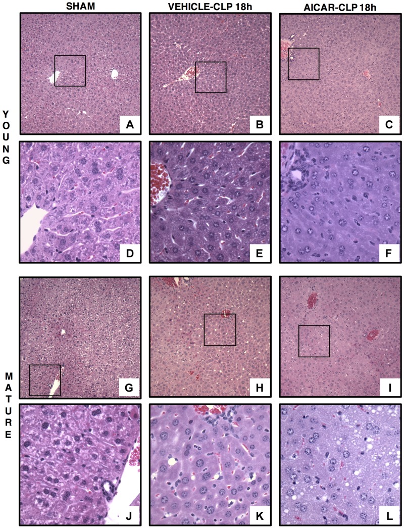
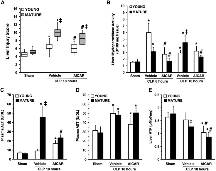
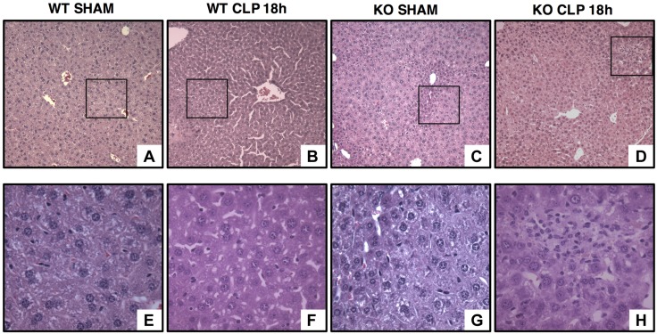
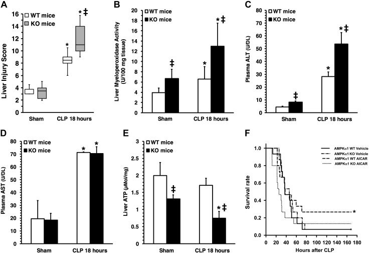
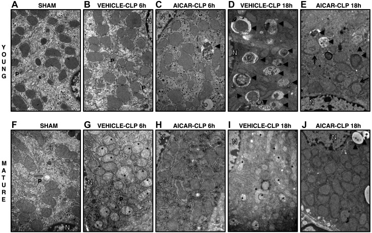
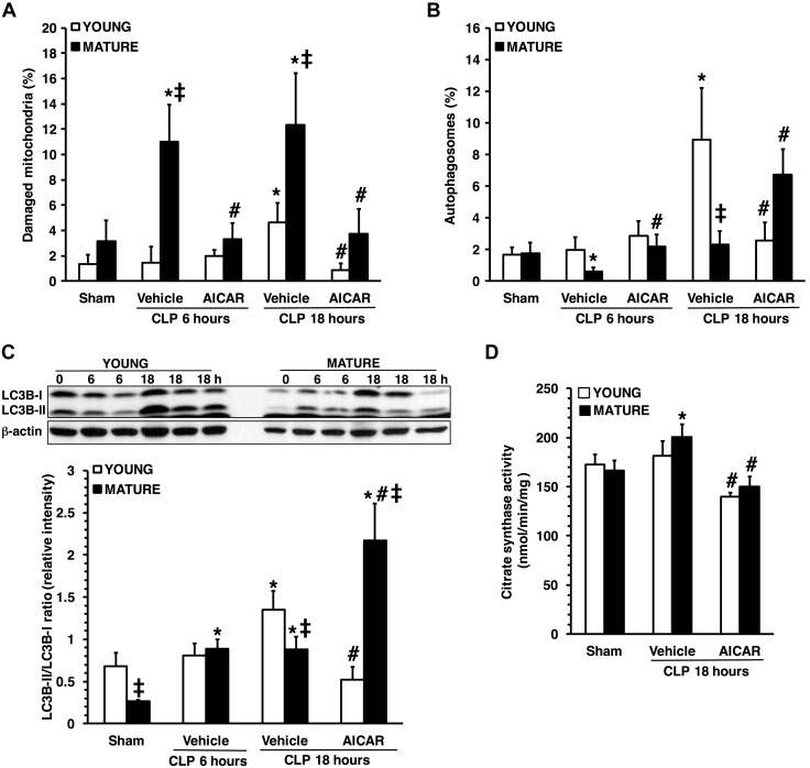
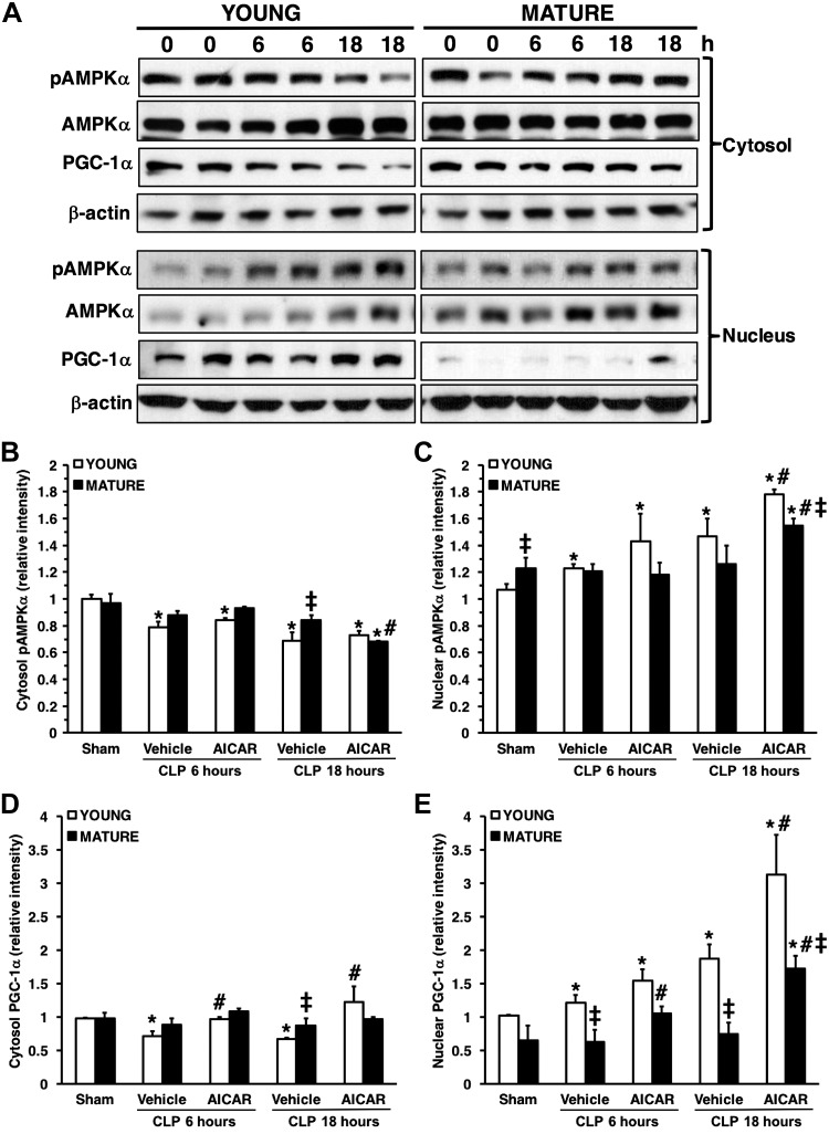
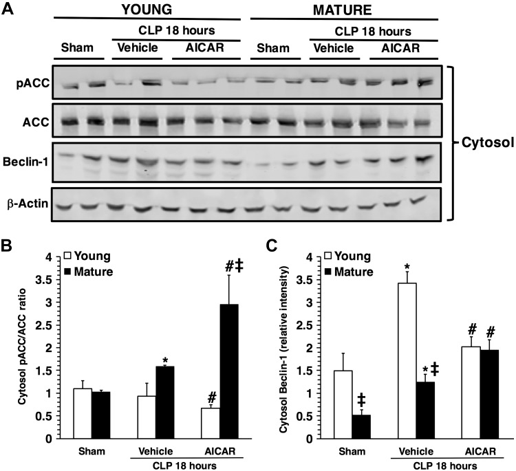
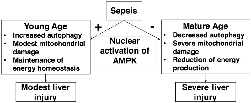
Age is an independent risk factor of multiple organ failure in patients with sepsis. However, the age-related mechanisms of injury are not known. AMPK is a crucial regulator of energy homeostasis, which controls mitochondrial biogenesis by activation of peroxisome proliferator-activated receptor-γ coactivator-α (PGC-1α) and disposal of defective organelles by autophagy. We investigated whether AMPK dysregulation might contribute to age-dependent liver injury in young (2-3 mo) and mature male mice (11-13 mo) subjected to sepsis. Liver damage was higher in mature mice than in young mice and was associated with impairment of hepatocyte mitochondrial function, structure, and biogenesis and reduced autophagy. At molecular analysis, there was a time-dependent nuclear translocation of the active phosphorylated catalytic subunits AMPKα1/α2 and PGC-1α in young, but not in mature, mice after sepsis. Treatment with the AMPK activator 5-amino-4-imidazolecarboxamide riboside-1-β-d-ribofuranoside (AICAR) improved liver mitochondrial structure in both age groups compared with vehicle. In loss-of-function studies, young knockout mice with systemic deficiency of AMPKα1 exhibited greater liver injury than did wild-type mice after sepsis. Our study suggests that AMPK is important for liver metabolic recovery during sepsis. Although its function may diminish with age, pharmacological activation of AMPK may be of therapeutic benefit.-Inata, Y., Kikuchi, S., Samraj, R. S., Hake, P. W., O'Connor, M., Ledford, J. R., O'Connor, J., Lahni, P., Wolfe, V., Piraino, G., Zingarelli, B. Autophagy and mitochondrial biogenesis impairment contribute to age-dependent liver injury in experimental sepsis: dysregulation of AMP-activated protein kinase pathway.

Keywords: AICAR; MODS; PGC-1α; cecal ligation and puncture.


Conflict of interest statement

The authors thank Dr. Benoit Viollet (INSERM and Cochin Institute, University Paris Descartes, Paris, France) for providing AMPK-α1 WT and KO mice. This work was supported by the U.S. National Institutes of Health (NIH) National Institute of General Medical Sciences Grant R01 GM-067202 (to B.Z.) and, in part, by NIH National Institute of Diabetes and Digestive and Kidney Diseases Grant P30 DK-078392 to the Digestive Research Core Center (Integrative Morphology Core). The content is solely the responsibility of the authors and does not necessarily represent the official views of the NIH. The authors declare no conflicts of interest.

Figures

Figure 1.
Figure 1.
Representative histology photomicrographs of liver sections. Normal liver architecture in control young [A, D (inset)] and mature [G, J (inset)] mice. Liver damage in vehicle-treated young (B) and mature (H) mice at 18 h after CLP with edema, inflammatory cell infiltration, and lipid vacuoles [E, K (insets)]. Amelioration of liver architecture in AICAR-treated young mice [(C, F (inset)]. Amelioration of liver architecture in AICAR-treated mature mice (I) with presence of small lipid vacuoles (L, inset) (n = 4–6 different tissue sections in each experimental group showed similar patterns). Original magnification: ×100 (AC, GI); ×400 (DF, JL).
Figure 2.
Figure 2.
Liver injury score (A), liver myeloperoxidase activity (B), plasma ALT levels (C), plasma AST levels (D) and liver ATP content (E) in young and mature mice subjected to CLP. Data are means ± sem (n = 5–19 mice for each group). Vehicle (distilled water) or AICAR (500 mg/kg) was administered i.p. at 1 and 6 h after CLP. *P < 0.05 vs. sham-treated mice of the same age; #P < 0.05 vs. vehicle-treated group of the same age; P < 0.05 vs. young group.
Figure 3.
Figure 3.
Representative histology photomicrographs of liver sections of young AMPKα1 WT and KO mice. Normal liver architecture in young WT (A, inset shown in E) and KO mice (C, inset shown in G). Liver damage in young WT (B) and KO (D) mice after sepsis with edema, necrosis, and inflammatory cell infiltration [F, H (insets)] (n = 4 different tissue sections in each experimental group showing a similar pattern). Original magnification: ×100 (AD); ×400 (EH).
Figure 4.
Figure 4.
Liver injury score (A), liver MPO activity (B), plasma ALT levels (C), plasma AST levels (D), and liver ATP content (E) in young AMPKα1 WT and KO mice subjected to CLP. Data represent means ± sem (n = 5–9 mice for each group). *P < 0.05 vs. sham-treated mice; P < 0.05 vs. WT mice. Survival rate of young AMPKα1 WT and KO mice subjected to CLP. Curves represent rate of 15 mice for each group. *P < 0.05 vs. vehicle-treated mice by χ2 test.
Figure 5.
Figure 5.
TEM of hepatocytes in sham-treated young (A) and mature (F) mice, in vehicle-treated young (B), and mature (G) mice at 6 h after CLP, in AICAR-treated young (C) and mature (H) mice at 6 h after CLP, in vehicle-treated young (D) and mature (I) mice at 18 h after CLP, in AICAR-treated young (E) and mature (J) mice at 18 h after CLP. Asterisks, damaged mitochondria presenting translucent matrix, disrupted membrane, and cristae; arrowheads, autophagic vesicles. N, nucleus; P, peroxisomes.
Figure 6.
Figure 6.
Quantification of damaged mitochondria (A) and autophagosomes (B) in hepatocytes in young and mature mice subjected to CLP. Damaged mitochondria and autophagosomes were determined by image-analysis software and expressed as a percentage of the total number of mitochondria in 9 consecutive cells. Data are means ± sem (n = 3–4 mice for each group. Western blot analysis of LC3B-I and -II and β-actin (used as the loading control) in liver cytosol extracts and image analyses of LC3B-II:LC3B-I ratio, as determined by densitometry (C). Data are means ± sem and are expressed as ratio of relative intensity units (n = 4–6 animals for each group). Citrate synthase activity in liver mitochondrial extracts (D). Data are means ± sem (n = 4–11 animals for each group). *P < 0.05 vs. age-matched control mice; #P < 0.05 vs. vehicle-treated group of the same age; P < 0.05 vs. young group.
Figure 7.
Figure 7.
A) Western blot analysis of pAMPKα1/α2, AMPKα1/α2, PGC-1α, and β-actin (used as the loading control) in liver cytosol and nuclear extracts. Image analyses of cytosol pAMPKα1/α2 (B), nuclear pAMPKα1/α2 (C), cytosol PGC-1α (D), and nuclear PGC-1α (E), as determined by densitometry. Data are means ± sem of 4–6 animals for each group and are expressed as relative intensity units. Expression of pAMPKα1/α2 was normalized toward total AMPKα1/α2 and β-actin. Expression of PGC-1α was normalized toward β-actin. *P < 0.05 vs. age-matched control mice; #P < 0.05 vs. vehicle-treated group of the same age; P < 0.05 vs. young group.
Figure 8.
Figure 8.
A) Western blot analysis of pACC, ACC, beclin-1 and β-actin (used as the loading control) in liver cytosol extracts. Image analyses of pACC/ACC ratio (B) and beclin-1 expression (C), as determined by densitometry. Data are means ± sem of 4–6 animals for each group and are expressed as relative intensity units. Expression of pACC was normalized toward total ACC and β-actin. Expression of beclin-1 was normalized toward β-actin. *P < 0.05 vs. age-matched control mice; #P < 0.05 vs. vehicle-treated group of the same age; P < 0.05 vs. young group.
Figure 9.
Figure 9.
A proposed overview of AMPK-dependent regulation of sepsis-induced liver injury in young and mature mice. During sepsis, AMPK is activated in the nucleus in young mice as a compensatory mechanism to maintain energy homeostasis and limit mitochondrial damage by increased autophagy. On the contrary, nuclear down-regulation of AMPK in mature mice is associated with energy failure and extensive mitochondrial damage with impaired autophagy resulting in severe liver damage.

References

    1. Singer M., Deutschman C. S., Seymour C. W., Shankar-Hari M., Annane D., Bauer M., Bellomo R., Bernard G. R., Chiche J. D., Coopersmith C. M., Hotchkiss R. S., Levy M. M., Marshall J. C., Martin G. S., Opal S. M., Rubenfeld G. D., van der Poll T., Vincent J. L., Angus D. C. (2016) The third international consensus definitions for sepsis and septic shock (sepsis-3). JAMA 315, 801–810 - PMC - PubMed
    1. Angus D. C., Linde-Zwirble W. T., Lidicker J., Clermont G., Carcillo J., Pinsky M. R. (2001) Epidemiology of severe sepsis in the United States: analysis of incidence, outcome, and associated costs of care. Crit. Care Med. 29, 1303–1310 - PubMed
    1. Milbrandt E. B., Eldadah B., Nayfield S., Hadley E., Angus D. C. (2010) Toward an integrated research agenda for critical illness in aging. Am. J. Respir. Crit. Care Med. 182, 995–1003 - PMC - PubMed
    1. GenIMS Investigators (2010) The effects of age on inflammatory and coagulation-fibrinolysis response in patients hospitalized for pneumonia. PLoS One 5, e13852. - PMC - PubMed
    1. Martin G. S., Mannino D. M., Moss M. (2006) The effect of age on the development and outcome of adult sepsis. Crit. Care Med. 34, 15–21 - PubMed

Publication types

MeSH terms

LinkOut - more resources