. 2017 Oct 3;8(5):e00976-17.
doi: 10.1128/mBio.00976-17.

Molecular and Physiological Logics of the Pyruvate-Induced Response of a Novel Transporter in Bacillus subtilis

Affiliations

Molecular and Physiological Logics of the Pyruvate-Induced Response of a Novel Transporter in Bacillus subtilis

Teddy Charbonnier et al. mBio. .

Abstract

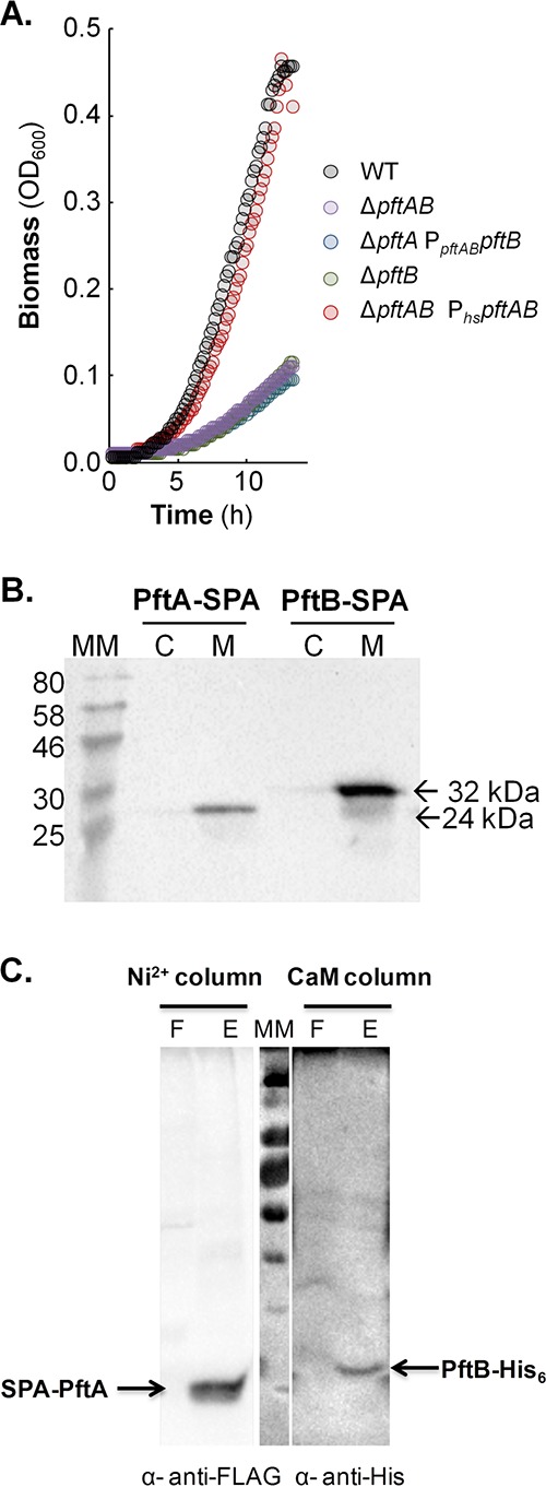
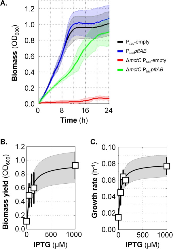
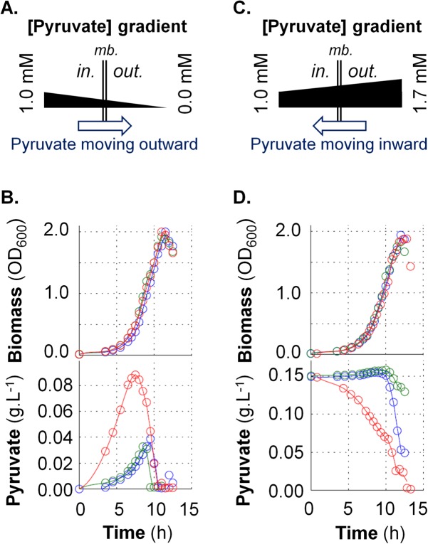
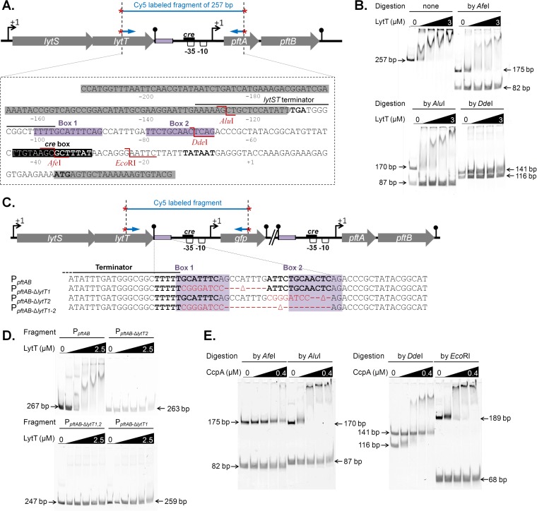
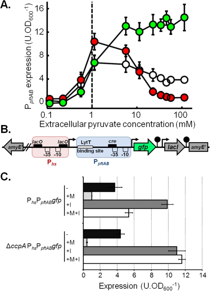
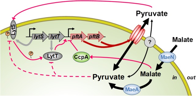
At the heart of central carbon metabolism, pyruvate is a pivotal metabolite in all living cells. Bacillus subtilis is able to excrete pyruvate as well as to use it as the sole carbon source. We herein reveal that ysbAB (renamed pftAB), the only operon specifically induced in pyruvate-grown B. subtilis cells, encodes a hetero-oligomeric membrane complex which operates as a facilitated transport system specific for pyruvate, thereby defining a novel class of transporter. We demonstrate that the LytST two-component system is responsible for the induction of pftAB in the presence of pyruvate by binding of the LytT response regulator to a palindromic region upstream of pftAB We show that both glucose and malate, the preferred carbon sources for B. subtilis, trigger the binding of CcpA upstream of pftAB, which results in its catabolite repression. However, an additional CcpA-independent mechanism represses pftAB in the presence of malate. Screening a genome-wide transposon mutant library, we find that an active malic enzyme replenishing the pyruvate pool is required for this repression. We next reveal that the higher the influx of pyruvate, the stronger the CcpA-independent repression of pftAB, which suggests that intracellular pyruvate retroinhibits pftAB induction via LytST. Such a retroinhibition challenges the rational design of novel nature-inspired sensors and synthetic switches but undoubtedly offers new possibilities for the development of integrated sensor/controller circuitry. Overall, we provide evidence for a complete system of sensors, feed-forward and feedback controllers that play a major role in environmental growth of B. subtilisIMPORTANCE Pyruvate is a small-molecule metabolite ubiquitous in living cells. Several species also use it as a carbon source as well as excrete it into the environment. The bacterial systems for pyruvate import/export have yet to be discovered. Here, we identified in the model bacterium Bacillus subtilis the first import/export system specific for pyruvate, PftAB, which defines a novel class of transporter. In this bacterium, extracellular pyruvate acts as the signal molecule for the LytST two-component system (TCS), which in turn induces expression of PftAB. However, when the pyruvate influx is high, LytST activity is drastically retroinhibited. Such a retroinhibition challenges the rational design of novel nature-inspired sensors and synthetic switches but undoubtedly offers new possibilities for the development of integrated sensor/controller circuitry.

Keywords: Bacillus subtilis; LytST; PftA PftB; YsbA YsbB; catabolite repression; malate; pyruvate transport; two-component regulatory systems.


Figures

FIG 1
FIG 1
Role and localization of pftAB. (A) Growth of the WT, ΔpftA PpftABpftB, ΔpftB, ΔpftAB, and ΔpftAB PhspftAB strains on M9P. (B) Cytoplasmic (C) versus membrane (M) localization of PftA-SPA (24 kDa) and PftB-SPA (32 kDa). Cells were grown in M9SE+P. Western blotting was performed using an anti-FLAG monoclonal antibody as the primary antibody and horseradish peroxidase-conjugated anti-mouse antibody as the secondary antibody. The positions of molecular mass markers (in kilodaltons) are indicated to the left of the gel. (C) Copurification of a N-terminal SPA-tagged PftA and of a C-terminal His-tagged version of PftB. The membrane fraction was first loaded onto a Ni2+ column to capture PftB-His6, and a Western blot using anti-FLAG antibodies revealed the presence of copurified SPA-PftA (F and E stand for flowthrough and eluate, respectively). The eluate was next loaded onto a CaM column to capture SPA-PftA, and a Western blot using anti-His antibodies revealed the presence of copurified PftB-His. A representative experiment is presented in each panel.
FIG 2
FIG 2
Functional complementation of the pyruvate uptake-deficient ΔmctC mutant of C. glutamicum by PftAB. (A) Growth in MM1 medium plus pyruvate (MM1+P) of the WT and ΔmctC C. glutamicum strains transformed with the empty pXMJ19 plasmid (Ptac-empty) or pXMJ19-pftAB plasmid (PtacpftAB, IPTG-inducible) in the presence of 1 mM IPTG. The 95% confidence intervals are shown by the shaded areas. (B) Biomass of the ΔmctC PtacpftAB strain upon entry into stationary phase after growth in MM1+P in the presence of different IPTG concentrations (0, 50, 100, 150, and 1,000 µM). (C) Growth rate of the ΔmctC PtacpftAB strain grown in MM1+P in the presence or absence of IPTG. In panels B and C, mean values ± standard deviations (error bars) from at least six independent experiments are presented. Data were fit with a Michaelis-Menten equation; the 95% confidence intervals of the fits are shown in gray. The data corresponding to the control strains are shown in Fig. S2 in the supplemental material.
FIG 3
FIG 3
Functional characterization of the PftAB pyruvate transporter in B. subtilis. (A and C) Schematic representation of a facilitated transport of pyruvate in the absence (A) or presence (C) of extracellular pyruvate. mb., membrane. (B and D) Growth of the B. subtilis WT (blue), ΔpftAB (green), and ΔpftAB PhspftAB (red) strains and corresponding concentrations of extracellular pyruvate. The results from a representative experiment are shown. (B) Cells were grown in M9G with 200 µM IPTG. (D) Cells were grown in M9G with 200 µM IPTG and 0.15 g ⋅ liter−1 pyruvate.
FIG 4
FIG 4
Mapping of the LytT and CcpA binding sites. (A) Genomic organization of the lytST pftAB region. At the top of panel A, the black box in the schematic representation indicates the putative binding site of CcpA. The violet boxes indicate the binding region of LytT. The AluI, DdeI, AfeI, and EcoRI restriction sites are indicated in red. (B) EMSAs using purified His6-LytT (0, 1, 1.5, 2, and 3 µM) and the Cy5-labeled PCR fragment of 257 bp (represented in panel A) either uncut or digested with AfeI, AluI, or DdeI. (C) The binding region of LytT is detailed for the unmodified (PpftAB), Box1-deleted (PpftABlytT1), Box2-deleted (PpftABlytT2), and Box1-Box2-deleted (PpftABlytT1,2) reporter strains. Black bold letters stand for the two putative LytT DNA binding sequences (Box1, Box2). The red letters indicate the DNA sequence that replaced the deleted region in each strain. The violet shaded regions are similar to the binding sites identified in E. coli for the LytT-like YpdA response regulator (35). (D) EMSAs using purified His6-LytT (0, 0.5, 1, 1.5, 2, and 2.5 µM) and the Cy5-labeled PCR fragment (represented in panel C) amplified from the PpftAB (267 bp), PpftABlytT1 (259 bp), PpftABlytT2 (263 bp), and PpftABlytT1,2 (247 bp) reporter strains. (E) EMSAs using purified His6-CcpA (0, 0.1, 0.2, 0.3, and 0.4 µM) and P-Ser-HPr (1:10 molar ratio) and the Cy5-labeled PCR fragment of 257 bp (represented in panel A) digested with AfeI (175  and 82 bp), AluI (170  and 87 bp), DdeI (141  and 116 bp), or EcoRI (189 and 68 bp).
FIG 5
FIG 5
Pyruvate influx tightly controls pftAB expression. (A) Expression of pftAB in the WT (white), ΔpftAB (green), and PhspftAB (red) strains grown in M9SE+P (at pyruvate concentrations ranging from 0.1 to 100 mM). The PhspftAB strain was grown with 1 mM IPTG. The dashed line represents the estimated Km of PftAB. (B) Genomic structure of the PhsPpftABgfp at the amyE locus. The red and blue boxes represent Phs and PpftAB, respectively. Black boxes indicate DNA binding sites: cre for CcpA and lacO for LacI. (C) Expression of PhsPpftABgfp in the WT and ΔccpA strains grown in M9SE+P in the presence (+) or absence (−) of malate (M) or 200 μM IPTG (I). Expression was estimated in the exponential phase of growth; mean values ± standard deviations from at least six experiments are presented in panels A and C.
FIG 6
FIG 6
Roles of PftAB and LytST in pyruvate homeostasis. The products of the pftAB operon form a hetero-oligomeric membrane complex encoding the major pyruvate import/export system in B. subtilis. The LytST TCS senses the extracellular pyruvate concentration and responds by inducing pftAB transcription. The accumulation of intracellular pyruvate (or of an intermediate of overflow metabolism) reduces the level of induction of pftAB via LytST. This accumulation results either from the uptake and metabolism of pyruvate or from the uptake of malate (by MaeN) and its consecutive transformation into pyruvate by the malic enzyme MaeA. Malate (and glucose) also triggers the catabolite repression of pftAB via CcpA. There is at least one other pyruvate transporter yet to be identified (gray filled circle). P, phosphate.

Comment in

References

    1. Bais HP, Weir TL, Perry LG, Gilroy S, Vivanco JM. 2006. The role of root exudates in rhizosphere interactions with plants and other organisms. Annu Rev Plant Biol 57:233–266. doi:10.1146/annurev.arplant.57.032905.105159. - DOI - PubMed
    1. Larsen PB, Degenhardt J, Tai CY, Stenzler LM, Howell SH, Kochian LV. 1998. Aluminum-resistant Arabidopsis mutants that exhibit altered patterns of aluminum accumulation and organic acid release from roots. Plant Physiol 117:9–18. doi:10.1104/pp.117.1.9. - DOI - PMC - PubMed
    1. Rudrappa T, Czymmek KJ, Paré PW, Bais HP. 2008. Root-secreted malic acid recruits beneficial soil bacteria. Plant Physiol 148:1547–1556. doi:10.1104/pp.108.127613. - DOI - PMC - PubMed
    1. Allard-Massicotte R, Tessier L, Lécuyer F, Lakshmanan V, Lucier JF, Garneau D, Caudwell L, Vlamakis H, Bais HP, Beauregard PB. 2016. Bacillus subtilis early colonization of Arabidopsis thaliana roots involves multiple chemotaxis receptors. mBio 7:e01664-16. doi:10.1128/mBio.01664-16. - DOI - PMC - PubMed
    1. Ahmad Z, Wu J, Chen L, Dong W. 2017. Isolated Bacillus subtilis strain 330-2 and its antagonistic genes identified by the removing PCR. Sci Rep 7:1777. doi:10.1038/s41598-017-01940-9. - DOI - PMC - PubMed

Publication types

MeSH terms

LinkOut - more resources