. 2017 Oct 3;8(1):777.
doi: 10.1038/s41467-017-00600-w.

Mechanistic understanding of in vivo protein corona formation on polymeric nanoparticles and impact on pharmacokinetics

Affiliations

Mechanistic understanding of in vivo protein corona formation on polymeric nanoparticles and impact on pharmacokinetics

Nicolas Bertrand et al. Nat Commun. .

Abstract

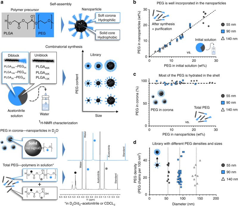
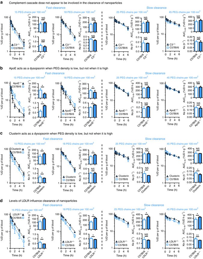
In vitro incubation of nanomaterials with plasma offer insights on biological interactions, but cannot fully explain the in vivo fate of nanomaterials. Here, we use a library of polymer nanoparticles to show how physicochemical characteristics influence blood circulation and early distribution. For particles with different diameters, surface hydrophilicity appears to mediate early clearance. Densities above a critical value of approximately 20 poly(ethylene glycol) chains (MW 5 kDa) per 100 nm2 prolong circulation times, irrespective of size. In knockout mice, clearance mechanisms are identified for nanoparticles with low and high steric protection. Studies in animals deficient in the C3 protein showed that complement activation could not explain differences in the clearance of nanoparticles. In nanoparticles with low poly(ethylene glycol) coverage, adsorption of apolipoproteins can prolong circulation times. In parallel, the low-density-lipoprotein receptor plays a predominant role in the clearance of nanoparticles, irrespective of poly(ethylene glycol) density. These results further our understanding of nanopharmacology.Understanding the interaction between nanoparticles and biomolecules is crucial for improving current drug-delivery systems. Here, the authors shed light on the essential role of the surface and other physicochemical properties of a library of nanoparticles on their in vivo pharmacokinetics.


Conflict of interest statement

O.C.F. and R.L. have financial interests in Tarveda Therapeutics, Selecta Biosciences and Placon Therapeutics. R.L. declares financial interests in Moderna. These biotechnology companies are developing nanoparticle technologies for medical applications. The remaining authors declare no competing financial interests.

Figures

Fig. 1
Fig. 1
Nanoparticles with different PEG densities and sizes prepared by combinatorial synthesis of PEG–PLGA and PLGA copolymers. a By self-assembly, polymer precursors form core-shell nanoparticles. Different sizes and PEG contents can be obtained by combining diblock and uniblock precursors. Analysis by 1H-NMR spectroscopy in different solvents can determine the PEG content in the nanoparticles and in the hydrated shell. b Most of the PEG used in the polymer precursor solution is incorporated in the nanoparticles. c For nanoparticles with diameters of 55, 90, and 140 nm, whose core is non-solvated (i.e., in D2O), most of the total PEG signal is detectable, suggesting that most of the hydrophilic polymer is hydrated in the shell. d Joining combinatorial synthesis and careful 1H-NMR characterization, a library of nanoparticles with different sizes and PEG densities can be prepared
Fig. 2
Fig. 2
A PEG density threshold of 20 PEG chains per 100 nm2 is necessary to avoid early clearance from the bloodstream. a The early circulation of nanoparticles with diameters of 55, 90, and 140 nm appears to be mainly affected by the density of PEG chains on their surface. Below a threshold around 20 PEG chains per 100 nm2, the nanoparticles are cleared rapidly; above this value, the nanoparticles exhibit roughly the same circulation profiles, irrespective of their diameter or extent of PEGylation. b Pharmacokinetic analysis of blood exposure (AUC0–6h) and elimination constants (k e) also highlight the presence of a threshold above which greater PEG coverage does not increase benefits on prolonging the circulation of nanoparticles. Values are means ± SD (n = 3–5); numbers in blue represent the PEG chains per 100 nm2 of surface
Fig. 3
Fig. 3
Circulation profiles in different mouse models highlight the role of different proteins on the clearance of nanoparticles. a For all nanoparticles, the circulation profiles between wildtype and C3−/− animals are similar. This suggests that the complement cascade is not involved in the clearance of nanoparticles, even those with very low steric protection and fast clearance. b The absence of ApoE accelerates the clearance of nanoparticles with fast intrinsic clearance. This suggests that when steric protection is low, interactions with ApoE prevent clearance-enhancing proteins from adsorbing on the nanoparticles. This effect is not observable for nanoparticles with higher PEG densities. c Similar to ApoE, pre-adsorption of clusterin on the surface of nanoparticles with low PEG densities decreases clearance. Clusterin does not appear to influence nanoparticles with slower intrinsic clearance rates. d In LDLR−/− animals circulation times are prolonged, with augmented blood exposures for all nanoparticles. This suggests direct involvement of LDLR on the clearance of nanoparticles. Values are means ± SD (n = 4–13). *p < 0.05 as determined by t-test, **p < 0.05 as determined by Mann–Whitney (non-parametric) test

References

    1. Tenzer S, et al. Rapid formation of plasma protein corona critically affects nanoparticle pathophysiology. Nat. Nanotechnol. 2013;8:772–781. doi: 10.1038/nnano.2013.181. - DOI - PubMed
    1. Monopoli MP, Aberg C, Salvati A, Dawson KA. Biomolecular coronas provide the biological identity of nanosized materials. Nat. Nanotechnol. 2012;7:779–786. doi: 10.1038/nnano.2012.207. - DOI - PubMed
    1. Saha K, et al. Regulation of macrophage recognition through the interplay of nanoparticle surface functionality and protein corona. ACS Nano. 2016;10:4421–4430. doi: 10.1021/acsnano.6b00053. - DOI - PMC - PubMed
    1. Akinc A, et al. Targeted delivery of RNAi therapeutics with endogenous and exogenous ligand-based mechanisms. Mol. Ther. 2010;18:1357–1364. doi: 10.1038/mt.2010.85. - DOI - PMC - PubMed
    1. Dong Y, et al. Lipopeptide nanoparticles for potent and selective siRNA delivery in rodents and nonhuman primates. Proc. Natl Acad. Sci. USA. 2014;111:3955–3960. doi: 10.1073/pnas.1322937111. - DOI - PMC - PubMed

Publication types

MeSH terms