. 2017 Dec;2(12):1648-1657.
doi: 10.1038/s41564-017-0029-y. Epub 2017 Oct 3.

Fluorescent D-amino-acids reveal bi-cellular cell wall modifications important for Bdellovibrio bacteriovorus predation

Affiliations

Fluorescent D-amino-acids reveal bi-cellular cell wall modifications important for Bdellovibrio bacteriovorus predation

Erkin Kuru et al. Nat Microbiol. 2017 Dec.

Erratum in

Abstract

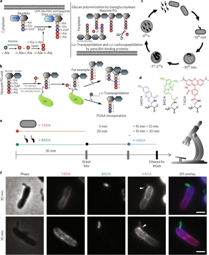
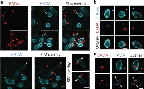
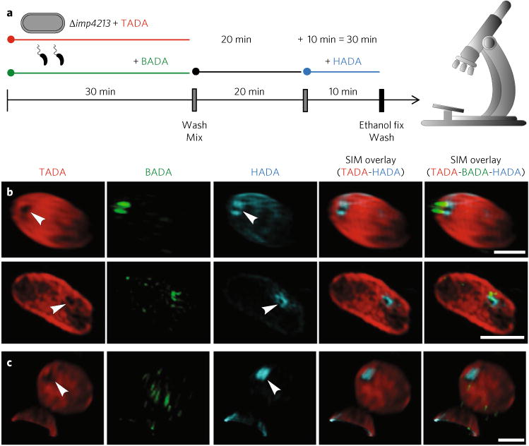
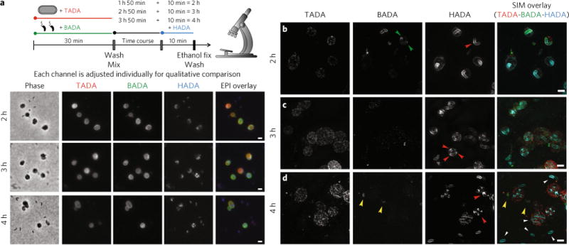
Modification of essential bacterial peptidoglycan (PG)-containing cell walls can lead to antibiotic resistance; for example, β-lactam resistance by L,D-transpeptidase activities. Predatory Bdellovibrio bacteriovorus are naturally antibacterial and combat infections by traversing, modifying and finally destroying walls of Gram-negative prey bacteria, modifying their own PG as they grow inside prey. Historically, these multi-enzymatic processes on two similar PG walls have proved challenging to elucidate. Here, with a PG-labelling approach utilizing timed pulses of multiple fluorescent D-amino acids, we illuminate dynamic changes that predator and prey walls go through during the different phases of bacteria:bacteria invasion. We show formation of a reinforced circular port-hole in the prey wall, L,D-transpeptidaseBd-mediated D-amino acid modifications strengthening prey PG during Bdellovibrio invasion, and a zonal mode of predator elongation. This process is followed by unconventional, multi-point and synchronous septation of the intracellular Bdellovibrio, accommodating odd- and even-numbered progeny formation by non-binary division.


Conflict of interest statement

Competing interests

The authors declare no competing financial interests.

Figures

Fig. 1
Fig. 1. Background and introduction to experimental procedures
a, Biosynthesis of PG starts in the cytoplasm by sequential addition of L-alanine, D-glutamic acid, a diamino acid and a dipeptide of D-alanine-D-alanine to disaccharide units. This subunit is then incorporated into the murein sacculus by glycan polymerization via transglycosylases. The D-alanine at position 5 can also be cleaved by the actions of D,D-carboxypeptidases. b, L,D-transpeptidases cleave the D-alanine from position 4 and utilize the energy from cleaving this bond to form a 3–3 crosslink with another acyl-acceptor stem peptide or replace the D-alanine with a free D-amino acid such as a fluorescent D-amino acid (FDAA). c, Timed stages of the predatory cycle of B. bacteriovorus (black) bacteria invading E. coli prey (grey). At 0–15 min post-mixing of B. bacteriovorus and prey, B. bacteriovorus attach and begin to enter the outer layers of the prey. At 30 min, most of the B. bacteriovorus have entered the prey periplasm, modifying the prey cell to form a rounded ‘bdelloplast’. At 1–3 h, B. bacteriovorus growth occurs at the expense of the prey cell contents in the form of elongation as a filament. At 4h, this filament fragments into smaller attack-phase cells that break out from the bdelloplast. d, FDAAs used in this study; colours are representative of emission maxima. e, Multi-coloured-FDAA labelling scheme with time points observed by wide-field epifluorescence microscopy. Predator and prey cells were pre-labelled separately with BADA and TADA, respectively, before being washed and then mixed. Samples of this mixed infection were then pulse-labelled with HADA for 10 min before each time point before being fixed, washed, and then microscopically observed. f, Phase contrast and epi-fluorescent microscopy images of the early stages of B. bacteriovorus predation The B. bacteriovorus are false-coloured in green, the E. coli prey cells are false-coloured in red and the pulsed HADA signal is false-coloured in blue. Each channel is displayed independently in white and with all three fluorescence channels merged in an epifluorescence (EPI) overlay. HADA fluorescence signal on the prey wall has an intense focus at each point of B. bacteriovorus contact and spreads from this point across the rest of the wall. Scale bars, 1 μm. The two images are representative of between 321 and 10,546 cells for each time point, detailed in Supplementary Table 1.
Fig. 2
Fig. 2. Three-dimensional structured illumination microscopy images of early predation by B. bacteriovorus (pre-labelled with BADA, false-coloured red) on prey E. coli cells after pulse labelling for 10 min with HADA (false-coloured cyan) to show early modification of cell walls
a, Predation 15min post-mixing reveals a ring of HADA-labelled prey cell wall modification at the point of B. bacteriovorus contact (arrowheads) and of similar width to the B. bacteriovorus cell (see Supplementary Table 2). Central pores in the labelled PG material can be seen where the B. bacteriovorus image is artificially removed from the overlay of the two channels. Such annuli may represent a thickened ring of PG modification. In the white inset, the lookup table for the BADA channel has been separately adjusted until all the BADA labelled predators are clearly visible. Three representative examples are displayed. b, Prey PG is deformed around the site of B. bacteriovorus invasion (arrowheads). c, The cells show HADA fluorescence at the end of the internal B. bacteriovorus cell (arrowheads), which probably represents transpeptidase activity re-sealing the hole in the prey PG after the B. bacteriovorus cell has entered. Images are representative of >100 3D-reconstructed cells in two independent experiments (Supplementary Table 2 for details of numbers analysed). Scale bars, 1 μm.
Fig. 3
Fig. 3. Three-dimensional structured illumination microscopy images of early predation by B. bacteriovorus (pre-labelled with BADA, false-coloured green) on prey E. coli imp4213 cells (which are more permeable and thus susceptible to the TADA pre-labelling, false coloured in red) after pulse labelling for 10min with HADA (false-coloured cyan) to show early modification of cell walls
a, FDAA labelling scheme (using excess B. bacteriovorus to promote synchronous invasion of E. coli Δimp4213 mutant prey) with time points observed by 3D-SIM fluorescence microscopy. Predator and prey cells were pre-labelled separately with BADA and TADA, respectively, before being washed and then mixed. Samples of this mixed infection were then pulse-labelled with HADA for 10min before the time points of 15 or 30min. The cells were then fixed, washed and microscopically observed. b, Predation 30 min post-mixing with this prey strain reveals a pore in the TADA signal coincident with the ring of HADA-labelled prey cell wall modification at the point of B. bacteriovorus contact (arrowheads) and of similar width to the B. bacteriovorus cell (Supplementary Table 3). c, In several cases (Supplementary Table 3) where the B. bacteriovorus cell had entered into the prey cell and established itself in the periplasm of the bdelloplast, the pore in the TADA was coincident with a patch of HADA labelling and thus is likely to represent the sealing of the pore through which the B. bacteriovorus had entered. Images are representative of two independent experimental repeats. Scale bars, 1 μm.
Fig. 4
Fig. 4. Quantitative and qualitative effects of two L,D-transpeptidases on prey cell wall modifications by FDAAs and their expression profiles
a, Plot of mean HADA fluorescent signal of cells against time throughout the predation cycle. Measurements are total mean background-corrected fluorescent signal from wild-type B. bacteriovorus cells (grey line), Δ2ldt mutant (yellow line) or invaded prey bdelloplast. Mean fluorescent signal was significantly lower in the bdelloplasts invaded by the Δ2ldt mutant (orange line) compared with those invaded by the wild type (blue line). Time is in minutes postmixing of predator and prey and fluorescence is in relative fluorescent units (RFU). Data are from at least two independent repeats (see Supplementary Table 1 for details of n). Error bars are s.e.m. The HADA signal differences between E. coli preyed on by WT or Δ2ldt mutant were significant in each of the time points (****p< 0.0001 for all time points except 240 min, for which *P= 0.016 by the Mann-Whitney test). b, RT-PCR showing the expression of the predicted L,D-transpeptidase genes bd0886 and bd1176 or the control gene dnaK, over the predatory cycle of B. bacteriovorus. L: 100 bp DNA ladder; AP: attack-phase cells; 15–45, 1h–4h: minutes or hours respectively since mixing of B. bacteriovorus and prey. Ec: E. coli S17-1 RNA (negative control: no B. bacteriovorus); NT, no-RNA control; Gen: B. bacteriovorus HD100 genomic DNA (positive control). The schematic diagram above represents the different stages of predation. Expression of both genes peaked at 15–30 min post-mixing predator and prey. Two independent repeats were carried out and showed the same transcription pattern. c,d, FDAA labelling of B. bacteriovorus wild-type HD100 (c) and Δ2ldt mutant (d) predation and bdelloplast establishment. The white arrowheads point to HADA modification of the bdelloplast and HADA polar foci visible on the mutant predators inside the bdelloplast. The B. bacteriovorus are false-coloured green, the E. coli prey cells are false-coloured red and the HADA pulse-labelling is false-coloured blue. HADA fluorescence of the prey cell during predation with the L,D-transpeptidase mutant is less than for predation by the wild type. Scale bars, 1 μm. Images are representative of five independent replicates for the wild-type and two independent replicates for the Δ2ldt mutant (see Supplementary Table 1 for details of n).
Fig. 5
Fig. 5. Plots showing HADA incorporation in the PG of prey E. coli mutants upon B. bacteriovorus predation and showing the damage by osmotic shock to bdelloplasts formed by B. bacteriovorus Ldt mutants
a, Chart of mean HADA fluorescent signal of prey strains preyed on by B. bacteriovorus (+Bd), and pulsed with HADA at 35–45 min post-mixing (the time point of maximal HADA incorporation for E. coli S17-1). Controls were in Ca/HEPES buffer without B. bacteriovorus predation, but pulsed with HADA at the same time point. Measurements are total mean background-corrected fluorescent signal of prey cells, reported in relative fluorescent units measured by MicrobeJ. Prey cells lacking all six L,D-transpeptidases (A6LDT) accumulated more HADA fluorescence following predation by B. bacteriovorus. Control samples without B. bacteriovorus predation accumulated considerably less HADA fluorescence. Controls of A6LDT prey cells without Bdellovibrio predation accumulated negligible HADA fluorescence. Data are from two (for the controls) or three independent repeats. Error bars are s.e.m. WT: E. coli BW25113 wild-type strain YB7421; A6LDT: E. coli BW25113 A6LDT strain deficient in all six L,D-transpeptidases; ΔdacA: E. coli BW25113 strain YB7423 deficient in dacΔ; Δ6LDTΔdacA: E. coli BW25113 Δ6LDTΔdacA strain YB7439 deficient in all six L,D-transpeptidases and dacA. NS, not significant; all other comparisons were significant P<0.0001, with the one exception shown, by the Mann-Whitney test. b, CPRG β-galactosidase assay measuring cytoplasmic leakage of shocked E. coli bdelloplasts formed by wild-type (E. coli + Bd WT) or bdelloplasts formed by Δ2ldt mutant B. bacteriovorus (E. coli + Bd Δ2ldt) with controls of uninvaded E. coli prey cells (E. coli alone) or B. bacteriovorus cells alone (Bd WT alone). Red colour from positive CPRG reaction was measured by spectrophotometry at 574nm and readings were normalized to each experiment. Bdelloplasts were harvested by centrifugation and shocked by resuspension in Ca/HEPES buffer for no shock, except centrifugation alone (Buffer), Ca/HEPES buffer supplemented with 750 mM NaCl (Upshock) or upshock followed by further centrifugation and resuspension in water (Downshock). Error bars are s.e.m. Statistical significance was determined by Student’s t-test (two-tailed) *P<0.05, **P< 0.01, ***P< 0.001. Data are the mean of seven independent repeats.
Fig. 6
Fig. 6. Epifluorescence and 3D-SIM images of the later stages of predation to show PG modification of the growing internal B. bacteriovorus
a–d, Phase-contrast (a) and epi-fluorescent microscopy and 3D-SIM (b–d) images of the later stages of B. bacteriovorus predation (after the peak of bdelloplast HADA labelling, by wild-type predator, has ended). The B. bacteriovorus were pre-labelled with BADA and are false-coloured in green; the E. coli prey cells were pre-labelled with TADA and are false-coloured in red. The cells were pulse-labelled for 10 min before each acquisition time point with HADA, which is false-coloured in cyan. Each channel is displayed independently and with all three fluorescence channels merged. The HADA fluorescence indicates synthesis of the B. bacteriovorus PG, which initiates at many points along the growing predator (2 h, b; red arrowhead) except the poles (2 h; b; green arrowheads), before developing into foci (3 h; c; red arrowheads), which become septa (4h; d; red arrowheads). After division, newly released B. bacteriovorus can be seen to modify their whole PG (4 h; d; white arrowheads). B. bacteriovorus that did not invade (there was an excess of B. bacteriovorus to ensure efficient predation) can be seen to have a strong BADA signal and low HADA signal (4h; d; yellow arrowheads). Images are representative examples from thousands of cells from five independent experiments (a) and of >100 3D-reconstructed cells in two independent experiments (b–d). See Supplementary Table 1 for numbers of cells analysed. Scale bars, 1 μm.

References

    1. Mainardi JL, et al. A novel peptidoglycan cross-linking enzyme for a β-lactam-resistant transpeptidation pathway. J Biol Chem. 2005;280:38146–38152. - PubMed
    1. Cava F, de Pedro MA, Lam H, Davis BM, Waldor MK. Distinct pathways for modification of the bacterial cell wall by non-canonical D-amino acids. EMBO J. 2011;30:3442–3453. - PMC - PubMed
    1. Magnet S, et al. Specificity of L,D-transpeptidases from Gram-positive bacteria producing different peptidoglycan chemotypes. J Biol Chem. 2007;282:13151–13159. - PubMed
    1. Fura JM, Kearns D, Pires MM. D-amino acid probes for penicillin binding protein-based bacterial surface labeling. J Biol Chem. 2015;290:30540–30550. - PMC - PubMed
    1. Gupta R, et al. The Mycobacterium tuberculosis protein LdtMt2 is a nonclassical transpeptidase required for virulence and resistance to amoxicillin. Nat Med. 2010;16:466–469. - PMC - PubMed

MeSH terms

LinkOut - more resources