. 2017 Oct 3;7(1):12567.
doi: 10.1038/s41598-017-12930-2.

Mechanism of mRNA-STAR domain interaction: Molecular dynamics simulations of Mammalian Quaking STAR protein

Affiliations

Mechanism of mRNA-STAR domain interaction: Molecular dynamics simulations of Mammalian Quaking STAR protein

Monika Sharma et al. Sci Rep. .

Abstract

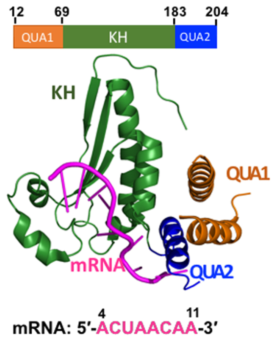
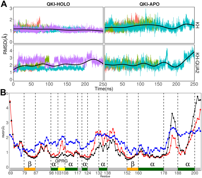
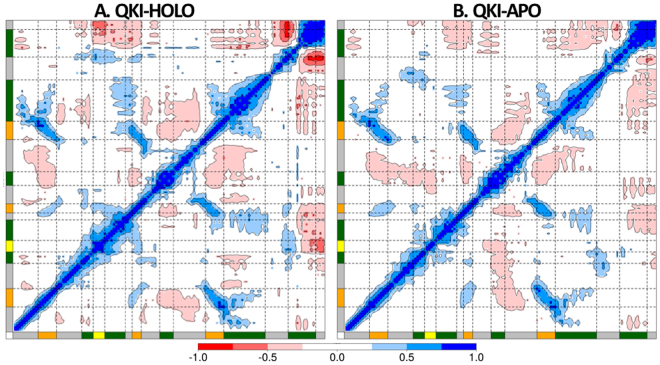
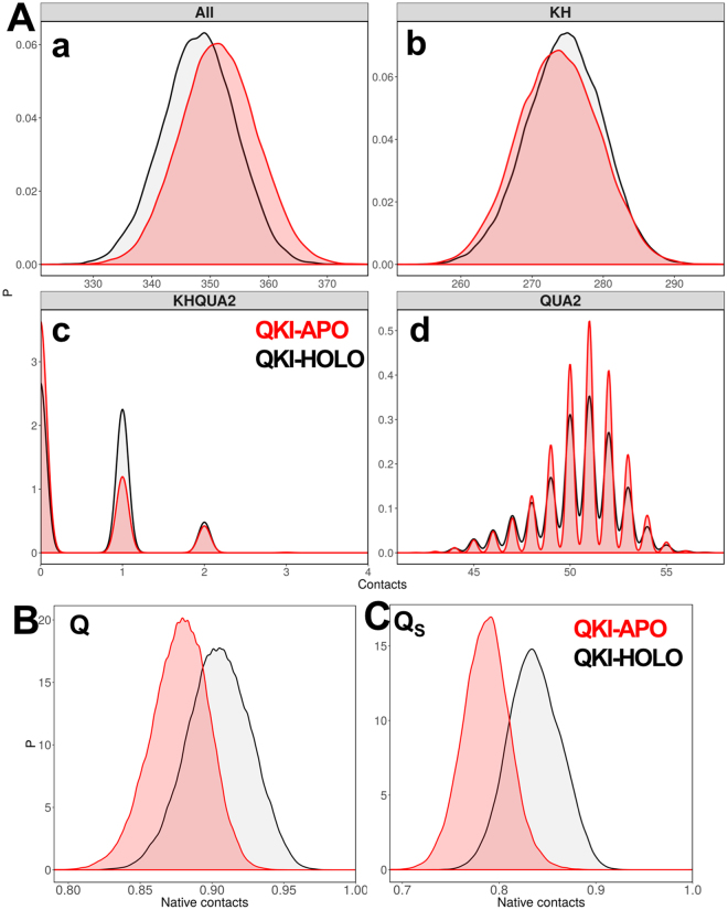
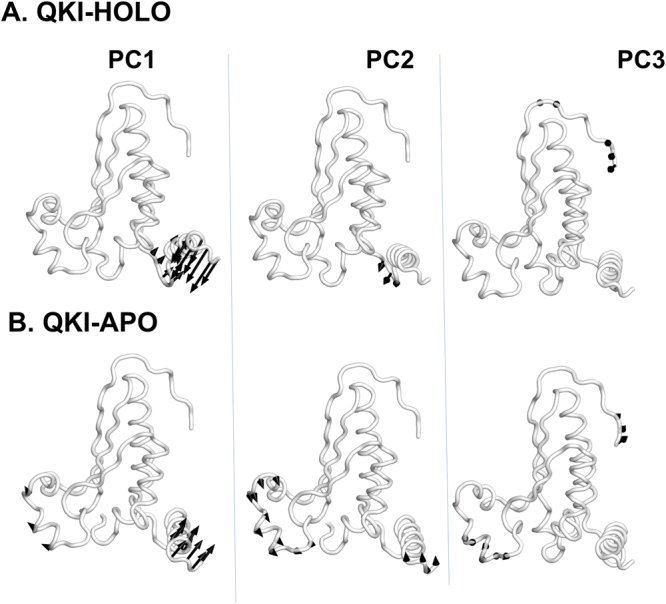
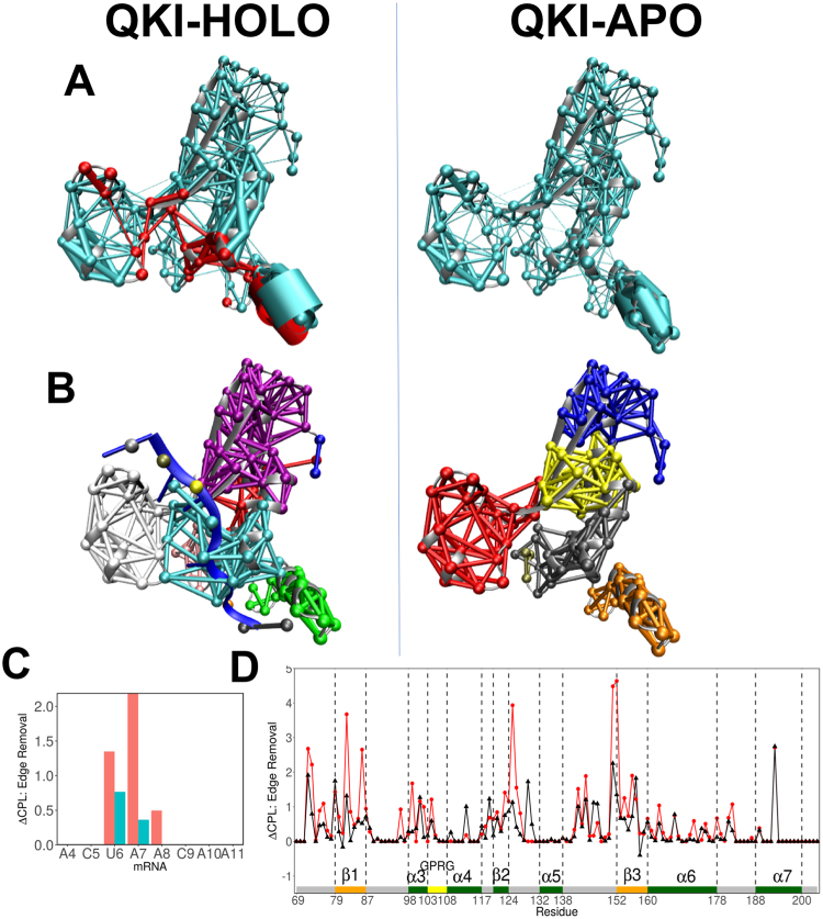
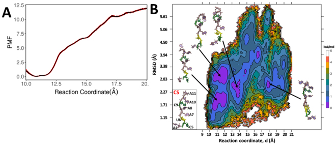
STAR proteins are evolutionary conserved mRNA-binding proteins that post-transcriptionally regulate gene expression at all stages of RNA metabolism. These proteins possess conserved STAR domain that recognizes identical RNA regulatory elements as YUAAY. Recently reported crystal structures show that STAR domain is composed of N-terminal QUA1, K-homology domain (KH) and C-terminal QUA2, and mRNA binding is mediated by KH-QUA2 domain. Here, we present simulation studies done to investigate binding of mRNA to STAR protein, mammalian Quaking protein (QKI). We carried out conventional MD simulations of STAR domain in presence and absence of mRNA, and studied the impact of mRNA on the stability, dynamics and underlying allosteric mechanism of STAR domain. Our unbiased simulations results show that presence of mRNA stabilizes the overall STAR domain by reducing the structural deviations, correlating the 'within-domain' motions, and maintaining the native contacts information. Absence of mRNA not only influenced the essential modes of motion of STAR domain, but also affected the connectivity of networks within STAR domain. We further explored the dissociation of mRNA from STAR domain using umbrella sampling simulations, and the results suggest that mRNA binding to STAR domain occurs in multi-step: first conformational selection of mRNA backbone conformations, followed by induced fit mechanism as nucleobases interact with STAR domain.


Conflict of interest statement

The authors declare that they have no competing interests.

Figures

Figure 1
Figure 1
Crystal structure of monomer of QKI STAR-RNA complex (PDBid: 4JVH). Domain architecture of QKI domain is shown, consisting of N-terminal QUA1 domain (in orange), mRNA-binding KH-domain (in green), and C-terminal QUA2 domain (in blue). RNA target is shown in magenta.
Figure 2
Figure 2
(A) Time-evolution of RMSD of STAR domain of QKI protein in presence of mRNA (QKI-HOLO state) and in absence of mRNA (QKI-APO state). RMSD values during independent runs are shown in varied colors, with averages shown in black thick line. (B) RMSF for C atoms of KH-QUA2 domain of QKI protein averaged over all the simulation runs. Data with black circles and black lines is for HOLO state, red colored triangles with red lines is for APO state, and blue colored squares with blues lines correspond to converted B-factors for crystal structure (4JVH). Secondary structure elements: alpha helices, beta sheets and loops are annotated with boxes of green color, orange color and grey color. ‘GPRG’ motif is annotated with yellow colored box.
Figure 3
Figure 3
Dynamic cross correlation maps calculated as time-averaged for Cα atoms of KH-QUA2 domain of QKI protein (A) in presence of bound mRNA, QKI-HOLO and (B) in absence of mRNA, QKI-APO. Secondary structure elements are shown as in Fig. 2.
Figure 4
Figure 4
(A) Distribution of native contacts within STAR domain of QKI protein during the simulations (a) within maxi KH (KH-QUA2) domain, (b) within KH domain only, (c) between KH domain and QUA2 domain, and (d) within QUA2 domain. Probability distribution functions for (B) fraction of native contacts, Q and (C) similarity of natively contacting residue pairs to their native distances, Qs for both HOLO (in black) and APO (in red) states.
Figure 5
Figure 5
Motions along the first three principal components are shown for (A) mRNA bound HOLO and (B) mRNA free APO state.
Figure 6
Figure 6
(A) Networks, weighed by correlation data, within KH-QUA2 domain of QKI protein is shown for mRNA-bound HOLO state and mRNA free APO state. Red colored nodes and edges correspond to interactions within 8 Å of mRNA. (B) Splitting of the networks into different communities are shown. (C) Change in CPL upon edge removal of nucleotide is shown for mRNA. Red and blue colored bars correspond to two nodes representing nucleotide base and sugar-phosphate backbone, respectively. (D) Change in CPL upon edge removal shown for KH-QUA2 domain for mRNA bound HOLO state (black colored) and mRNA free APO state (red colored). Structural elements are depicted as in Fig. 2B.
Figure 7
Figure 7
(A) Spatial organization of various mutations (reported for STAR proteins) in QKI protein. (B) Time evolution plots depicting interactions between mRNA nucleotide and its amino acid partner in STAR domain. Here interactions present in crystal structure are considered. Different colors indicate four different runs for HOLO state.
Figure 8
Figure 8
(A) 1-D Potential of mean force (PMF) profile along the biased reaction coordinate as obtained from 15 umbrella sampling simulations within range of 10.5–20 Å. Errors in the PMF profile are shown in red. (B) Two-dimensional free energy profile along the biased reaction coordinate, d and rmsd values determined from umbrella sampling simulations. The representative structures are shown for each minima observed by violet-colored islands. Crystal structure of bound mRNA is shown for comparison.

Similar articles

Cited by

References

    1. Vernet C, Artzt K. STAR, a gene family involved in signal transduction and activation of RNA. Trends Genet. 1997;13:479–484. doi: 10.1016/S0168-9525(97)01269-9. - DOI - PubMed
    1. Arning S, Grüter P, Bilbe G, Krämer A. Mammalian splicing factor SF1 is encoded by variant cDNAs and binds to RNA. RNA. 1996;2:794–810. - PMC - PubMed
    1. Liu Z, et al. Structural basis for recognition of the intron branch site RNA by splicing factor 1. Science. 2001;294:1098–1102. doi: 10.1126/science.1064719. - DOI - PubMed
    1. Matter N, Herrlich P, König H. Signal-dependent regulation of splicing via phosphorylation of Sam68. Nature. 2002;420:691–695. doi: 10.1038/nature01153. - DOI - PubMed
    1. Reddy TR, et al. Inhibition of HIV replication by dominant negative mutants of Sam68, a functional homolog of HIV-1 Rev. Nat. Med. 1999;5:635–642. doi: 10.1038/9479. - DOI - PubMed

Publication types

LinkOut - more resources